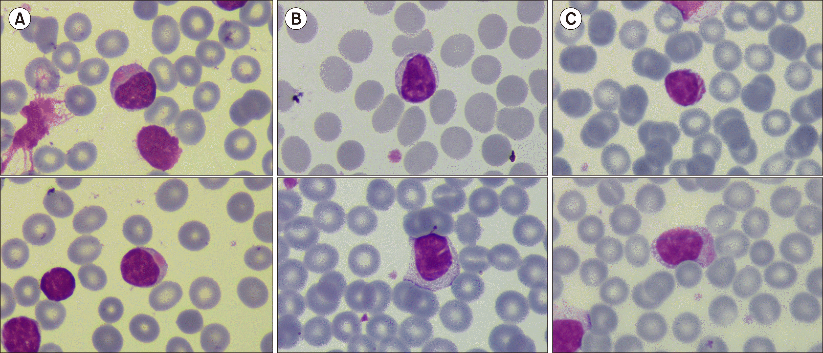
(A) Globular inclusions in peripheral blood lymphocytes in a 67-year-old woman with 5-year history of CLL managed with a “watch and wait” approach. Other findings included increased lymphocyte count of 270×109/L, hemoglobin of 110 g/L, and platelets of 78×109/L. Serum lactate dehydrogenase level was normal. Positron emission tomography/computed tomography revealed multiple adenopathies with low SUV and no evidence of transformation. (B) Multiple crystalline inclusions in peripheral blood lymphocytes in a 60-year-old asymptomatic woman with axillary lymphadenopathies. Laboratory evaluations revealed the following: hemoglobin of 132 g/L, platelets of 212×109/L, and lymphocytes of 3.7×10⁹/L. B-cells revealed clonal CLL phenotype on flow cytometry. Lymph node biopsy revealed lymphoid infiltrate with low Ki67 and positivity for CD20, CD23, and CD5. A diagnosis of SLL was established. (C) Azurophilic needle-shaped inclusions (“Auer rod-like”) in a 45-year-old asymptomatic man with the following laboratory parameters: hemoglobin of 163 g/L, platelets of 287×109/L, and lymphocytes of 10.08×10⁹/L. These atypical lymphocytes had CLL phenotype (positive for CD19, CD20, CD5, CD23, CD200, and CD43; negative for CD10 and kappa and lambda surface chains) on flow cytometry (all images, May–Grünwald Giemsa staining, ×100). The presence of inclusions is an uncommon finding in B-cell lymphoproliferative disorders; however, they can aid in arriving at the diagnosis. They are more frequently described in CLL with an approximate incidence of 5%. On light microscopy, the inclusions may have different forms and density. They may appear at diagnosis or during disease evolution.


 PDF
PDF Citation
 Print
Print


 XML Download