Journal List > Korean J Orthod > v.52(6) > 1516080231

Mattos, Gonçalves, Basso, Ferraz, Zeigelboim, Stechman-Neto, Santos, Araujo, and Guariza-Filho: Periodontal parameters in orthodontically tractioned teeth: A systematic review and meta-analysis

Abstract

Objective

This systematic review aimed to evaluate periodontal parameters in orthodontically tractioned teeth compared with the respective non-tractioned contralateral teeth.

Methods

Search strategies were developed for six electronic databases and gray literature. Random-effects meta-analyses were performed for the outcomes of interest. Furthermore, the certainty of the evidence was assessed using the GRADE (Grading of Recommendations Assessment, Development, and Evaluation) tool.

Results

Overall, 2,082 articles were identified, of which 24 were selected for the qualitative synthesis. A significant difference was observed between the impacted and contralateral teeth (mean difference [MD] = 0.25; 95% confidence interval [CI] = 0.10–0.40; I2 = 0%) when the gingival index was evaluated. Additionally, impacted teeth showed a greater probing depth, with a significant mean difference between the groups (MD = 0.14; 95% CI = 0.07–0.20; I2 = 6%). Most studies had a low risk of bias; however, the certainty of the evidence was very low owing to the design of existing studies.

Conclusions

The evidence in the literature indicated that tractioned teeth might show worsening of periodontal parameters related to the gingival index and probing depth; however, the evidence remains uncertain about this outcome. Furthermore, probing depth should be considered regarding its clinical significance because of the small effect size observed.

INTRODUCTION

Dental anomalies such as ectopic eruptions can be observed during the diagnosis of orthodontic patients.1 Notably, the lack of early detection of impacted teeth can cause severe damage, such as resorption of adjacent teeth, aesthetic problems, and periodontal changes that can lead to the loss of dental element.2,3 Some orthodontic mechanisms can be used for spontaneous eruption; however, if this does not occur even after space is acquired, orthodontic traction can be employed.4 The function of orthodontic treatment involves aligning the impacted tooth on the dental arch, reducing potential periodontal complications, and maintaining the integrity of supporting tissues.5 This loss of support is associated with forming periodontal pockets, which can cause gingival recession.6
Dental impaction is a condition that affects between 0.8 and 3.6% of the general population,7,8 and it is estimated that in the age group of 15–19 years, excluding third molars, 1.65% of patients have an impacted tooth.9 The most recurrent impacted teeth are the third molars (16.7%), upper canines (2.8%), and lower premolars and upper central incisors (2.6%), which are more frequent in females.9-13 The principal causes of impactions are related to primary factors, including the degree of root resorption of the deciduous tooth, trauma to the tooth germ, changes following an eruption, reduced space in the dental arch, rotation of the impacted tooth, and premature closure of the root apexes. Additionally, secondary factors are associated with the causes of impactions, such as abnormal muscle pressure, vitamin D deficiency, endocrine disturbances, and fever.6
Periodontal status is an indicator of effectiveness in the treatment of impacted teeth;14 however, some complications may be observed following treatment, including gingival recession, periodontal disease, gingival inflammation, alveolar bone loss, and/or inserted gingiva.15 The anatomical structure of the soft tissue overlying the impacted tooth is one of the main factors determining whether the surgical exposure method will work, as the orthodontic-surgical treatment should trigger a natural eruption pattern of the impacted tooth through the gum inserted.16 The correction of this disorder in the posterior or anterior teeth is vital for establishing correct occlusion. The development of malocclusion can even generate functional asymmetry during mastication.17 Posterior teeth are of fundamental importance in crushing the food bolus; similarly, adequate occlusal contact in the anterior region is essential for efficient mastication.18 Early detection of impacted teeth can help prevent the establishment of malocclusion, thus aiding occlusal and aesthetic functional gain.19 Therefore, it is crucial to understand the behavior of the periodontal support structures of impacted teeth after orthodontic traction.
A meta-analysis evaluated periodontal aspects of probing depth and gingival recession between palatally impacted and contralateral canines and found no statistical difference.20 However, to date, no systematic review has been found that addressed periodontal parameters involving various groups of traction teeth and evaluated whether there is heterogeneity between the findings according to the group of teeth included in the analysis. Since teeth other than canines may be affected by impaction, it is justified to conduct a systematic review of the subject. Therefore, the objective of this systematic review was to evaluate the periodontal parameters in orthodontically tractioned teeth when compared with the respective non-tractioned contralateral teeth.

MATERIALS AND METHODS

This systematic review was performed according to the Preferred Reporting Items for Systematic Reviews and Meta-Analysis (PRISMA) checklist.21 The study protocol was registered on the PROSPERO website (International Prospective Register of Systematic Review, Center for Reviews and Dissemination University of York, CRD42020205104).

Eligibility criteria

To consider the eligibility of studies to be included or excluded from this review, the acronym “PICOS” was employed to answer the following focused question: "What are the periodontal parameters in orthodontically tractioned teeth?"
  • Population (P) – Patients with impacted teeth

  • Intervention (I) – Orthodontic traction

  • Comparison (C) – Teeth with normal eruption (contralateral)

  • Outcomes (O): Periodontal parameters (gingival recession, probing depth, plaque index, gingival index, and width of keratinized mucosa)

  • Study design (S): Randomized clinical studies, pseudo-randomized or non-randomized, cross-sectional observational studies, and cohort or case-control

Inclusion criteria

Studies in which the sample comprised patients undergoing treatment for orthodontic-surgical traction of impacted teeth regardless of the tooth group were included. There were no restrictions on the malocclusion type, sex, or age. The study language or publication time also had no restrictions.
Additionally, studies that evaluated at least one of the following periodontal parameters were included: gingival recession, probing depth, periodontal attachment level, plaque index, gingival index, gingival bleeding index, and width of the keratinized mucosa.
  • a) Gingival recession: distance between the cementoenamel junction (CEJ) and the gingival margin, where the gingival margin found apically at the CEJ is positive, and the gingival margin coronally at the CEJ is negative.

  • b) Probing depth: periodontal pockets were measured from the level of the free gingival margin to the bottom of the pocket.

  • c) Periodontal attachment level: measurement of the probing depth and the distance between the gingival margin and the CEJ. In cases where the gingival recession was present, the clinical attachment level was calculated as follows: clinical attachment level = depth of periodontal probing + gingival recession.

  • d) Plaque index: the tooth surfaces were classified with a score between 0 and 3, according to the method described by Silness and Löe.22

  • e) Gingival index: the tooth surfaces were classified with a score between 0 and 3, similar to the plaque index.22

  • f) Gingival bleeding index: the presence or absence of bleeding was checked after periodontal probing using the method described by Carter and Barnes.23

  • g) Width of keratinized mucosa: measurement of the distance between the gingival margin and mucogingival junction.

Moreover, this study included randomized and non-randomized controlled clinical trials, cohort, case-control studies, and cross-sectional studies.

Exclusion criteria

The following exclusion criteria were used:
  • Studies on animals or patients with associated syndromes.

  • Studies in which tractioned tooth or a tooth with normal eruption underwent surgical periodontal treatment.

  • Studies in which at least one of the periodontal parameters stated above was not evaluated.

  • Reviews, letters, conference abstracts, expert opinions, case reports, and case series.

Information sources and search strategy

Appropriate word combinations and truncations were selected and modified for each database search. Search strategies were developed specifically for each of the following electronic databases: PubMed/Medline, LILACS, Scopus, Web of Science, Embase, and Cochrane Library. Additionally, a search of grey literature through Google Scholar, Proquest, and Open Grey was conducted (Supplementary Table 1).
Finally, a manual search of the references of the included studies was performed, and an expert on the subject was consulted to verify any possible publications on the subject. Duplicated references were managed and eliminated using the EndNote® software (EndNote® X7; Thomson Reuters, Philadelphia, PA, USA).

Selection process

Articles were selected in two phases. In phase 1, two reviewers independently reviewed the titles and abstracts of all the references, and those that did not meet the inclusion criteria were excluded. In phase 2, the same reviewers independently read the selected articles. A third author was involved in the final decision in a case of disagreement that could not be resolved through discussion between the first and second reviewers.
Furthermore, to ensure blind reading of references and guarantee independence and confidentiality in both phases, the Rayyan website (http://rayyan.qcri.org) was used, where the reviewers were blinded in all evaluations and a member of the team, who was not involved in the selection process, served as a moderator.

Data collection process

Two reviewers collected and discussed information from the included studies. The data collected included study characteristics (authors' names, year of publication, country, and study design), population characteristics (sample size and age), evaluation characteristics (composition of control and experimental groups, surgical technique, number of evaluators, parameters of interest, outcome assessment method, and index used for assessment), characteristics of the results (results presented relating to the outcome), and the main conclusions.
Additionally, if any data were missing or incomplete, attempts were made to contact the authors to obtain pertinent unpublished information. Three attempts were made to contact the article’s first, corresponding, and last authors, with a one-week interval between each attempt. The article was appropriately excluded in the absence of a response.

Data items

The mean, standard deviation, and sample size values for each group were extracted from the studies included in the synthesis when they were available. For the probing depth variable, in the absence of the mean value, the mean value between the measured sites was calculated when more than one site was measured. In cases when the standard deviation values were not reported and there was no description of any measure of variability that would allow its calculation, the value based on the study with the highest variance within the analysis was then imputed; thus producing a more conservative result since it decreased study weight and generated a broader confidence interval.

Assessment of risk of bias

The methodology of the selected observational studies was assessed using the risk of bias tool Meta-Analysis of Statistics Assessment and Review Instrument (MASTARI).24 The risk of bias was categorized as “high,” “moderate,” and “low” when the study had a “yes” score greater than 49%, between 50% and 69%, and more than 70%, respectively, for the bias risk questions.
Additionally, the tool “Cochrane Collaboration tool for assessing the risk of bias” was used for the interventional studies.25 This tool covered seven domains: random sequence generation, allocation concealment, blinding of participants and professionals, blinding of outcome evaluators, incomplete outcomes, selective outcome reporting, and other biased sources. Based on the information extracted from the study, an assessment of the potential risk of bias in each of these domains was made, and they were categorized as either “high risk” or “low risk” of bias. If insufficient details were reported in the study, the risk of bias was judged to be “not clear,” and the authors of the original research were contacted for more information. These assessments were performed by two independent reviewers. Notably, disagreements were first resolved by discussion, and in the case of non-consensus, a third reviewer was consulted for the tie-breaking vote.

Effect measurement

Since the analyzed outcomes were continuous and presented using the same measurement scale, the difference between means (MD) was determined.

Synthesis method

The studies were weighted using the inverse variance method and a random-effects meta-analysis approach using the statistical software RStudio version 1.2.1335 (Rstudio Inc., Boston, MA, USA) and Stata version 16.0 (Stata Corp LLC, College Station, TX, USA) was performed. The DerSimonian–Laird method was used to estimate the inconsistency index (I2) and the variance by Tau2 to determine heterogeneity. Additionally, 95% confidence intervals (95% CI) were generated, with the significance level set at 5%, and prediction intervals of 95% (95% PI) were calculated to evaluate the influence of heterogeneity of the analysis on interval estimates.
When extreme effect sizes were observed as a source of heterogeneity within the analysis, the leave-one-out method was then performed, recalculating the global effect estimate k - 1 times, with the respective CI and I2 values, omitting one study at a time, thus assessing whether the influence of any study distorted the combined effect estimate.26

Reporting bias assessment

A graphic evaluation of the existence of publication bias was performed using a funnel plot and the Egger test to assess the presence of asymmetry in the funnel. Additionally, sensitivity analysis was conducted for analyzes that included studies categorized as having a high risk of bias.

Certainty of evidence

The certainty of the evidence was evaluated using the Grading of Recommendations Assessment, Development, and Evaluation (GRADE) tool27 across all domains of risk of bias, consistency, openness, accuracy, and publication bias. The certainty of the evidence was rated as high, moderate, low, or very low.

RESULTS

Study selection

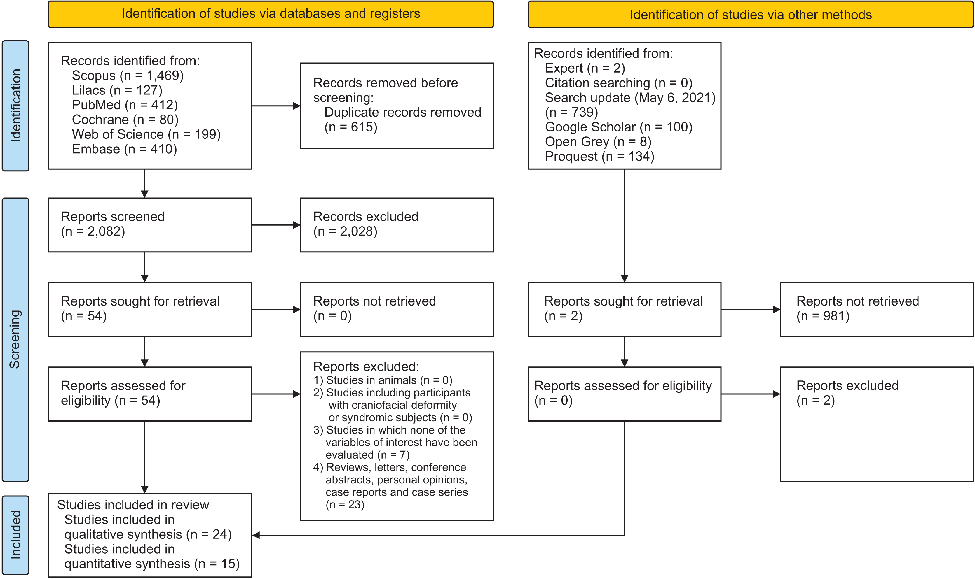
A search was conducted in the databases through a complex research strategy, totaling 2,697 articles. Overall, 2,082 articles were selected for the title and abstract reading after excluding duplicate articles (phase 1). Of these articles, 54 were selected for full reading (phase 2), of which 30 were excluded (Supplementary Table 2), resulting in 24 articles to carry out the qualitative synthesis (Figure 1).

Study characteristics

Among the articles included, 23 were published in English and only one in Portuguese, with the following countries as sources: Brazil, Belgium, Canada, Korea, Spain, Israel, Italy, Lithuania, the United Kingdom, Sweden, and Turkey, with the year of publication ranging from 1978 to 2019.
Furthermore, the sample sizes ranged from 11 to 271 patients, while ages ranged from 11 to 52 years. The included studies that reported the sex of the samples showed a predominance of the female sample.
Regarding the study design, 19 articles were classified as observational because of the absence of manipulation of the exposure factor, of which three longitudinal studies were a prospective cohort, and 16 were cross-sectional. In addition, five clinical trials were included.
Twenty-one articles evaluated specific groups of dental elements treated orthodontically and subjected to traction. Of these, 18 and three studies assessed the upper canines and upper incisors, respectively; three articles evaluated the following groups of teeth: maxillary incisors and canines, mandibular canines and premolars, canines and second premolars, and maxillary incisors and canines.
Moreover, regarding the type of surgical technique used in the articles, 12 and six articles used the closed and the open techniques, respectively, and six articles compared the closed and open techniques. Supplementary Table 3 presents all characterization data from the included studies.

Risk of bias

Among the 19 observational studies included, 12 had a low risk of bias, five articles had a moderate risk of bias,28-32 and two had a high risk of bias.33,34 Of the 5 interventional studies included, two articles presented more than 50% of the domains assessed, which were at high risk or lacking information that would provide adequate judgment (Supplementary Table 4).6,35

Results of individual studies

The probing depth was significantly higher in the tractioned teeth when considering only the periodontal parameters of the upper incisors.28,36,37 However, there was no difference in the gingival and plaque indices (p > 0.05). Only one study reported higher values for gingival and plaque indices in the central incisors under traction.36
Similarly, when considering the maxillary canines,6,14,29-31,33,35,38-43 the pulled teeth showed greater probing depth14,29,41-44 in at least one face of the tooth. However, the literature showed divergences in this outcome, with studies indicating no differences observed between these teeth when compared to contralateral ones,38 or that there was only a difference in this variable on the surfaces of teeth adjacent to the canine.6 There was a predominance of non-significant differences between the groups of treated and untreated canines for plaque, gingival bleeding, and gingival recession parameters. However, only one study showed a significant difference in plaque and gingival bleeding rates, which were higher in the treated group.41 Moreover, there was a discrepancy in the literature regarding the width of keratinized mucosa, of which some studies showed lower values in treated canines14,30,31,39,41 or even larger,40 and others showed no difference between groups.14,43
Furthermore, in the studies that evaluated mixed groups of teeth, there was a predominance among the included studies regarding the non-statistical significance of periodontal parameters, plaque index, and gingival index,9,32 and the probing depth and gingival recession parameters had higher values in the treated group.32

Synthesis of results

The quantitative synthesis included 15 studies, making evaluating the following outcomes possible: plaque index, gingival index, keratinized tissue width, and periodontal probing depth.
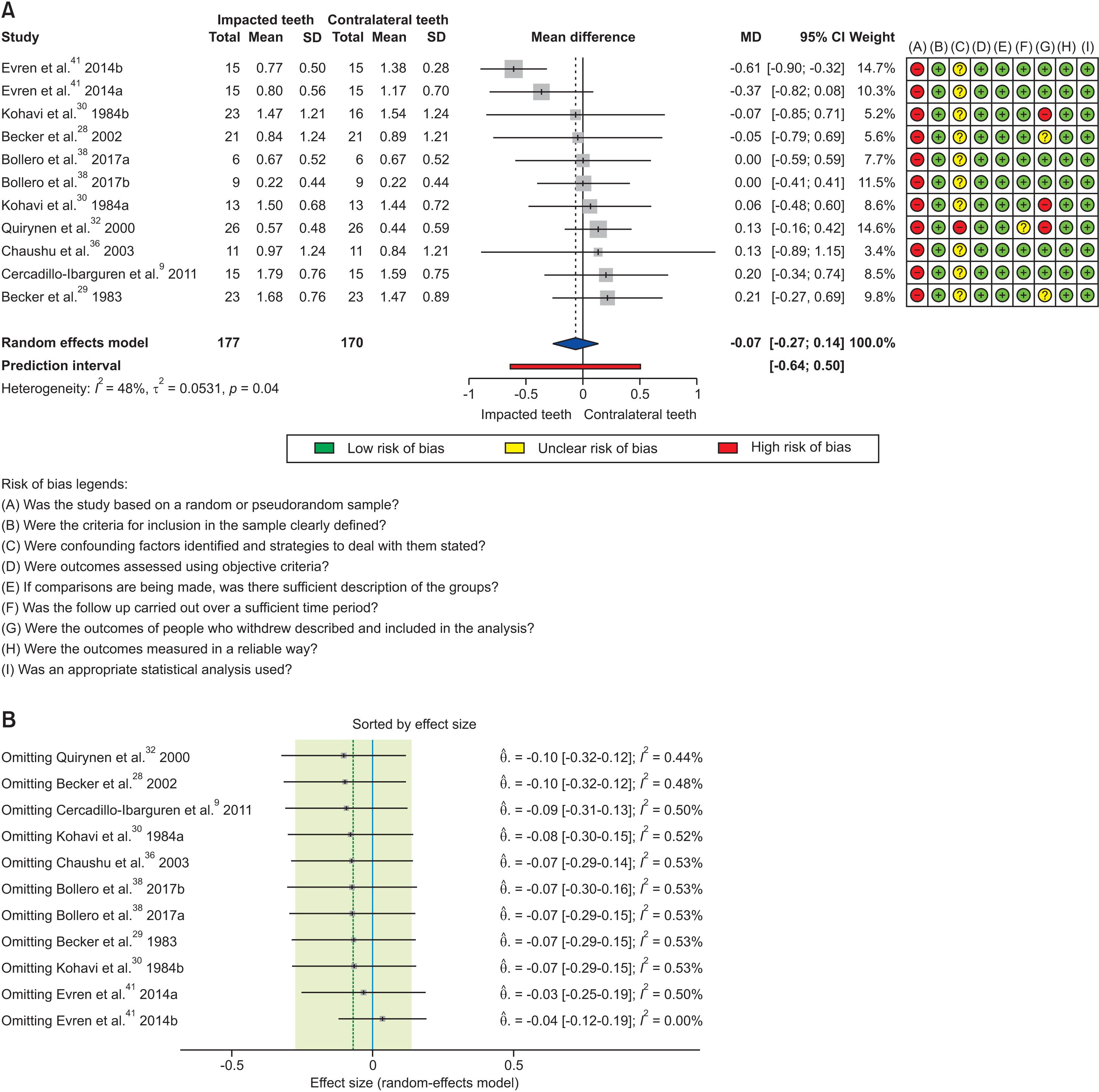
Dental impaction and plaque index showed no correlation (MD = –0.07; 95% PI = –0.64 to 0.50; I2 = 48%), and no difference was observed in the plaque index between the impacted teeth and the contralateral teeth (Figure 2A). Additionally, through the influence analysis, it was possible to verify that the study by Evren et al.41 was responsible for the observed heterogeneity (I2 = 48%); however, even after excluding this study from the analysis, the values remained without significance (p > 0.05) (Figure 2B), indicating the robustness of the analysis.
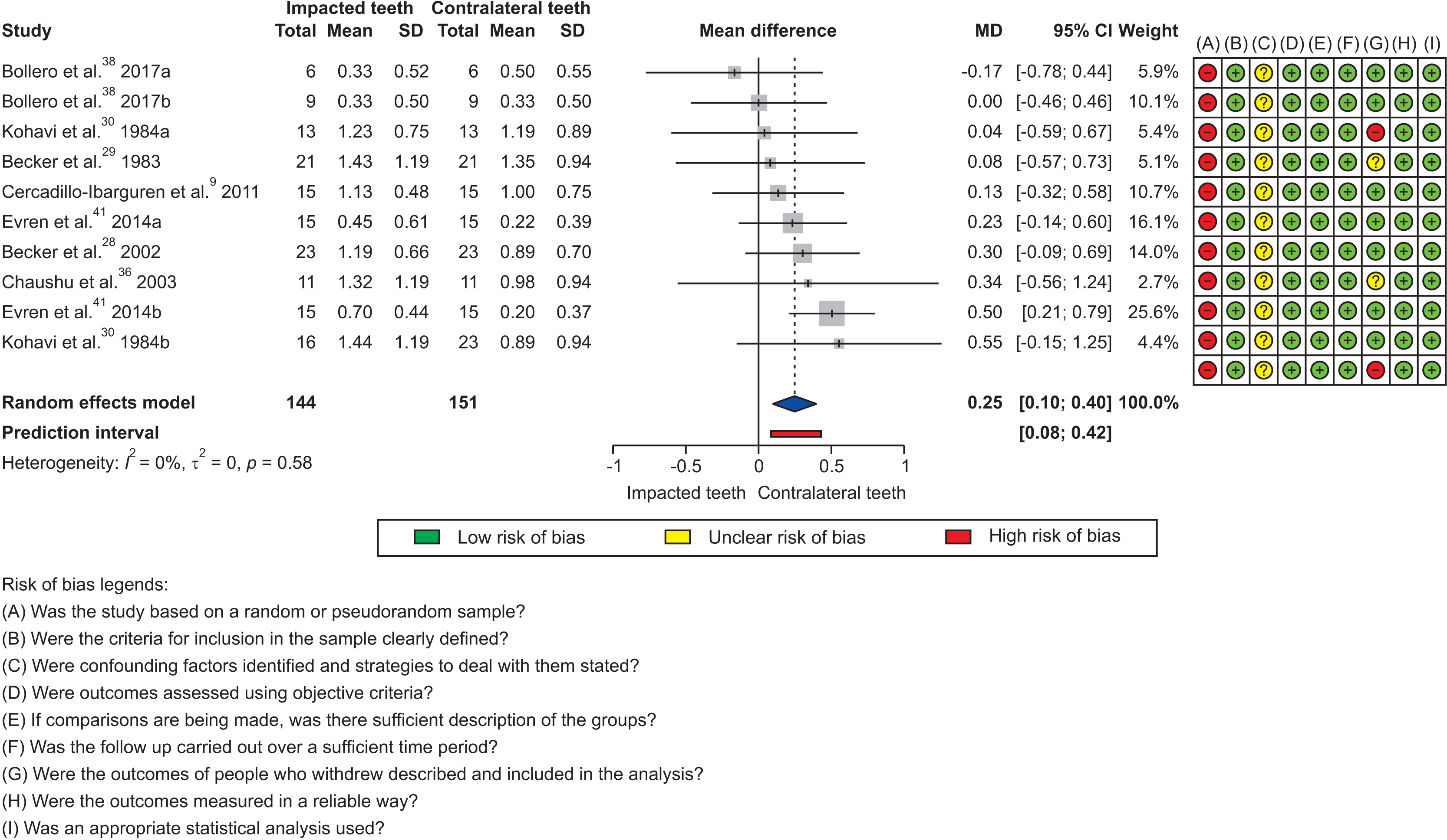
Furthermore, a significant difference was observed between the impacted and contralateral teeth (MD = 0.25; 95% CI = 0.10 to 0.40; I2 = 0%) when evaluating the gingival index. There was no heterogeneity between the observed effect sizes, demonstrating similar values regardless of the tooth type (p = 0.58; Tau2 = 0.00) even when more than one group of teeth was subjected to the traction procedure. Thus, impacted teeth had higher gingival index scores than contralateral teeth, which denotes a worse gingival condition (Figure 3). The analysis showed robustness with a narrow prediction interval (95% PI = 0.08 to 0.42), even with the inclusion of studies that examined different groups of teeth, demonstrating that this was not a confounding factor capable of altering the effect size.
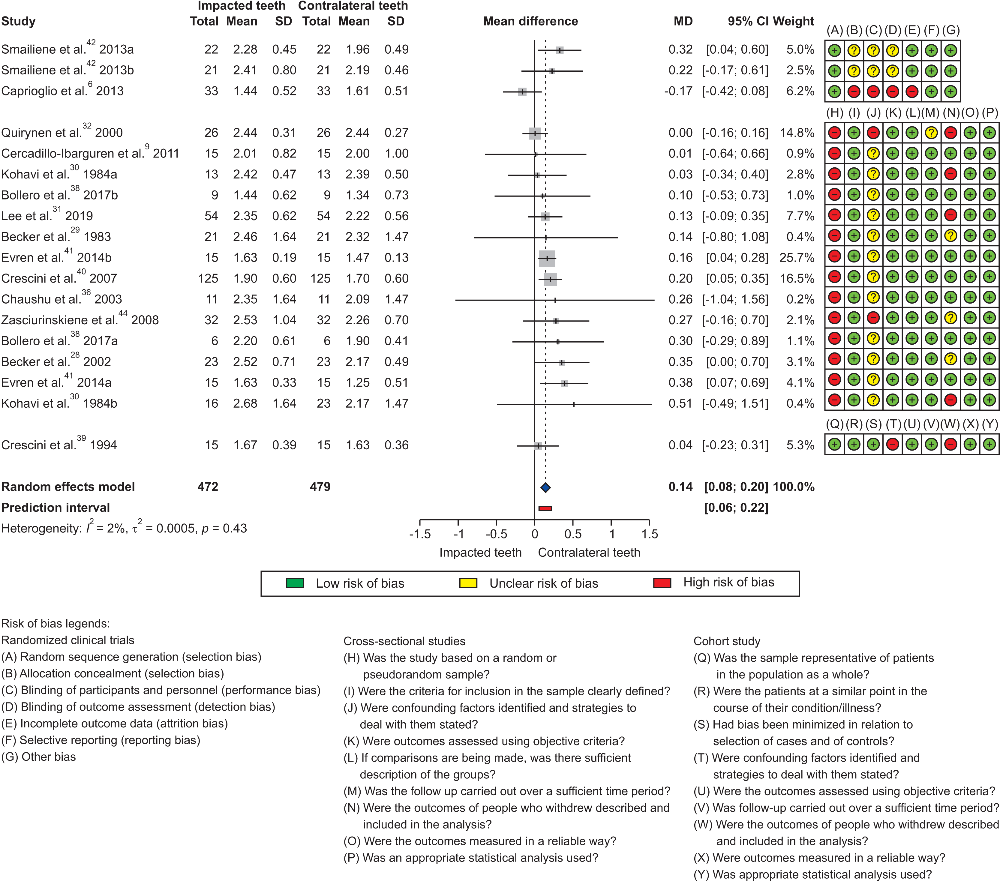
Similarly impacted teeth showed greater probing depth, with a significant mean difference between the groups (MD = 0.14; 95% CI = 0.08 to 0.20; I2 = 2%), when the periodontal probing depth was evaluated. Likewise, the variance between the effects was close to zero, with no significant heterogeneity, regardless of the type of teeth included in the analysis (p = 0.43; Tau2 = 0.00) and with a narrow prediction range (Figure 4). One of the articles included in the analysis presented a high risk of bias; however, even after the sensitivity analysis with the exclusion of this article, the estimates remained close (MD = 0.16; 95% PI = 0.09 to 0.23; I2 = 0%).
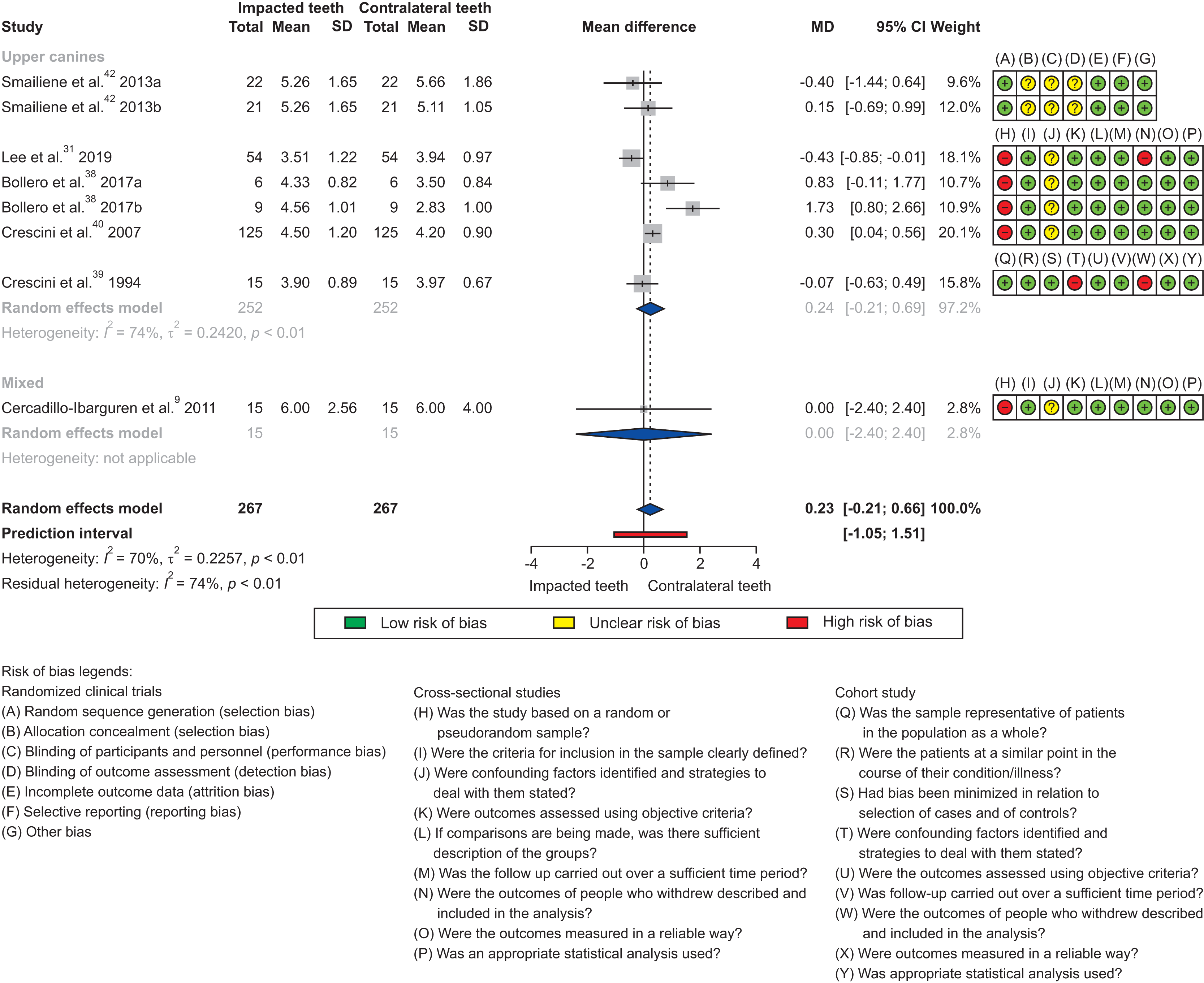
Additionally, the evaluation of the width of the keratinized tissue revealed no significant difference between the two groups (MD = 0.23; 95% CI = –0.21 to 0.66; I2 = 70%), with no difference between the width of the impacted teeth and the respective contralateral teeth (Figure 5). Subgroup analysis was performed to assess the heterogeneity; however, the type of teeth was not a predictive factor that explained the heterogeneity between the observed effects.

Reporting biases

There was no significance (p > 0.05) when funnel asymmetry was evaluated using the Egger test, indicating the absence of publication bias (Supplementary Figure 1).

Certainty of evidence

Since most of the included studies were cross-sectional, the certainty of the evidence was reduced, with low evidence for the following outcomes: plaque index, gingival index, and probing depth. Furthermore, the heterogeneity in assessing the width of keratinized tissue persisted even after the subgroup analysis. Therefore, reducing the certainty of the evidence regarding its imprecision is considered a very low level of evidence certainty (Supplementary Table 5).
Supplementary data is available at https://doi.org/10.4041/kjod.22.123.

DISCUSSION

The present study analyzed the available evidence on the periodontal parameters of orthodontically tractioned teeth compared with the respective non-tractioned contralateral teeth. The findings indicated that orthodontic traction was associated with worsening of the gingival index and a slight increase in periodontal probing depth, with no difference in the plaque index and width of keratinized tissue.
Dental plaque is considered a primary cause of periodontal disease and requires mechanical cleaning of all sides of the tooth to maintain good oral health.45 Bollero et al.,38 observed no difference between impacted canines and contralateral teeth, regardless of the arch in which they were positioned. In contrast, Evren et al.41 observed an increase in the plaque index and the gingival index scores in lower canines under traction when compared with the respective contralateral ones; though, it was not possible to elucidate the causes of the worsening of periodontal health. In this current systematic review, the mean plaque index scores showed no statistical difference (p > 0.05), with the exception of one study41 which did not touch the null line; however, this study showed no influence on the analysis to the point of changing the significance in relation to this outcome.
Patients undergoing orthodontic treatment require periodontal evaluations at each visit because of possible periodontal implications during orthodontic therapy.46 The current literature data indicated that tractioned teeth had higher gingival index scores and greater probing depth (p < 0.05). Lee et al.31 reported that the periodontal tissues of canines impacted after orthodontic traction might not have the same characteristics as a canine with normal eruption. Factors involved in the surgical procedure, including the need for more extensive bone removal in closed flaps, may be related to the increase in pocket depth and gingival level after treatment.41 In this review, both outcomes demonstrated low heterogeneity in the effect sizes of the articles included in the analysis, thus indicating that the type of tooth did not interfere with the observed effect. Additionally, the effect of the worsening of these parameters was similar regardless of the type of impacted tooth. Despite demonstrating statistical significance, data related to probing depth should be considered in relation to the clinical significance of these findings since the difference between the groups had a small effect size.
In clinical practice, factors including the dimensions of soft and hard tissues and oral tissues are vital parameters that can influence diagnosis and periodontal treatment.47 Following the orthodontic-surgical traction procedure, the findings showed different data for determining whether the width of keratinized tissue in impacted teeth differs from that in teeth with normal eruption. Lee et al.31 observed a significant decrease in the width of keratinized tissue in canines compared with their non-tractioned contralateral counterparts, with the minimum width considered for the maintenance of periodontal health as 2 mm, and these values were less than 2 mm for this parameter in treated teeth, which predisposes to recurrent inflammation and loss of periodontal support. Smailiene et al.43 did not observe significant differences between the treated teeth and their non-tractioned contralateral teeth. Conversely, Bollero et al.,38 observed a greater amount of keratinized tissue in tractioned canines than in contralateral teeth. Although the meta-analysis did not show statistical significance for this outcome (p > 0.05), there was heterogeneity among the observed effects that persisted even after the subgroup analysis. The certainty of evidence for this outcome was reduced because the source of heterogeneity could not be identified.
The difficulty of surgical access in impacted third molars requires the patient to remain with the jaw open for a long period, which can cause muscle exhaustion, trauma, and even overload of the temporomandibular joint (TMJ). Therefore, the location and severity of the third molar impaction may contribute to the development of temporomandibular disorders.48 Additionally, the individual’s systemic health is a crucial factor to consider, especially in patients with a predisposition to TMJ problems. Patients with systemic sclerosis may have difficulties in mandibular excursion and mouth opening, making it challenging to perform the dental procedure, and may have more signs and symptoms of temporomandibular disorders compared with healthy individuals.49 None of the included studies examined the influence of dental impaction and orthodontic-surgical traction on the development of temporomandibular disorders, hence demonstrating a gap in the literature on this topic.
The severity of the impaction of tractioned canines can play a significant role in cases of greater severity, with the possibility of periodontal damage at the end of the treatment.50 Notably, this present review included only one study that evaluated the impact of impaction severity on periodontal parameters. The impossibility of assessing the influence of this variable through meta-analysis was because primary studies did not evaluate it, reducing the certainty of the evidence generated, thus acting as a confounding factor for the estimate. Further studies on this topic should consider the degree of initial impaction of the tractioned teeth.
This systematic review and meta-analysis revealed important data on periodontal parameters in impacted teeth undergoing orthodontic-surgical traction. These findings help the orthodontist and/or surgeon stipulate the risks, understand the predictability of the techniques, and communicate with the patient efficiently. Some limitations should be pointed out, including the study design. Notably, most of the included studies were cross-sectional observational studies; thus, different confounding factors can be influenced by the impossibility of randomization.

CONCLUSIONS

Tractioned teeth may exhibit worsening in the periodontal parameters related to the gingival index and probing depth; nevertheless, the probing depth should be weighed as to its clinical significance because of the small effect size observed. These estimates should be viewed with caution since the severity of the impacted tooth could not be considered in the analyses, and the traction methods were not standardized. Finally, the observed effect sizes remained similar across the examined groups regardless of the group of teeth analyzed.

Notes

CONFLICTS OF INTEREST

No potential conflict of interest relevant to this article was reported.

REFERENCES

1. Thilander B, Myrberg N. 1973; The prevalence of malocclusion in Swedish schoolchildren. Scand J Dent Res. 81:12–21. DOI: 10.1111/j.1600-0722.1973.tb01489.x. PMID: 4510864. PMID: https://www.scopus.com/inward/record.uri?partnerID=HzOxMe3b&scp=84984483775&origin=inward.
2. Campbell CM, DiBiase A, Fleming PS. 2014; Concomitant dilaceration, transposition, and intraosseous migration: report of a patient treated with maxillary canine-central incisor substitution. Am J Orthod Dentofacial Orthop. 146:514–21. DOI: 10.1016/j.ajodo.2013.11.024. PMID: 25263154. PMID: https://www.scopus.com/inward/record.uri?partnerID=HzOxMe3b&scp=84908014602&origin=inward.
3. Cavuoti S, Matarese G, Isola G, Abdolreza J, Femiano F, Perillo L. 2016; Combined orthodontic-surgical management of a transmigrated mandibular canine. Angle Orthod. 86:681–91. DOI: 10.2319/050615-309.1. PMID: 26502299. PMCID: PMC8601488. PMID: https://www.scopus.com/inward/record.uri?partnerID=HzOxMe3b&scp=84976412689&origin=inward.
4. Kokich VG. 2004; Surgical and orthodontic management of impacted maxillary canines. Am J Orthod Dentofacial Orthop. 126:278–83. DOI: 10.1016/j.ajodo.2004.06.009. PMID: 15356485. PMID: https://www.scopus.com/inward/record.uri?partnerID=HzOxMe3b&scp=4444226191&origin=inward.
5. Stellzig A, Basdra EK, Komposch G. 1994; The etiology of canine tooth impaction--a space analysis. Fortschr Kieferorthop. 55:97–103. German. DOI: 10.1007/BF02341481. PMID: 8045486. PMID: https://www.scopus.com/inward/record.uri?partnerID=HzOxMe3b&scp=0028456980&origin=inward.
6. Caprioglio A, Vanni A, Bolamperti L. 2013; Long-term periodontal response to orthodontic treatment of palatally impacted maxillary canines. Eur J Orthod. 35:323–8. DOI: 10.1093/ejo/cjs020. PMID: 22531665. PMID: https://www.scopus.com/inward/record.uri?partnerID=HzOxMe3b&scp=84878558810&origin=inward.
7. Chu FC, Li TK, Lui VK, Newsome PR, Chow RL, Cheung LK. 2003; Prevalence of impacted teeth and associated pathologies--a radiographic study of the Hong Kong Chinese population. Hong Kong Med J. 9:158–63. PMID: 12777649. PMID: https://www.scopus.com/inward/record.uri?partnerID=HzOxMe3b&scp=0038352250&origin=inward.
8. Hattab FN, Rawashdeh MA, Fahmy MS. 1995; Impaction status of third molars in Jordanian students. Oral Surg Oral Med Oral Pathol Oral Radiol Endod. 79:24–9. DOI: 10.1016/S1079-2104(05)80068-X. PMID: 7614155. PMID: https://www.scopus.com/inward/record.uri?partnerID=HzOxMe3b&scp=0029186007&origin=inward.
9. Cercadillo-Ibarguren I, Gargallo-Albiol J, Abad-Sánchez D, Echeverría-García JJ, Berini-Aytés L, Gay-Escoda C. 2011; Periodontal health and esthetic results in impacted teeth exposed by apically positioned flap technique. Med Oral Patol Oral Cir Bucal. 16:e89–95. DOI: 10.4317/medoral.16.e89. PMID: 20711131. PMID: https://www.scopus.com/inward/record.uri?partnerID=HzOxMe3b&scp=78751661441&origin=inward.
10. Hashemipour MA, Tahmasbi-Arashlow M, Fahimi-Hanzaei F. 2013; Incidence of impacted mandibular and maxillary third molars: a radiographic study in a Southeast Iran population. Med Oral Patol Oral Cir Bucal. 18:e140–5. DOI: 10.4317/medoral.18028. PMID: 23229243. PMCID: PMC3548634. PMID: https://www.scopus.com/inward/record.uri?partnerID=HzOxMe3b&scp=84871888774&origin=inward.
11. Hugoson A, Kugelberg CF. 1988; The prevalence of third molars in a Swedish population. An epidemiological study. Community Dent Health. 5:121–38. PMID: 3165039. PMID: https://www.scopus.com/inward/record.uri?partnerID=HzOxMe3b&scp=0024026048&origin=inward.
12. Aydin U, Yilmaz HH, Yildirim D. 2004; Incidence of canine impaction and transmigration in a patient population. Dentomaxillofac Radiol. 33:164–9. DOI: 10.1259/dmfr/15470658. PMID: 15371316. PMID: https://www.scopus.com/inward/record.uri?partnerID=HzOxMe3b&scp=7944224758&origin=inward.
13. Oliver RG, Mannion JE, Robinson JM. 1989; Morphology of the maxillary lateral incisor in cases of unilateral impaction of the maxillary canine. Br J Orthod. 16:9–16. DOI: 10.1179/bjo.16.1.9. PMID: 2923851. PMID: https://www.scopus.com/inward/record.uri?partnerID=HzOxMe3b&scp=0024616613&origin=inward.
14. Crescini A, Nieri M, Buti J, Baccetti T, Pini Prato GP. 2007; Pre-treatment radiographic features for the periodontal prognosis of treated impacted canines. J Clin Periodontol. 34:581–7. DOI: 10.1111/j.1600-051X.2007.01094.x. PMID: 17555411. PMID: https://www.scopus.com/inward/record.uri?partnerID=HzOxMe3b&scp=34250008744&origin=inward.
15. Axelsson P, Lindhe J. 1978; Effect of controlled oral hygiene procedures on caries and periodontal disease in adults. J Clin Periodontol. 5:133–51. DOI: 10.1111/j.1600-051X.1978.tb01914.x. PMID: 275282. PMID: https://www.scopus.com/inward/record.uri?partnerID=HzOxMe3b&scp=0017875739&origin=inward.
16. Pearson MH, Robinson SN, Reed R, Birnie DJ, Zaki GA. 1997; Management of palatally impacted canines: the findings of a collaborative study. Eur J Orthod. 19:511–5. DOI: 10.1093/ejo/19.5.511. PMID: 9386337. PMID: https://www.scopus.com/inward/record.uri?partnerID=HzOxMe3b&scp=0031255497&origin=inward.
17. Cutroneo G, Piancino MG, Ramieri G, Bracco P, Vita G, Isola G, et al. 2012; Expression of muscle-specific integrins in masseter muscle fibers during malocclusion disease. Int J Mol Med. 30:235–42. DOI: 10.3892/ijmm.2012.986. PMID: 22552408. PMID: https://www.scopus.com/inward/record.uri?partnerID=HzOxMe3b&scp=84863558404&origin=inward.
18. Piancino MG, Isola G, Merlo A, Dalessandri D, Debernardi C, Bracco P. 2012; Chewing pattern and muscular activation in open bite patients. J Electromyogr Kinesiol. 22:273–9. DOI: 10.1016/j.jelekin.2011.12.003. PMID: 22236764. PMID: https://www.scopus.com/inward/record.uri?partnerID=HzOxMe3b&scp=84857030884&origin=inward.
19. Alamri A, Alshahrani N, Al-Madani A, Shahin S, Nazir M. 2020; Prevalence of impacted teeth in Saudi patients attending dental clinics in the Eastern province of Saudi Arabia: a radiographic retrospective study. ScientificWorldJournal. 2020:8104904. DOI: 10.1155/2020/8104904. PMID: 32952455. PMCID: PMC7481944. PMID: https://www.scopus.com/inward/record.uri?partnerID=HzOxMe3b&scp=85091266390&origin=inward.
20. de Araujo CM, Trannin PD, Schroder AGD, Stechman-Neto J, Cavalcante-Leão BL, Mattos NHR, et al. 2020; Surgical-periodontal aspects in orthodontic traction of palatally displaced canines: a meta-analysis. Jpn Dent Sci Rev. 56:164–76. DOI: 10.1016/j.jdsr.2020.10.001. PMID: 33294059. PMCID: PMC7701265. PMID: https://www.scopus.com/inward/record.uri?partnerID=HzOxMe3b&scp=85097434912&origin=inward.
21. Page MJ, Moher D, Bossuyt PM, Boutron I, Hoffmann TC, Mulrow CD, et al. 2021; PRISMA 2020 explanation and elaboration: updated guidance and exemplars for reporting systematic reviews. BMJ. 372:n160. DOI: 10.1136/bmj.n160. PMID: 33781993. PMCID: PMC8005925. PMID: https://www.scopus.com/inward/record.uri?partnerID=HzOxMe3b&scp=85103235741&origin=inward.
22. Silness J, Loe H. 1964; Periodontal disease in pregnancy. II. Correlation between oral hygiene and periodontal condtion. Acta Odontol Scand. 22:121–35. DOI: 10.3109/00016356408993968. PMID: 14158464. PMID: https://www.scopus.com/inward/record.uri?partnerID=HzOxMe3b&scp=75949163484&origin=inward.
23. Carter HG, Barnes GP. 1974; The Gingival Bleeding Index. J Periodontol. 45:801–5. DOI: 10.1902/jop.1974.45.11.801. PMID: 4547795. PMID: https://www.scopus.com/inward/record.uri?partnerID=HzOxMe3b&scp=0016296301&origin=inward.
24. The Joanna Briggs Institute. 2014. Joanna Briggs Institute reviewers' manual. The Joanna Briggs Institute;Adelaide: PMID: https://www.scopus.com/inward/record.uri?partnerID=HzOxMe3b&scp=0024026048&origin=inward.
25. Higgins JP, Altman DG, Gøtzsche PC, Jüni P, Moher D, Oxman AD, et al. 2011; The Cochrane Collaboration's tool for assessing risk of bias in randomised trials. BMJ. 343:d5928. DOI: 10.1136/bmj.d5928. PMID: 22008217. PMCID: PMC3196245. PMID: https://www.scopus.com/inward/record.uri?partnerID=HzOxMe3b&scp=84859001212&origin=inward.
26. Viechtbauer W, Cheung MW. 2010; Outlier and influence diagnostics for meta-analysis. Res Synth Methods. 1:112–25. DOI: 10.1002/jrsm.11. PMID: 26061377.
27. Manheimer E. 2012; Summary of findings tables: presenting the main findings of Cochrane complementary and alternative medicine-related reviews in a transparent and simple tabular format. Glob Adv Health Med. 1:90–1. DOI: 10.7453/gahmj.2012.1.1.015. PMID: 24278805. PMCID: PMC3833472. PMID: https://www.scopus.com/inward/record.uri?partnerID=HzOxMe3b&scp=85115900710&origin=inward.
28. Becker A, Brin I, Ben-Bassat Y, Zilberman Y, Chaushu S. 2002; Closed-eruption surgical technique for impacted maxillary incisors: a postorthodontic periodontal evaluation. Am J Orthod Dentofacial Orthop. 122:9–14. DOI: 10.1067/mod.2002.124998. PMID: 12142887. PMID: https://www.scopus.com/inward/record.uri?partnerID=HzOxMe3b&scp=0036633860&origin=inward.
29. Becker A, Kohavi D, Zilberman Y. 1983; Periodontal status following the alignment of palatally impacted canine teeth. Am J Orthod. 84:332–6. DOI: 10.1016/S0002-9416(83)90349-4. PMID: 6578683. PMID: https://www.scopus.com/inward/record.uri?partnerID=HzOxMe3b&scp=0021073065&origin=inward.
30. Kohavi D, Zilberman Y, Becker A. 1984; Periodontal status following the alignment of buccally ectopic maxillary canine teeth. Am J Orthod. 85:78–82. DOI: 10.1016/0002-9416(84)90125-8. PMID: 6581729. PMID: https://www.scopus.com/inward/record.uri?partnerID=HzOxMe3b&scp=0021300914&origin=inward.
31. Lee JY, Choi YJ, Choi SH, Chung CJ, Yu HS, Kim KH. 2019; Labially impacted maxillary canines after the closed eruption technique and orthodontic traction: a split-mouth comparison of periodontal recession. J Periodontol. 90:35–43. DOI: 10.1002/JPER.18-0034. PMID: 30001475. PMID: https://www.scopus.com/inward/record.uri?partnerID=HzOxMe3b&scp=85059383279&origin=inward.
32. Quirynen M, Op Heij DG, Adriansens A, Opdebeeck HM, van Steenberghe D. 2000; Periodontal health of orthodontically extruded impacted teeth. A split-mouth, long-term clinical evaluation. J Periodontol. 71:1708–14. DOI: 10.1902/jop.2000.71.11.1708. PMID: 11128918. PMID: https://www.scopus.com/inward/record.uri?partnerID=HzOxMe3b&scp=0034324086&origin=inward.
33. Landim FS, de Freitas GB, Rocha NS, Caubi AF, Vasconcellos RJH. 2010; Clinical-radiographic analysis of canines after orthodonthic-surgical treatment. Rev Cir Traumatol Buco Maxilo Fac. 10:103–10. Português. PMID: https://www.scopus.com/inward/record.uri?partnerID=HzOxMe3b&scp=0024026048&origin=inward.
34. Odenrick L, Modéer T. 1978; Peridontal status following surgical-orthodontic alignment of impacted teeth. Acta Odontol Scand. 36:233–6. DOI: 10.3109/00016357809004673. PMID: 280118. PMID: https://www.scopus.com/inward/record.uri?partnerID=HzOxMe3b&scp=0017869506&origin=inward.
35. Caminiti MF, Sandor GK, Giambattistini C, Tompson B. 1998; Outcomes of the surgical exposure, bonding and eruption of 82 impacted maxillary canines. J Can Dent Assoc. 64:572–4. 576–9. PMID: 9785687. PMID: https://www.scopus.com/inward/record.uri?partnerID=HzOxMe3b&scp=0032150081&origin=inward.
36. Chaushu S, Brin I, Ben-Bassat Y, Zilberman Y, Becker A. 2003; Periodontal status following surgical-orthodontic alignment of impacted central incisors with an open-eruption technique. Eur J Orthod. 25:579–84. DOI: 10.1093/ejo/25.6.579. PMID: 14700263. PMID: https://www.scopus.com/inward/record.uri?partnerID=HzOxMe3b&scp=1542577976&origin=inward.
37. Chaushu S, Dykstein N, Ben-Bassat Y, Becker A. 2009; Periodontal status of impacted maxillary incisors uncovered by 2 different surgical techniques. J Oral Maxillofac Surg. 67:120–4. DOI: 10.1016/j.joms.2008.08.012. PMID: 19070757. PMID: https://www.scopus.com/inward/record.uri?partnerID=HzOxMe3b&scp=57349113198&origin=inward.
38. Bollero P, Danesi C, Ricchiuti MR, Milazzo A, Mampieri G, Agrestini C, et al. 2017; Long-term periodontal status of palatally and buccally impacted canines after closed surgical-orthodontic approach. Oral Implantol (Rome). 10:162–71. DOI: 10.11138/orl/2017.10.2.162. PMID: 29876041. PMCID: PMC5965064. PMID: https://www.scopus.com/inward/record.uri?partnerID=HzOxMe3b&scp=85030634264&origin=inward.
39. Crescini A, Clauser C, Giorgetti R, Cortellini P, Pini Prato GP. 1994; Tunnel traction of infraosseous impacted maxillary canines. A three-year periodontal follow-up. Am J Orthod Dentofacial Orthop. 105:61–72. DOI: 10.1016/S0889-5406(94)70100-8. PMID: 8291494. PMID: https://www.scopus.com/inward/record.uri?partnerID=HzOxMe3b&scp=0028058603&origin=inward.
40. Crescini A, Nieri M, Buti J, Baccetti T, Mauro S, Prato GP. 2007; Short- and long-term periodontal evaluation of impacted canines treated with a closed surgical-orthodontic approach. J Clin Periodontol. 34:232–42. DOI: 10.1111/j.1600-051X.2006.01042.x. PMID: 17257160. PMID: https://www.scopus.com/inward/record.uri?partnerID=HzOxMe3b&scp=33846925506&origin=inward.
41. Evren AD, Nevzatoğlu Ş, Arun T, Acar A. 2014; Periodontal status of ectopic canines after orthodontic treatment. Angle Orthod. 84:18–23. DOI: 10.2319/041513-290.1. PMID: 23841541. PMCID: PMC8683066. PMID: https://www.scopus.com/inward/record.uri?partnerID=HzOxMe3b&scp=84891957738&origin=inward.
42. Smailienė D, Kavaliauskienė A, Pacauskienė I. 2013; Posttreatment status of palatally impacted maxillary canines treated applying 2 different surgical-orthodontic methods. Medicina (Kaunas). 49:354–60. DOI: 10.3390/medicina49080055. PMID: 24509145. PMID: https://www.scopus.com/inward/record.uri?partnerID=HzOxMe3b&scp=84897664576&origin=inward.
43. Smailiene D, Kavaliauskiene A, Pacauskiene I, Zasciurinskiene E, Bjerklin K. 2013; Palatally impacted maxillary canines: choice of surgical-orthodontic treatment method does not influence post-treatment periodontal status. A controlled prospective study. Eur J Orthod. 35:803–10. DOI: 10.1093/ejo/cjs102. PMID: 23349422. PMID: https://www.scopus.com/inward/record.uri?partnerID=HzOxMe3b&scp=84890477800&origin=inward.
44. Zasciurinskiene E, Bjerklin K, Smailiene D, Sidlauskas A, Puisys A. 2008; Initial vertical and horizontal position of palatally impacted maxillary canine and effect on periodontal status following surgical-orthodontic treatment. Angle Orthod. 78:275–80. DOI: 10.2319/010907-8.1. PMID: 18251594. PMID: https://www.scopus.com/inward/record.uri?partnerID=HzOxMe3b&scp=40749133303&origin=inward.
45. Worthington HV, MacDonald L, Poklepovic Pericic T, Sambunjak D, Johnson TM, Imai P, et al. 2019; Home use of interdental cleaning devices, in addition to toothbrushing, for preventing and controlling periodontal diseases and dental caries. Cochrane Database Syst Rev. 4:CD012018. DOI: 10.1002/14651858.CD012018.pub2. PMID: 30968949. PMCID: PMC6953268. PMID: https://www.scopus.com/inward/record.uri?partnerID=HzOxMe3b&scp=85064427986&origin=inward.
46. Cerroni S, Pasquantonio G, Condò R, Cerroni L. 2018; Orthodontic fixed appliance and periodontal status: an updated systematic review. Open Dent J. 12:614–22. DOI: 10.2174/1745017901814010614. PMID: 30369970. PMCID: PMC6182882. PMID: https://www.scopus.com/inward/record.uri?partnerID=HzOxMe3b&scp=85055743238&origin=inward.
47. Vlachodimou E, Fragkioudakis I, Vouros I. 2021; Is there an association between the gingival phenotype and the width of keratinized gingiva? A systematic review. Dent J (Basel). 9:34. DOI: 10.3390/dj9030034. PMID: 33806934. PMCID: PMC8004949. PMID: 116b2d9e3ec846d09bc6a6d4a022041f. PMID: https://www.scopus.com/inward/record.uri?partnerID=HzOxMe3b&scp=85107790527&origin=inward.
48. Damasceno YSS, Espinosa DG, Normando D. 2020; Is the extraction of third molars a risk factor for the temporomandibular disorders? A systematic review. Clin Oral Investig. 24:3325–34. DOI: 10.1007/s00784-020-03277-6. PMID: 32776170. PMID: https://www.scopus.com/inward/record.uri?partnerID=HzOxMe3b&scp=85089142235&origin=inward.
49. Matarese G, Isola G, Alibrandi A, Lo Gullo A, Bagnato G, Cordasco G, et al. 2016; Occlusal and MRI characterizations in systemic sclerosis patients: a prospective study from Southern Italian cohort. Joint Bone Spine. 83:57–62. DOI: 10.1016/j.jbspin.2015.04.014. PMID: 26552635. PMID: https://www.scopus.com/inward/record.uri?partnerID=HzOxMe3b&scp=84959194802&origin=inward.
50. Caprioglio A, Comaglio I, Siani L, Fastuca R. 2019; Effects of impaction severity of treated palatally displaced canines on periodontal outcomes: a retrospective study. Prog Orthod. 20:5. DOI: 10.1186/s40510-018-0256-7. PMID: 30714076. PMCID: PMC6361313. PMID: 703b64983db04a47a6119585152ad8b6. PMID: https://www.scopus.com/inward/record.uri?partnerID=HzOxMe3b&scp=85061067551&origin=inward.

Figure 1
Flowchart of literature search and selection criteria.
kjod-52-6-420-f1.tif
Figure 2
Comparison of the plaque index between tractioned teeth and their respective contralateral. A, Forest plot showing the risk of bias for each included study. B, Influence analysis. The letters 'a, b' were placed when more than one estimate was extracted from the same study.
SD, standard deviation; MD, mean difference; CI, confidence interval.
kjod-52-6-420-f2.tif
Figure 3
Comparison of the gingival index between tractioned teeth and their respective contralateral, showing the risk of bias for each included study. The letters 'a, b' were placed when more than one estimate was extracted from the same study.
SD, standard deviation; MD, mean difference; CI, confidence interval.
kjod-52-6-420-f3.tif
Figure 4
Comparison of the periodontal probing depth between tractioned teeth and their respective contralateral, showing the risk of bias for each included study. The letters 'a, b' were placed when more than one estimate was extracted from the same study.
SD, standard deviation; MD, mean difference; CI, confidence interval.
kjod-52-6-420-f4.tif
Figure 5
Comparison of the width of the keratinized tissue between tractioned teeth and their respective contralateral, showing the risk of bias for each included study. The letters 'a, b' were placed when more than one estimate was extracted from the same study.
SD, standard deviation; MD, mean difference; CI, confidence interval.
kjod-52-6-420-f5.tif
TOOLS
Similar articles