

 PDF
PDF Citation
 Print
Print


INTRODUCTION
Calcitonin is a 32 amino acid polypeptide hormone produced by parafollicular C cells [1]. The disulfide bridges at the N- and C-terminals play a functionally essential role in biological activity [2]. The development of an immunoradiometric assay using two different monoclonal antibodies—each recognizing the N- and C-terminals—allowed more specific and sensitive assays to detect the mature 32 amino acid monomer of calcitonin compared with the radioimmunoassay of the past using polyclonal antibodies which detect not only the mature calcitonin but also other circulating forms, such as precursors or degradation products [2]. This two-site immunoradiometric assay reports calcitonin levels below 10 pg/mL for the normal healthy population [3,4].
Calcitonin is a key marker for monitoring the disease course in patients with medullary thyroid carcinoma (MTC) [5,6]. Despite the improvement of diagnostic techniques, its usefulness in initial screening or diagnosing MTC is an issue of debate [7,8]. Although several studies reported that measuring serum calcitonin when evaluating thyroid nodules can detect MTC at an earlier stage and consequently improve the overall survival [9–13], the guidelines do not recommend either for or against its routine measurement in patients with thyroid nodules [7,14]. One of the major obstacles to adopting serum calcitonin as a routine test is the lack of an established cutoff value. Calcitonin is not specific for MTC and can be elevated in various conditions such as C cell hyperplasia, neuroendocrine tumors of nonthyroidal organs (lung, pancreas, and prostate), chronic kidney disease (CKD), autoimmune thyroid disease, hypergastrinemias, presence of heterophilic antibodies, and use of certain drugs (proton pump inhibitors, H2 blockers, or steroid) [15–19]. Moreover, the calcitonin level is affected by age, gender, and cigarette smoking [20–22], which makes determining the precise reference range more difficult. Previous studies defining the reference level of calcitonin are mostly derived from western countries, focused on the young population, or now outdated using the assays of the past. As aforementioned, calcitonin levels under 10 pg/mL are typically regarded as normal with a two-site immunoradiometric assay. However, this range remains wide, raising the need for redefining its reference level with a lower threshold.
To the best of our knowledge, there are no established normal range for serum calcitonin in Asian adults. Therefore, this study aimed to assess the reference range of serum calcitonin in normal males and females by taking into account the various factors affecting its level by using the data from the routine health checkup in the Republic of Korea.
METHODS
Study design and subjects
This cross-sectional study initially included 13,622 subjects aged ≥20 years with available serum calcitonin and thyroid ultrasonography (US) data from a routine health checkup performed at the Health Screening and Promoting Center in Asan Medical Center, Seoul, Korea, from January 2011 to December 2015. Patients with serum calcitonin level ≥65 pg/mL were excluded to minimize the effects of extreme values because this level has been previously examined as a meaningful cutoff value for detecting MTC in Korean subjects [19]. Moreover, subjects with abnormal corrected serum calcium levels (<8.6 or >10.2 mg/dL) were also excluded because it can affect serum calcitonin levels [23]. Other exclusion criteria were previous history of thyroid cancer and any history of thyroid surgery or radiofrequency ablation for thyroid nodules. Medical records of age, gender, body mass index (BMI), smoking status, kidney function, thyroid autoantibodies, previous medical history, and family history were reviewed. Informed consent was waived, and the Institutional Review Board of Asan Medical Center (No. 2016-0410) approved the data collection and subsequent analyses.
Laboratory assay
Serum calcitonin level was measured by immunoradiometric assay–human calcitonin (IRMA-hCT; CisBio International, Codolet, France) with a functional sensitivity, detection limit, and reference upper limit of 4.1, 1.3, and 10 pg/mL [19]. The intra- and inter-assay coefficients of variation of the serum calcitonin assay was 1.2% to 6.7% and 4.3% to 5.2%, respectively. The same calcitonin assay using IRMA-hCT was used during the whole study period.
Serum free thyroxine levels were measured using a radioimmunoassay kit (Beckman Coulter/IMMUNOTECH, Prague, Czech Republic). Serum thyroid-stimulating hormone (TSH) levels were measured with a TSH-CTK-3 kit (DiaSorin S.p.A., Saluggia, Italy) that had a functional sensitivity and an inter-assay variation coefficient of 0.07 mU/mL and 20%, respectively. The thyroglobulin antibody levels were determined using a radioimmunoassay (BRAHMS anti-Tgn RIA), and ≥60 U/mL was designated as the minimum threshold denoting positivity. The functional sensitivity of this assay was below 20 U/mL, while the analytical sensitivity from the optimal curve was 5.5 U/mL. Thyroid peroxidase antibody levels were measured using a radioimmunoassay (BRAHMS anti-TPOn RIA), and ≥60 U/mL was again considered positive. The functional sensitivity of this assay was below 30 U/mL, and analytical sensitivity was 5.5 U/mL.
Grouping of subjects
Subjects were categorized into groups 1, 2, and 3 which included total eligible subjects in group 1; those without thyroid nodules, thyroid dysfunction, family history of thyroid cancer, thyroid hormone, or antithyroid drug administration for any reason in group 2; and, in addition to group 2 criteria, those without CKD (defined as estimated glomerular filtration rate <60 mL/min by Modification of Diet in Renal Disease equation), autoimmune thyroid disease (defined as positive thyroid autoantibody with diffuse parenchymal heterogeneity on thyroid US), medication of proton pump inhibitor/H2 blocker/steroid, or other malignancies in group 3, which are all possible factors that can elevate serum calcitonin levels [15–18,20,24,25].
To assess the association between serum calcitonin levels and BMI, the subjects were classified as either underweight (BMI <18.5 kg/m2), normal (BMI 18.5 to 22.9 kg/m2), overweight (BMI 23 to 24.9 kg/m2), or obese (BMI ≥25 kg/m2) based on the Asian-Pacific criteria [26].
Statistical analysis
The R version 3.4.0 was used for data analysis (R Foundation for Statistical Computing, Vienna, Austria; http://www.R-project.org). Continuous variables were presented as median with interquartile range (IQR) or mean with standard deviation (SD), and analyzed using Mann-Whitney U test or Student’s t test, respectively, to compare baseline characteristics between male and female subjects. Categorical variables were presented as numbers with percentages and compared using Pearson’s chi-square test. The analysis of variance was used when comparing the mean value of the calcitonin level among the three groups. The differences in P values <0.05 were regarded as significant.
RESULTS
A total of 10,566 subjects were eligible for analyses and were classified as group 1. After excluding subjects with thyroid nodules, thyroid dysfunction, family history of thyroid cancer, thyroid hormone, or antithyroid drug administration for any reason, 5,152 subjects were categorized as group 2. Group 3 included 4,638 subjects by further excluding other factors associated with hypercalcitoninemia.
Baseline characteristics
Table 1 summarizes the baseline clinical characteristics of group 1 subjects. The median age was 55 years (IQR, 49 to 60) with a median BMI of 23.5 (IQR, 21.6 to 25.5). About 50%, 28.0%, and 22.5% of the subjects were non-, ex-, and current smokers, respectively. Thyroid dysfunction was present in 4.6% of the total subjects, and 1.6% were taking thyroid hormone or antithyroid drugs. The mean calcitonin level was 2.3±1.9 pg/mL. When compared according to gender, a higher portion of male subjects were ex- or current smokers (P<0.001) and had more thyroid nodule(s) (P=0.001) than female subjects. However, thyroid dysfunction was more prevalent in females (P<0.001). The mean calcitonin level was higher in males than females (2.7 vs. 1.9, P<0.001). The baseline characteristics in groups 1, 2, and 3 are shown in Supplemental Table S1.
Serum calcitonin level according to gender in three groups
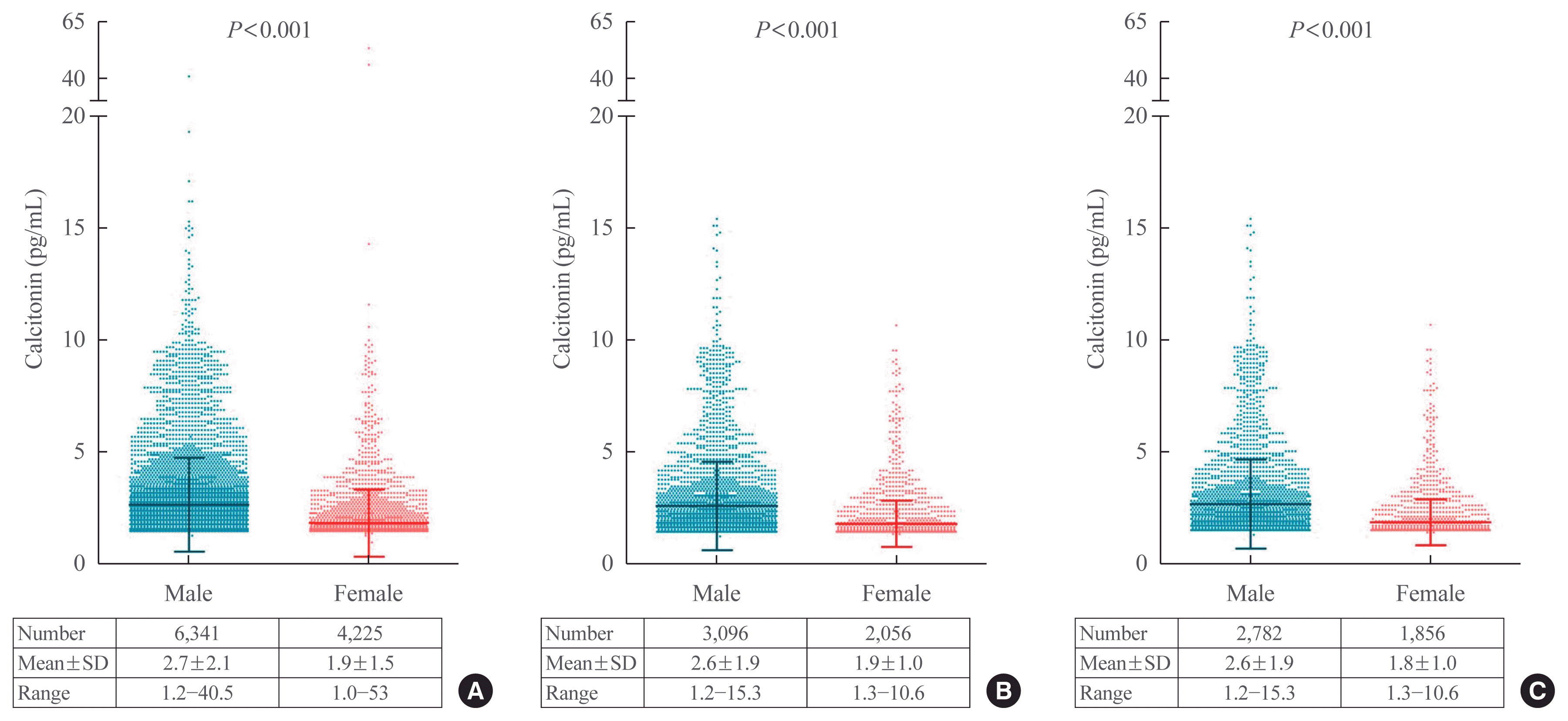
Fig. 1 depicts the serum calcitonin levels in male and female subjects by groups. The mean calcitonin level was higher in males than females in all three groups along with larger SD in males: 2.7±2.1 vs. 1.9±1.5, 2.6±1.9 vs. 1.9±1.0, and 2.6±1.9 vs. 1.8±1.0 pg/mL in males vs. females in groups 1 (P<0.001), 2 (P<0.001), and 3 (P<0.001), respectively. The mean calcitonin level was similar in three groups for each gender, but the range was narrower in the subjects in groups 2 and 3 compared with group 1. None of the subjects in groups 2 or 3 had a calcitonin level >20 pg/mL.
Serum calcitonin level according to age and BMI
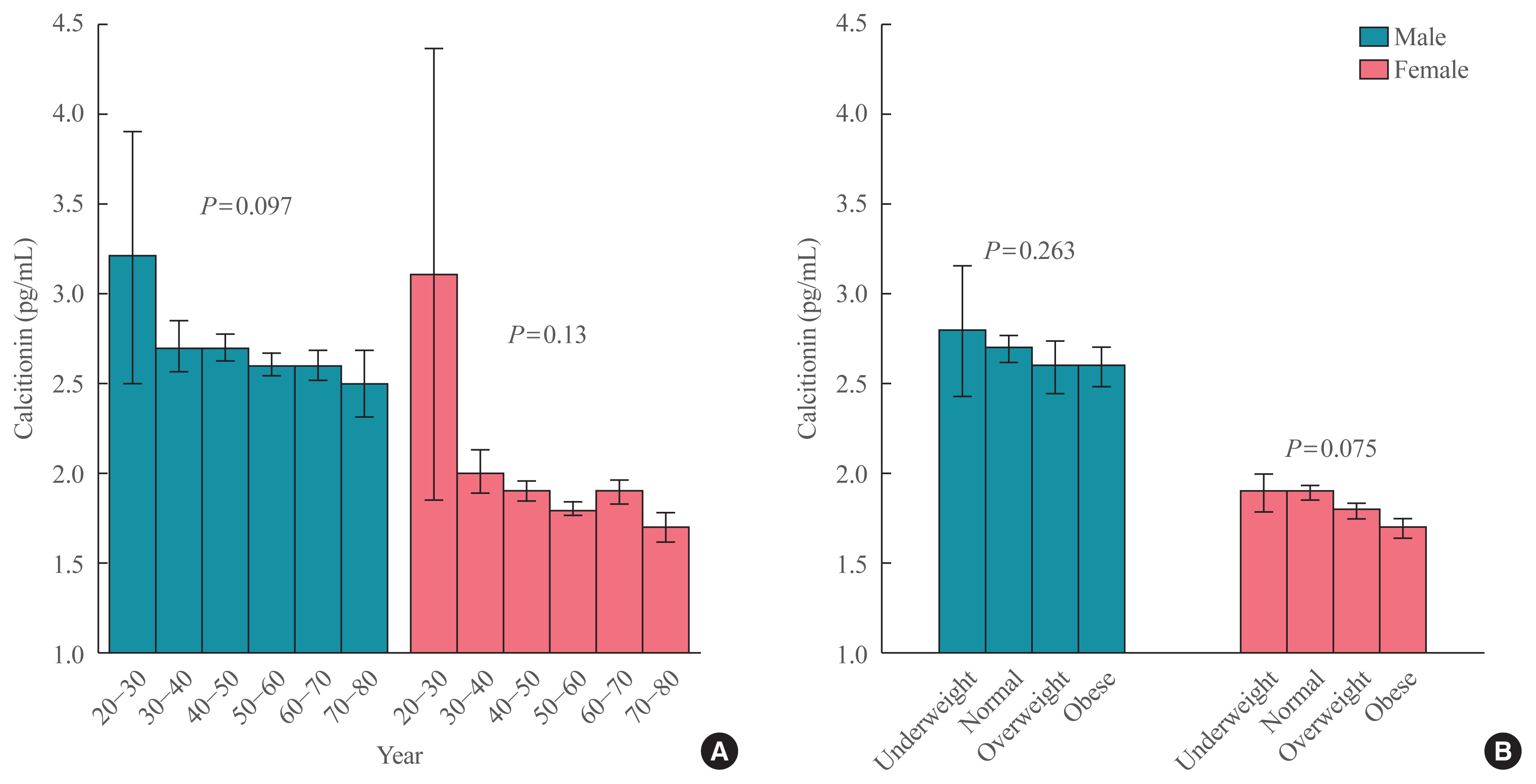
The association between serum calcitonin level and age or BMI was assessed in group 3 (Fig. 2). Fig. 2A shows the calcitonin level according to 10-year age groups. In the older age groups, a trend for lower serum calcitonin levels exists, but the statistical significance was not achieved in both male and female subjects (P=0.097 and P=0.13 in males and females, respectively). In addition, Fig. 2B shows that no significant relationship between BMI range and calcitonin levels exists in both genders (P= 0.263 and P=0.075 in males and females, respectively).
Association between smoking status and serum calcitonin level
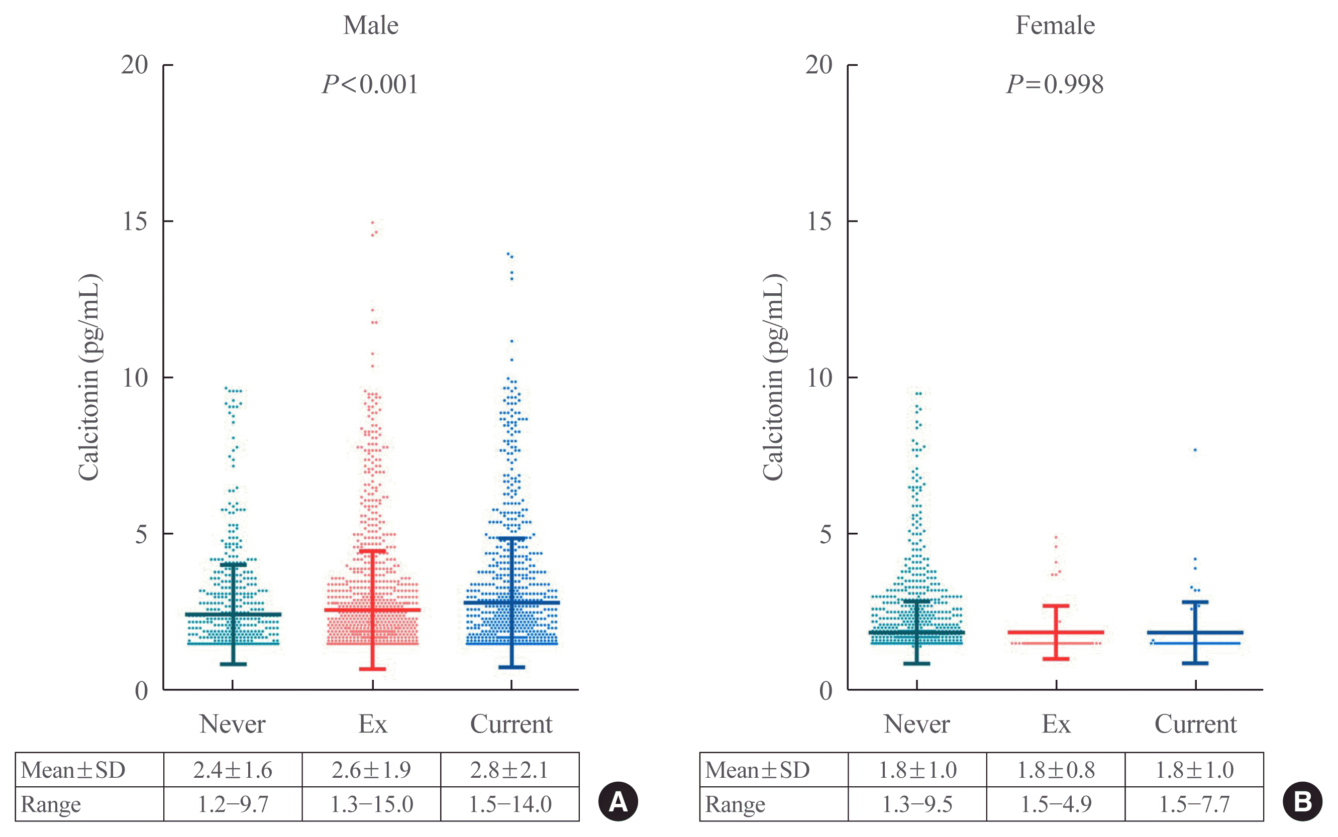
Serum calcitonin levels according to smoking status were assessed in each gender (Fig. 3). In male subjects, the mean calcitonin levels were 2.4±1.6, 2.6±1.9, and 2.8±2.1 pg/mL in non-, ex-, and current smokers, respectively. Moreover, the difference between the three groups was statistically significant (log-rank, P<0.001). However, no difference in mean calcitonin values exists in female subjects according to their smoking status (1.8± 1.0, 1.8±0.8, and 1.8±1.0 in non-, ex-, and current smokers, respectively; P=0.998).
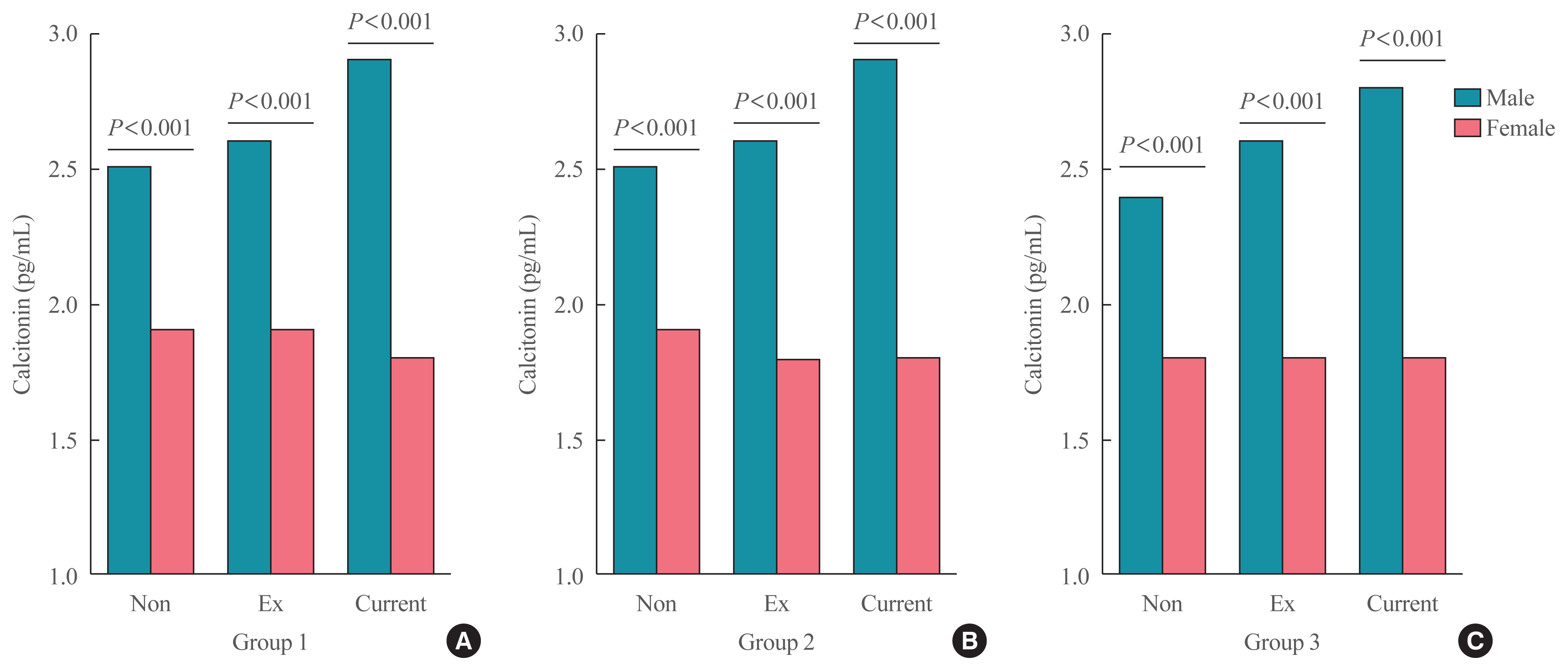
The difference in mean serum calcitonin level according to gender within the same smoking status category was evaluated (Fig. 4). In group 1, nonsmoker males had significantly higher mean serum calcitonin levels than nonsmoker females (P<0.001) (Fig. 4A). This was similar in the ex- and current smokers in the male and female subjects (all P<0.001). Identical differences—males having higher mean serum calcitonin levels than females despite the same smoking status—were observed in groups 2 (Fig. 4B) and 3 (Fig. 4C).
Distribution of serum calcitonin levels
The distribution of serum calcitonin concentrations as 95th percentile is summarized in Table 2 according to gender and smoking status. In group 1, non-, ex-, and current smoker males had 95th percentile ranges of 1.5 to 6.5, 1.5 to 7.0, and 1.5 to 8.1 pg/mL, respectively. In the same group, non-, ex-, and current smoker females had 95th percentile ranges of 1.5 to 3.5, 1.5 to 4.1, and 1.5 to 3.3 pg/mL, respectively. Generally, the 95th percentile range was similar among the three groups within the same gender and smoking status except for the nonsmoking males where the upper 95th percentile value was higher in group 1 (6.5 pg/mL) than those in groups 2 (5.8 pg/mL) or 3 (5.7 pg/mL).
Factors associated with hypercalcitoninemia
As aforementioned, the upper reference limit of serum calcitonin was 10 pg/mL. Although the majority of the subjects (99.3%, n=10,497) in group 1 had a calcitonin level of ≤10 pg/mL, 69 subjects had a calcitonin level higher than the upper reference limit. The possible factors that can elevate serum calcitonin levels in these 69 subjects are shown in Table 3. Autoimmune thyroid disease (eight subjects), CKD (two subjects), use of proton pump inhibitors (14 subjects), use of H2 blocker (two subjects), routine steroid administration (two subjects), and other malignancies (three subjects; two lung cancer and one myelodysplastic syndrome) were present. Among the 38 remaining subjects without any of these factors, 33 were ex- or current smokers. However, the reason for hypercalcitoninemia is unknown in five subjects.
DISCUSSION
This large-scale study including 10,566 healthy adults observed significant correlations of serum calcitonin levels with gender in which males had higher serum calcitonin levels than females. Smoking can further elevate calcitonin levels in males, but no such finding was found in females. While subjects aged 20 to 30 years had higher serum calcitonin levels than those aged ≥30 years, no significant association exists between serum calcitonin levels and age in subjects aged 30 to 80 years in both genders. Moreover, BMI did not affect calcitonin levels either. The results of our study suggest the following reference intervals:
Males: nonsmoker <5.7 pg/mL; ex-smoker <7.1 pg/mL; smoker <7.9 pg/mL
Females: <3.6 pg/mL (irrespective of smoking status)
Various factors can increase the concentration of calcitonin as listed in Table 3. These factors should be taken into account when interpreting elevated serum calcitonin levels than the suggested reference levels, especially when >10 pg/mL.
In this study, males can have higher calcitonin levels than females and this finding is supported by previous studies [20,21]. However, it is unclear why males have normally high calcitonin levels. This gender-dependent pattern cannot be explained by the difference of physique between males and females because BMI did not affect its level, and even under- or normal-weight males exhibited higher calcitonin levels than females in the same BMI range (Fig. 2B). Also, nonsmoking males had significantly higher calcitonin levels compared to nonsmoking females (Fig. 4) which leads to the conclusion that smoking in either gender was not the reason for the difference. A previous study of 57 normal autopsy thyroid glands revealed that maximum C cell surface area was twice as high in men as in women [27]. Moreover, 14 (33%) adult subjects fulfilled C cell hyperplasia criteria, and 12 out of these 14 adults were men. These suggest that normal adults can have C cell hyperplasia quite frequently and higher prevalence was observed in males than in females. Some researchers suggested the possible role of androgens in C cell growth regulation and proposed that testosterone may partially explain the higher serum calcitonin levels in men compared to women [28]. Taken together, it is clear that the reference range of serum calcitonin should be separately provided according to gender.
Smoking—either in the past or currently—evidently elevated calcitonin concentration in males. However, its effect on calcitonin levels in females was not significant. This finding was first reported by d’Herbomez et al. [20]. They reported significantly higher serum calcitonin levels in smoking than nonsmoking men, but no such difference was found in women. A large body of evidence suggests that cigarette smoking alters thyroid function and metabolism [29–31]. However, its specific effect on C cells is unclear. One study observed nicotine-dependent elevation of calcitonin levels in human serum and urine, and a similar effect was noted in thyroidectomized patients [32]. This phenomenon suggests that the source of calcitonin secreted by smoking is not the thyroid gland but rather the lung, possibly from the pulmonary neuroendocrine cells. The small number of female subjects with smoking habits (2.9% and 3.5% ex- and current smokers, respectively) may have limited this subgroup in reaching statistical significance.
This study classified the subjects from groups 1 to 3 by excluding possible factors associated with hypercalcitoninemia in phases. Groups 1, 2, and 3 included all eligible subjects; those without any self or family history of thyroid disease; and had no evident CKD, medication of proton pump inhibitor/H2 blocker/steroid, or other malignancies, respectively. The mean serum calcitonin levels were similar in the three groups but the ranges of its level were narrower in groups 2 (1.2 to 15.3 pg/mL) and 3 (1.2 to 15.3 pg/mL) compared with group 1 (1.0 to 53.0 pg/mL). Although the range still remains wide in group 3, about 97% of the total subject with serum calcitonin levels higher than the manufacturer’s upper reference limit (10 pg/mL) can be explained by factors other than MTC as shown in Table 3. Increased serum calcitonin release can occur in autoimmune thyroid disease probably due to benign C cell hyperplasia [33,34]. Nonthyroidal conditions for elevated calcitonin levels include chronic renal failure, various neuroendocrine tumors, hypergastrinemias, presence of heterophilic antibodies, and use of certain drugs (proton pump inhibitors, H2 blockers, or steroid), although each physiological significance and the underlying mechanism remains unclear [15–19,25].
In conclusion, males and smokers have normally higher serum calcitonin levels than their counterparts according to the results of this large sample-sized study of healthy Korean adults. Taking into account the several variables known to induce hypercalcitoninemia can help to interpret moderately elevated serum calcitonin levels and reduce further unnecessary examinations by suspecting MTC in these subjects. Specific calcitonin reference range should be provided considering gender and smoking status, which may ameliorate the role of serum calcitonin in screening for MTCs.
 XML Download