Abstract
Median nerve is formed by lateral root from lateral cord and medial root from medial cord of brachial plexus. Formation of median nerve occur in front or lateral to axillary artery in axilla. In the present study we observed anatomical variations of median nerve formation in the brachial plexus. We examined formalin fixed 60 upper limbs from 30 adult cadavers (15 males and 15 females) which were above the age 40 years from the department of Anatomy. All the cadavers were dissected on both sides according to Cunningham’s Manual of Practical Anatomy. Normal formation of median nerve by two roots noted in 42 (70.0%) of upper limb specimen. Variation of median nerve formation noted in 18 (30.0%) upper limb specimen. Three roots taking part in the formation of median nerve in 13 (21.7%) upper limb specimen where additional root coming from lateral cord of brachial plexus. Four roots taking part in formation of median nerve in 3 (5.0%) upper limb specimen, where additional roots coming from lateral cord and posterior cord of brachial plexus. Lateral root crossed the axillary artery anteriorly to join with medial root lying medial to axillary artery. The median nerve formed medial to third part of axillary artery. Additional communication with musculocutaneous nerve with median nerve seen in 2 (3.3%) upper limb specimen. Knowledge of such anatomical variations is of interest to the anatomist and clinician alike. Surgeons who perform procedures involving neoplasm or repairing trauma need to be aware of these variations. Median nerve variation may lead to confusions in surgical procedures and axillary brachial plexus nerve block anesthesia.
The brachial plexus supplies the upper limb and is formed by union of the ventral rami of the lower four cervical and first thoracic spinal nerves (C5-T1). It consists of roots, trunk, division, and cords. The fourth cervical ventral ramus frequently gives a branch to the fifth as pre-fixed type and the first thoracic ventral ramus often receives a contribution from the second thoracic ventral ramus as post fixed type [1, 2]. The ventral rami of C5 and C6 join to form the upper trunk, C7 becomes the middle trunk, and C8 and T1 form the lower trunk. These three trunks just above or behind the clavicle bifurcate into anterior and posterior divisions. All of the posterior divisions form the posterior cord. The lateral cord is formed by the union of the anterior divisions of the upper and middle trunk. The medial cord is formed as a continuation of the anterior division of the lower trunk [3, 4]. Median nerve is formed by union of two roots, lateral root and medial root coming from lateral and medial cord of brachial plexus respectively. Formation of median nerve occur in front or lateral to axillary artery in axilla [5]. The medial root of the nerve crosses from lateral to medial anterior to third part of the axillary artery superficially. In the arm the median nerve crosses anterior to the brachial artery from lateral to medial side, and enters the cubital fossa along with the brachial artery. Formations and anomalies of the nerves of the upper limb have been described by many authors [6]. Nerve variations of the upper limb are very important in routine surgery and during radical neck dissections where these variations are more prone to injury [7]. These variations may also help in interpretation of a nervous compression having unexplained clinical symptoms.
However, in the present study we are reporting multiple variations of median nerve during the routine dissection. Understanding these anatomical variations of the brachial plexus, will append to existing knowledge explaining its morphological and clinical significance.
During routine dissection of axilla of embalmed adult cadavers in the department of Anatomy, we studied 60 upper limbs in 30 cadavers (15 males and 15 females) which were above the age 40 years. All the cadavers were dissected on both sides according to Cunningham’s Manual of Practical Anatomy [8]. Skin, superficial fascia and deep fascia were separated and various muscles were reflected, to visualize the formation of median nerve and its variation. The cadavers studied did not show any gross anomalies or evidence of surgical procedures on the skin around the area dissected. Visual observations were preserved with digital photography and were recorded. The study was approved by the ethical committee of the institute (Id no: KMC/IEC/2021-35) and the informed consent was waived.
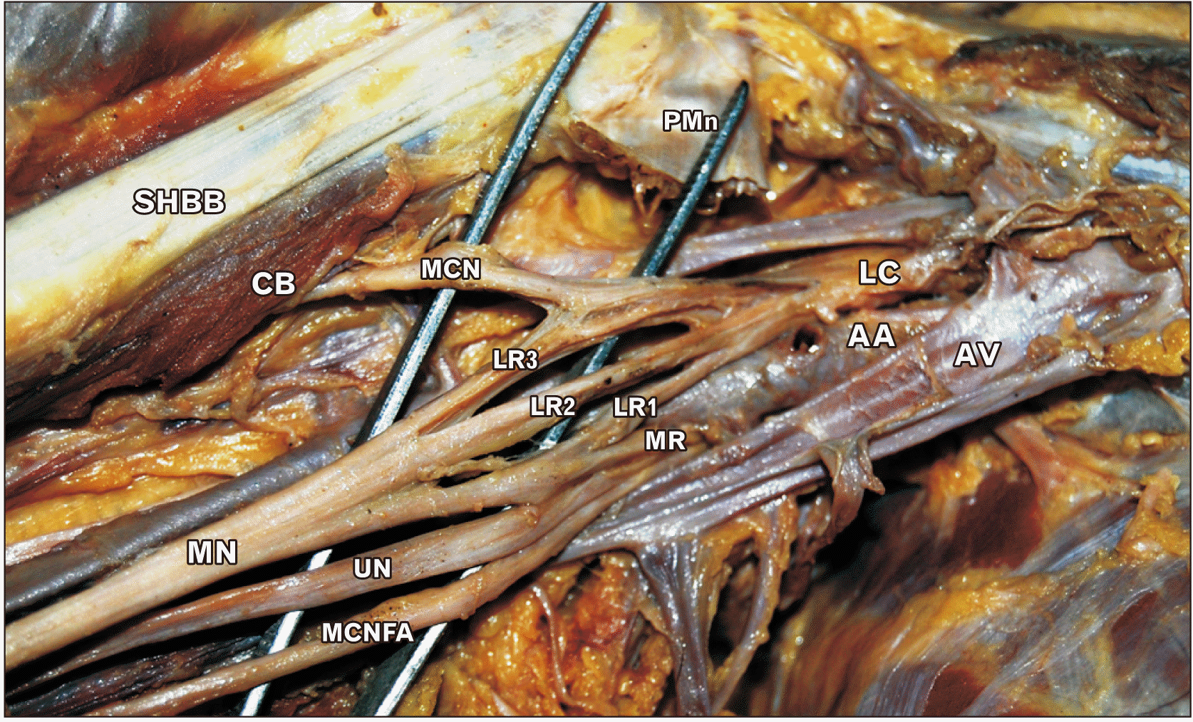
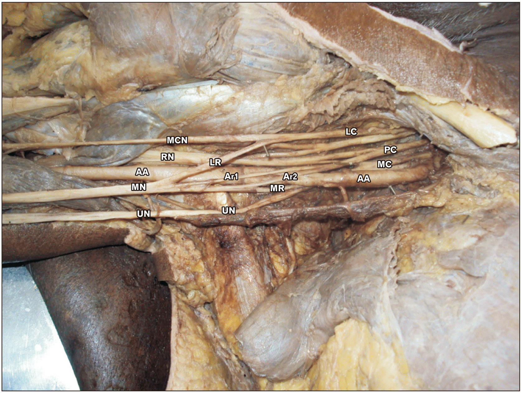
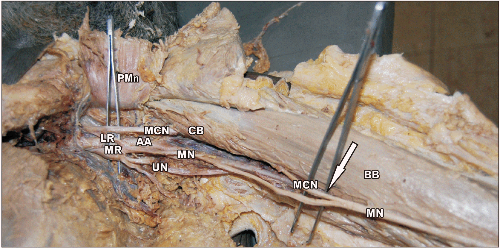
The macroscopic examination of brachial plexus for the presence of median nerve variation was done among the 60 upper limbs. Normal formation of median nerve by two roots noted in 42 (70.0%) upper limb specimen seen in Fig. 1. One root coming from lateral cord and another root coming from medial cord of brachial plexus. Variation of median nerve formation noted in 18 (30.0%) upper limb specimen. Three roots taking part in the formation of median nerve in 13 (21.7%) upper limb specimen where one root coming from medial cord and two roots coming from lateral cord of brachial plexus seen in Fig. 2. Four roots taking part in formation of median nerve in 3 (5.0%) upper limb specimen, where additional three roots coming from lateral cord of brachial plexus seen in Fig. 3 and additional roots coming from posterior cord of brachial plexus seen in Fig. 4. Lateral root crossed the axillary artery anteriorly to join with medial root lying medial to axillary artery. The median nerve formed medial to third part of axillary artery. Communication with musculocutaneous nerve with median nerve in 2 (3.3%) upper limb specimen seen in Fig. 5.
Brachial plexus variation may occur in the formation of trunks, division, cords and terminal branches [9]. Formations and anomalies of the nerves of the upper limb have been described by many authors [10-12]. Formation of median nerve by two lateral and one medial root is reported earlier [13, 14]. In the present study we found unusual formation of median nerve by three and four roots from medial and lateral cord of brachial plexus. We also find additional roots taking part in formation of median nerve from posterior cord of brachial plexus. No previous study reported this kind of variation of median nerve formation. Such cases of origin of median nerve may lead to confusions in surgical procedures and nerve block anesthesia. Median nerve was formed by three roots where two roots came from lateral cord and one from medial cord. The formation of median nerve noted in the axilla and also in the arm [15]. Pais et al. [16] reported that median nerve formation by three roots in the axilla. In the present study it has been observed that three roots taking part in the formation of median nerve in 13 (21.7%) upper limb specimen where one root coming from medial cord and two roots coming from lateral cord of brachial plexus. Uzun and Seelig (2001) [17] reported that median nerve formed by four roots where three roots coming from lateral cord and one root coming from medial cord of brachial plexus. In the present study four roots taking part in formation of median nerve in 3 (5.0%) upper limb specimen where additional root coming from lateral cord and posterior cord of brachial plexus.
Present study also reported three roots from lateral cord and one root from medial cord of brachial plexus. We also find two additional roots from the posterior cord of brachial plexus along with lateral root and medial root of median nerve formation. No previous study found this kind of median nerve formation. Normally median nerve runs on the lateral side of axillary artery. Medial root crosses the axillary artery to join the lateral root. Haviarova et al. (2001) [18] reported that formation of median nerve was posterior to axillary artery. Pandey and Shukla (2007) [19] reported median nerve formation medial to third part of axillary artery in 4.7% cases. In the present study the lateral root crossed the axillary artery anteriorly to join with medial root lying medial to axillary artery. The median nerve formed medial to third part of axillary artery. Knowledge of such variation has clinical importance especially in post-traumatic evaluations and peripheral nerve repair. The communications between musculocutaneous and median nerve are also well documented [20, 21]. In the present study communication was present between the median nerve and musculocutaneous nerves. The interpretation of the nerve anomaly of the arm requires consideration of the phylogeny and development of the nerves of the upper limb. Communication between the median nerve and musculocutaneous nerves is considered as a remnant from the phylogenetic or comparative point of view [13]. Embryologically upper limb buds lie opposite to the lower five cervical and upper two thoracic segments. During the buds formation ventral rami of the spinal nerve penetrate into the mesenchyme of the limb buds and establish intimate contact with differentiating mesodermal condensations. The early contact between nerve and muscle cell is a prerequisite for their complete functional differentiation [22, 23]. The variations could arise from circulatory factors at the time of fusion of brachial plexus cord. In human, the forelimb muscles develop from the mesenchyme of the para-axial mesoderm during fifth week of embryonic life [22]. The axon of spinal nerve grows distally to reach the limb bud mesenchyme. The peripheral process of the motor and sensory neurons grows in the mesenchyme in different directions. Once formed, any developmental differences would obviously persist postnatally [23]. As the guidance of the developing axons is regulated by expression of chemo-attractants and chemo-repellants in a highly coordinated site specific fashion, any alteration in signaling between mesenchymal cells and neuronal growth cones can lead to significant variations [24]. Variation of median nerve is very common and clinician and anatomist must know about these variations.
In conclusion, median nerve formation may arise from more than two roots in the brachial plexus. Contribution of three roots for median nerve formation are more common. Median nerve variation is essential to anatomist and clinician during surgical exploration in the axilla and nerve block anesthesia. It also helps in diagnosis of peripheral neuropathy in traumatic injuries.
Acknowledgements
The authors wish to thankfully acknowledge the technician support received from the department of Anatomy to carry out the work.
Notes
References
1. Ghosh B, Parashar V, Gaur N, Thakur B, Yadav S. 2015; Variation in median nerve formation associated with anomalous higher origin of profunda brachi artery. Int J Recent Sci Res. 6:5654–6.
2. Datta AK. 2017. Essentials of human anatomy. 5th ed. Vol 3:Current Books International;Kolkata: p. 41.
3. Standring S. 2016. Gray's anatomy: the anatomical basis of clinical practice. 41st ed. Elsevier;Philadelphia: p. 781–3.
4. Taams KO. 1997; Martin-Gruber connections in South Africa. An anatomical study. J Hand Surg Br. 22:328–30. DOI: 10.1016/S0266-7681(97)80396-8. PMID: 9222911.
5. Drake RL, Vogl W, Mitchell AWM. 2005. Gray's anatomy for students. Elsevier/Churchill Livingstone;Philadelphia: p. 661–3.
6. Vollala VR, Raghunathan D, Rodrigues V. 2005; Nerve compressions in upper limb: a case report. Neuroanatomy. 4:35–6.
7. Gacek RR. 1990; Neck dissection injury of a brachial plexus anatomical variant. Arch Otolaryngol Head Neck Surg. 116:356–8. DOI: 10.1001/archotol.1990.01870030120023. PMID: 2306357.

8. Romanes GJ. 1986. Cunningham's manual of practical anatomy. 15th ed. Upper and lower limb. Volume 1:Oxford University Press;Oxford: p. 28–30. DOI: 10.1001/archotol.1990.01870030120023.
9. Gupta M, Goyal N, Harjeet . 2005; Anomalous communications in the branches of brachial plexus. J Anat Soc India. 54:22–5.
10. Budhiraja V, Rastogi R, Asthana AK. 2011; Anatomical variations of median nerve formation: embryological and clinical correlation. J Morphol Sci. 28:283–6.
11. Nayak S. 2007; Absence of musculocutaneous nerve associated with clinically important variations in the formation, course and distribution of the median nerve- a case report. Neuroanatomy. 6:49–50.
12. Uzun A, Bilgic S. 1999; Some variations in the formation of the brachial plexus in infants. Tr J Med Sci. 29:573–7.
13. Chauhan R, Roy TS. 2002; Communication between the median and musculocutaneous nerve - a case report. J Anat Soc India. 51:72–5.
14. Saeed M, Rufai AA. 2003; Median and musculocutaneous nerves: variant formation and distribution. Clin Anat. 16:453–7. DOI: 10.1002/ca.10096. PMID: 12903070.

15. Sontakke BR, Tarnekar AM, Waghmare JE, Ingole IV. 2011; An unusual case of asymmetrical formation and distribution of median nerve. Int J Anat Var. 4:57–60.
16. Pais D, Casal D, Santos A, O'Neill JG. 2010; A variation in the origin of the median nerve associated with an unusual origin of the deep brachial artery. J Morphol Sci. 27:35–8.
17. Uzun A, Seelig LL Jr. 2001; A variation in the formation of the median nerve: communicating branch between the musculocutaneous and median nerves in man. Folia Morphol (Warsz). 60:99–101. PMID: 11407150.
18. Haviarova Z, el Falougy HA, Killingerova A. 2001; Atypical course of the median nerve. Bratisl Lek Listy. 102:372–3. PMID: 11763668.
19. Pandey SK, Shukla VK. 2007; Anatomical variations of the cords of brachial plexus and the median nerve. Clin Anat. 20:150–6. DOI: 10.1002/ca.20365. PMID: 16795062.

20. Choi D, Rodríguez-Niedenführ M, Vázquez T, Parkin I, Sañudo JR. 2002; Patterns of connections between the musculocutaneous and median nerves in the axilla and arm. Clin Anat. 15:11–7. DOI: 10.1002/ca.1085. PMID: 11835538.

21. Yang ZX, Pho RW, Kour AK, Pereira BP. 1995; The musculocutaneous nerve and its branches to the biceps and brachialis muscles. J Hand Surg Am. 20:671–5. DOI: 10.1016/S0363-5023(05)80289-8. PMID: 7594300.

22. Larsen WJ. 1997. Human embryology. 2nd ed. Churchill Livingstone;New York: p. 311–39.
23. Brown MC, Hopkins WG, Keynes RI. 1991. Essentials of neural development. Cambridge University Press;Cambridge: p. 46–66.
24. Sanes DH, Reh TA, Harris WA. 2000. Development of the nervous system. Academic Press;San Diego: p. 189–97.
Fig. 1
Photograph of formation of median nerve (MN) by two roots. LC, lateral cord; LR, lateral root; MR, medial root; MCN, musculocutaneous nerve; AA, axillary artery; BB, biceps brachii muscle; CB, coracobrachialis muscle; UN, ulnar nerve.
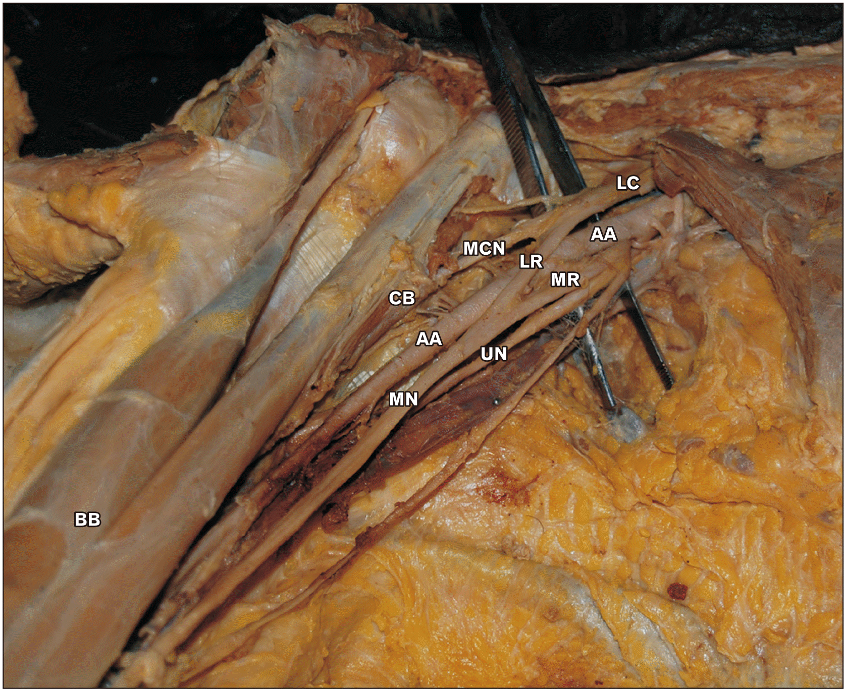
Fig. 2
Photograph of formation of median nerve (MN) by three roots. LC, lateral cord; AA, axillary artery; AV, axillary vein; CB, coracobrachialis; MCN, musculocutaneous nerve; LR1, lateral root first; LR2, lateral root second; MR, medial root; UN, ulnar nerve.
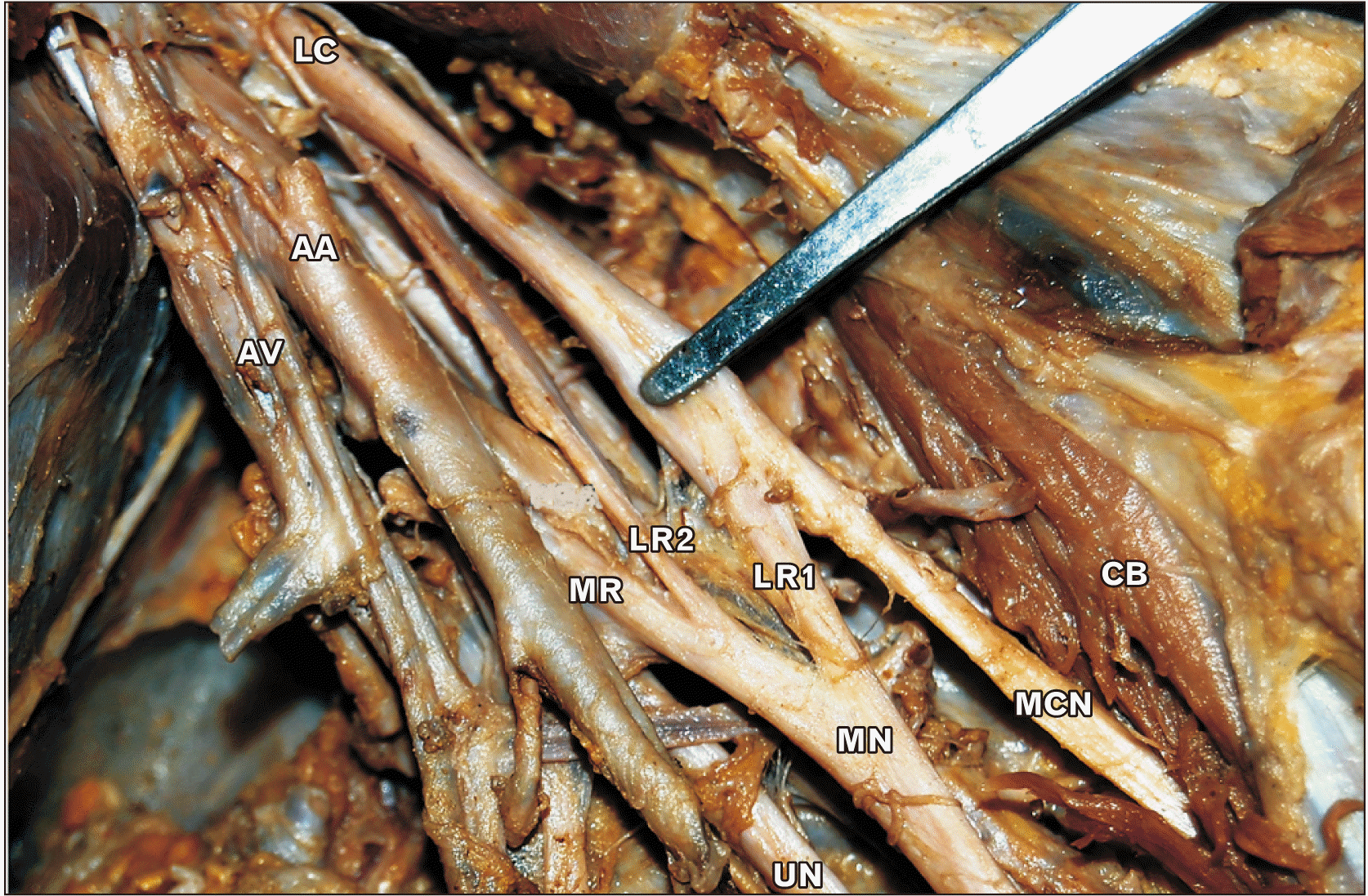
Fig. 3
Photograph of formation of median nerve (MN) by four roots. LC, lateral cord; AA, axillary artery; AV, axillary vein; CB, coracobrachialis; SHBB, small head of biceps brachii muscle; PMn, pectoralis minor muscle cut end; MCN, musculocutaneous nerve; LR1, lateral root first; LR2, lateral root second; LR3, lateral root third; MR, medial root; UN, ulnar nerve; MCNFA, musculocutaneous nerve of forearm.


 PDF
PDF Citation
 Print
Print


 XML Download