

 PDF
PDF ePub
ePub Citation
 Print
Print


INTRODUCTION
Ganglion cell tumors (GCT) are divided into two sub-types : gangliocytoma and ganglioglioma. Although intramedullary gangliocytoma are extremely rare, intramedullary gangliogliomas are more frequent. Only ten cases of intramedullary gangliocytoma have been reported in the literature. Only one gangliocytoma case located at cervicomedullary junction has been published15). Although association with scoliosis and Von Recklinghausen's disease were previously reported in the literature, no gangliocytoma cases concomitant with neuroendocrine tumor of lung have been published. In this report, we aim to present a gangliocytoma case at cervicomedullary junction in a patient with neuroendocrine tumor of lung and discuss diagnosis and treatment options with literature.
CASE REPORT
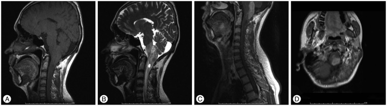
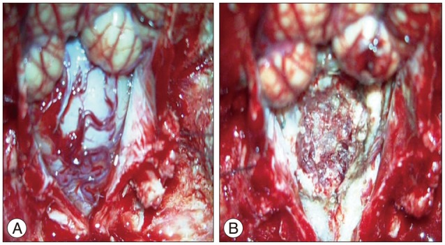
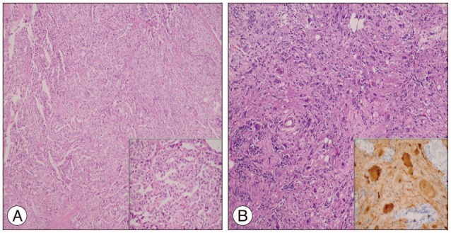
A 20-year-old male patient admitted with pain of neck and head. The patient underwent 2 surgeries previously 6 and 3 year ago due to neuroendocrine tumor of lung and had received octreotide therapy. Cervical magnetic resonance (MR) imaging revealed a 45×14 mm sized mass lesion mixed with cystic and solid components, calcification foci extending from medulla oblongata to C2 level, with edema or infiltration areas extending to C6 level, and the mass was heterogeneous on T1-weighted MR imaging, hyperintense on T2-weighted MR imaging and solid components showed intense contrast-enhancing following injection of Gadolinium (Gd) (Fig. 1). Metastasis was considered in the first place in preliminary diagnosis. The patient was scheduled for surgery. Suboccipital craniectomy and C1 laminectomy were performed following brainstem mapping with neurophysiological monitoring. The dura was opened in the midline. Cervicomedullary junction was found to be enlarged. After midline myelotomy, a stiff-structured gray tumor with unclear boundaries was encountered. Partial resection was performed with the help of ultrasonic aspirator (Fig. 2). Operation was terminated upon detection of increase in temporary somatosensory evoked potential latencies and decrease in motor evoked potential amplitudes during the operation. Postoperative neurological examination was intact. On microscopic examination, intense inflammation, lots of ganglion cells with focal calcification that stained positively with S100 and synaptophysin. No obvious glial component was detected with glial fibrillary acidic protein (GFAP). Ki67 was 1%. In the light of these findings, the case was reported as grade 1 gangliocytoma pathology (Fig. 3). No adjuvant therapy was given. The patient had no neurologic deficits at 3-month follow-up and radiographic progression was not detected.
DISCUSSION
Ganglioglioma accounts for 0.4–7.6% of pediatric central nervous system (CNS) neoplasms and 1.3% of those in adults8). They are slowly growing benign tumors and malignant transformation may be seen in less than 10%16). Generally, they are located in the supratentorial region and most commonly involve temporal lobe. Brain stem and spinal location are rare. Spinal location has been reported to be associated with scoliosis. Gangliocytoma are 0.1% to 0.5% of all CNS tumors and children and young adults constitute 60% of all patients5). Intracranial location is usually seen in temporal lobe, frontoparietal region and floor of 3rd ventricle, gangliocytoma involving cerebellum is described as Lhermitte-Duclos disease5). Association with scoliosis and Von Recklinghausen's disease was reported5). Intramedullary spinal cord and cervicomedullary junction gangliocytomas are extremely rare. Up to date, nine cases for spinal cord and only one case cervicomedullary junction gangliocytomas were reported in the literature1234567111315).
Clinical symptoms usually vary depending on spinal cord compression or infiltration of located region. They may present with neurological symptoms ranging from pain, as in our case, radiculopathy, paraparesis and paraplegia. Although there are no significant radiological imaging features, they are usually hypointense on T1-weighted MR imaging and hyperintense on T2-weighted MR imaging and are enhancing lesions at different rates15). Astrocytoma, brain stem gliomas, ependymomas and metastases should be considered in differential diagnosis.
Ganglioglioma consist of dysplastic neurons and neoplastic glial cells, whereas gangliocytoma have dysplastic neural cells and normal glial cells212). Immunohistochemical study aids the confirmation of diagnosis of ganglion cell tumor. The glial populations are reactive for GFAP, S-100 protein, and vimentin, but the neurons are for synaptophysin and Chromogranin A91014).
Primary treatment of GCT is surgical excision and they are considered resistant to other adjuvant therapies, as they are well-differentiated slow-growing tumors. However, malignant transformations have been reported in the literature5). Radiological follow-up is done for patients who underwent total excision, but adjuvant therapy remains controversial in patients who underwent subtotal excision. Total resection is difficult in reported cervical-junction GCTs. As published in the literature, only four of 10 cases of intramedullary gangliocytoma underwent gross total resection671315). Gangliocytoma cases published in the literature do not have enough follow-up periods. Jacob et al5). reported no clinical and radiographic progression in a partially resected case in 3-year follow-up.
In our case, because of pre-existing neuroendocrine tumor of lung, metastases were considered in the first line and operation has been decided although there were no neurological deficits. Although association with scoliosis and Von Recklinghausen's disease were previously reported in the literature, no GCT cases concomitant with neuroendocrine tumor of lung have been published.
With increasingly reported GCTs in recent years, they should be kept in mind in the differential diagnosis of intramedullary tumors especially in children and young adults. Pathological study is the most important diagnostic method for GCTs. Surgical excision is the primary treatment, but difficulty in total surgical tumor resection is the most important problem.
 XML Download