This article has been
cited by other articles in ScienceCentral.
Abstract
An ultrasonogram is very useful for the diagnosis of thyroid cancer and determining the preoperative stage. Accurate staging before surgery using an ultrasonogram is essential for determining the extent of surgery. This review summarizes the use of ultrasonography in the preoperative staging of thyroid cancer.
Keywords: Ultrasonography, Thyroid carcinoma, Stage
서 론
국가암정보센터의 암등록통계 자료에 따르면 2017년 현재 갑상선암은 전체 성별에서 4번째, 남자에서는 6번째, 여자에서는 두 번째로 많이 발생하는 암종으로 보고되고 있다.(
1) 특히 우리나라에서는 1 cm 미만의 비교적 작은 크기에서 발견되는 비율이 많은 것으로 보고되고 있다.(
2) 비교적 크기가 작고 갑상선 내에 한 쪽 엽에만 국한되어 있으며 림프절이나 전신전이가 없는 갑상선암의 경우 갑상선엽절제술을 시행할 수 있으며 이는 갑상선전절제술을 시행한 경우에 비해 환자의 삶의 질을 향상시키는데 크게 도움이 된다.(
3,
4) 따라서 초음파를 이용해 정확한 수술 전 병기 결정 후 수술범위와 치료를 결정하는 것이 필수적이다.
이에 문헌 고찰을 통해 최근 개정된 AJCC Cancer Staging Manual 8판을 기준으로 갑상선암에서 수술 전 초음파를 이용한 병기 결정에 대해 알아보고자 한다.(
5)
본 론
갑상선 내에만 국한된 작은 크기의 갑상선암은 추가적인 검사 없이 초음파만으로도 병기 결정이 충분하다. T 병기는 종양의 크기와 갑상선 외부로의 침범여부에 따라 결정이 된다.
T1은 갑상선 내에 종양이 국한되어 있으면서 크기가 2 cm 이하로 정의되며, 이중 미세갑상선유두암(micropapillary carcinoma)에 해당하는 1 cm 이하는 T1a로(
Fig. 1A), 1 cm보다는 크며 2 cm 이하인 경우를 T1b로 세분한다(
Fig. 1B). T2는 종양이 갑상선 내에 국한되어 있으며 크기가 2 cm보다는 크고 4 cm 이하인 경우로 정의한다(
Fig. 2). 여전히 논란의 여지는 있지만 2015년에 개정된 미국갑상선학회(American Thyroid Association)의 권고안에 따르면 한쪽 엽에만 국한되어 있으면서 림프절 전이가 없는 T2까지에 해당하는 분화갑상선암은 갑상선엽절제술의 대상으로 권고하고 있다.(
3) 하지만, 크기가 작아도 양쪽 엽에 다발성으로 존재하는 경우 초음파유도 세침 흡인 세포검사(fine needle aspiration cytology; FNAC)를 시행하여 악성으로 판명되는 경우 갑상선전절제술의 대상이 되므로 수술 전 세심한 평가가 필수적이라 하겠다.
T3는 4 cm보다 크거나 크기에 상관없이 육안적으로 띠근육(strap muscle)을 침범한 경우로 정의하며(
Fig. 3), T4는 종양의 크기에 상관없이 주변의 기관이나 되돌이후두신경(recurrent laryngeal nerve) 또는 혈관의 육안적인 침범이 관찰되는 경우로 정의한다. 이러한 소견은 수술 전 초음파를 통해 평가 가능하며, 큰 갑상선암의 전체 범위를 평가하거나 특히 T4에 해당하는 갑상선외 침범이 관찰되는 경우 경부 전산화단층촬영이나 자기공명영상을 통해 추가적으로 병기설정을 하는데 도움을 줄 수 있다. 특히 T4에 해당하는 경우에는 이러한 추가 검사를 통해 재건수술이 필요한지 혹은 수술이 불가능한 상태인지를 판단할 수 있다.
갑상선암이 경부림프절로 전이되는 빈도는 갑상선유두암에서 30-90%로 가장 흔하게 보고되고 있으며 수질암에서 50%, 역형성암에서 40% 정도로 보고되며 이에 반해 혈행성 전이를 주로 한다고 알려져 있는 여포암에서도 10% 정도에서 림프절 전이를 하는 것으로 보고되고 있다.(
6) 초음파는 이러한 림프절 전이를 평가하는 일차 도구로 이용되고 있으며, 림프절 전이를 의심할 수 있는 소견이 관찰되면 FNAC를 통해 확인을 하는데 역시 초음파가 이용되고 있다.
수술 전 초음파를 통해 림프절 전이 여부를 평가하는 것은 수술의 범위를 결정하는데 필수이다. 중심구획(Level VI)이나 상종격동구획(Level VII)에 전이가 있는 상태를 N1a로 정의하며(
Fig. 4), 이 구획의 림프절은 수술 전 초음파로 확인을 하는데 해부학적으로 한계가 있지만 초음파의 탐색자(probe)를 흉골 뒤편 아래로 기울여 관찰할 수 있다. 그에 반해 갑상선암이 있는 편측, 양측 혹은 반대측 경부림프절(Level I, II, III, IV 또는 V)로 전이가 있는 상태를 N1b로 정의하며(
Fig. 5), 보통 갑상선암으로 수술하는 경우 중심구획림프절(Level VI)과 달리 통상적으로 림프절절제술을 시행하는 부위가 아니므로 수술 전 세심하고 정확한 평가가 필수적이며 전이가 확인된 경우 변형근치림프절절제술(modified radical neck dissection)을 계획하고 시행해야 한다. 특히 목의 어느 구획이든 수술 전 림프절 전이가 확인된 경우에는 갑상선의 수술 범위도 갑상선전절제술을 시행해야 하므로 수술 전 림프절 전이 여부를 확인하는 것은 아무리 강조해도 지나치지 않는다. 수술 전 림프절전이 평가를 하는데 초음파와 더불어 필요한 경우 경부 전산화단층촬영이나 자기공명영상이 도움을 줄 수 있다.
결 론
초음파는 갑상선암의 수술 전 병기 설정에 매우 유용하게 이용이 되고 있다. 수술 전 초음파를 이용한 정확한 병기 설정이 수술의 범위를 결정하는데 매우 필수적이며, 이를 통해 수술 후 환자의 삶의 질 향상에도 기여할 수 있다.
ACKNOWLEDGEMENTS
이 논문은 2019학년도 조선대학교 연구지원금의 지원을 받아 연구되었습니다.
REFERENCES
1. 2019. National Cancer Information Center [Internet]. National Cancer Information Center;Goyang: Available from:
https://www.cancer.go.kr. cited 2020 Jan 22.
2. Cho BY, Choi HS, Park YJ, Lim JA, Ahn HY, Lee EK, et al. 2013; Changes in the clinicopathological characteristics and outcomes of thyroid cancer in Korea over the past four decades. Thyroid. 23:797–804. DOI:
10.1089/thy.2012.0329. PMID:
23427907. PMCID:
PMC3704118.

3. Haugen BR, Alexander EK, Bible KC, Doherty GM, Mandel SJ, Nikiforov YE, et al. 2016; 2015 American Thyroid Association management guidelines for adult patients with thyroid nodules and differentiated thyroid cancer: the American Thyroid Association Guidelines task force on thyroid nodules and differentiated thyroid cancer. Thyroid. 26:1–133. DOI:
10.1089/thy.2015.0020. PMID:
26462967. PMCID:
PMC4739132.
4. Kebebew E, Clark OH. 2000; Differentiated thyroid cancer: "complete" rational approach. World J Surg. 24:942–51. DOI:
10.1007/s002680010165. PMID:
10865038.
5. Amin MB, Edge SB, Greene FL, Byrd DR, Brookland RK, Washington MK, et al. 2017. AJCC Cancer Staging Manual. 8th ed. Springer International Publishing;New York: DOI:
10.1007/978-3-319-40618-3_2.
Fig. 1
Ultrasonographic image of T1 (≤2 cm) papillary thyroid carci-noma confined to thyroid gland. (A) T1a (≤1 cm) papillary thyroid carci-noma showing taller than wide appearance with internal microcalci-fication. (B) T1b (>1 cm but ≤2) papillary thyroid carcinoma showing irregular margin with internal micro-calcification.
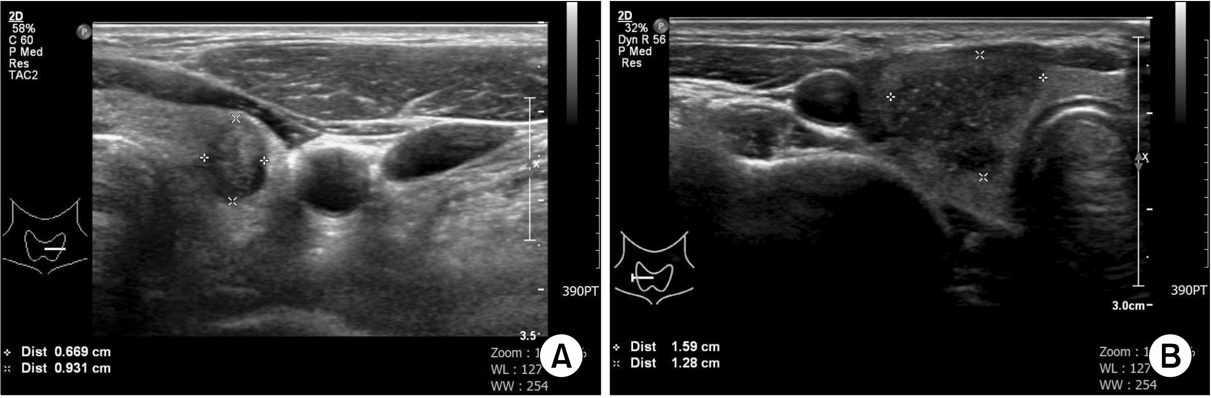
Fig. 2
Ultrasonographic image of T2 (>2 cm but ≤4 cm) papillary thyroid carcinoma confined to thyroid gland.
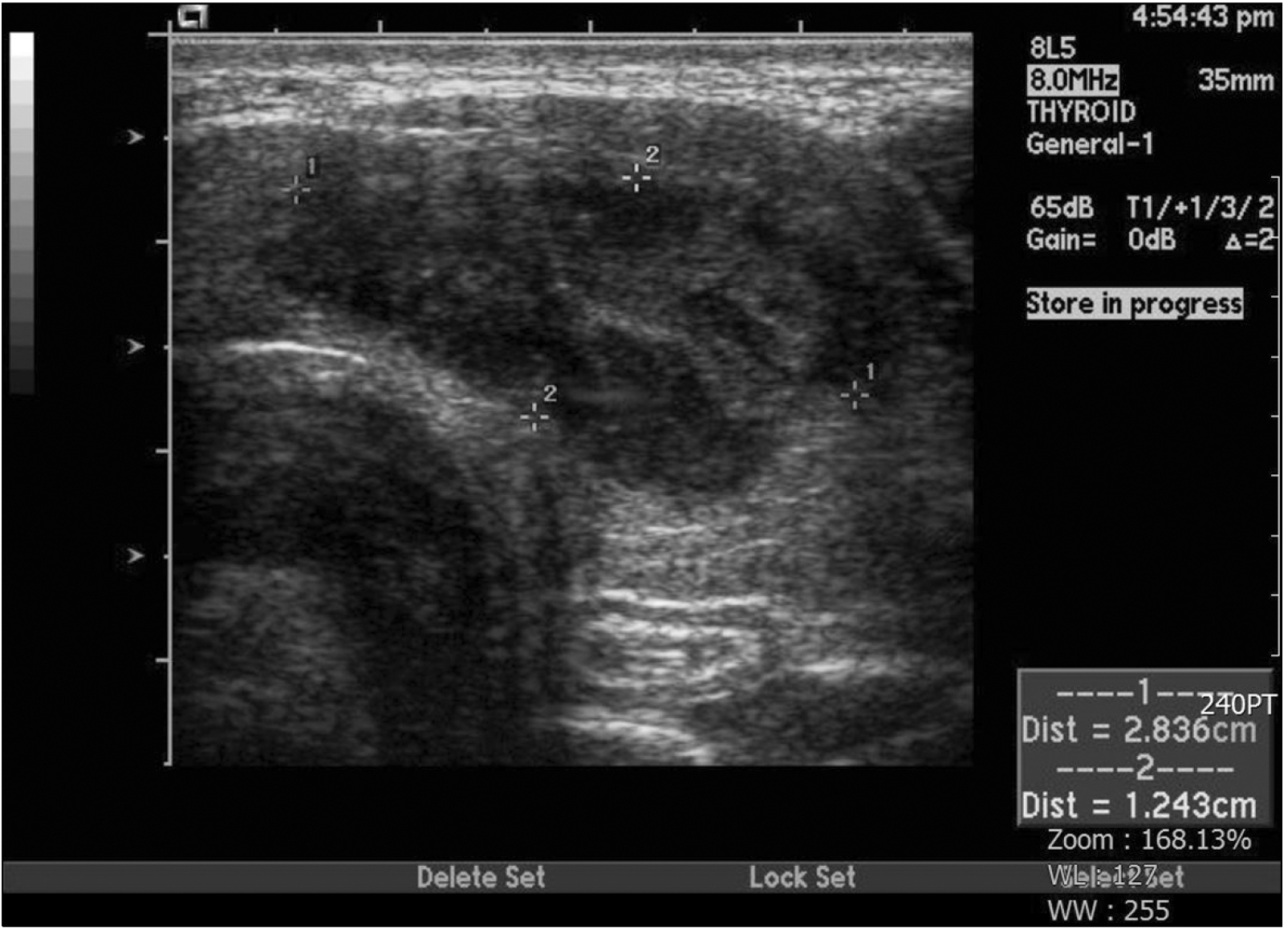
Fig. 3
Ultrasonographic image of T3 papillary thyroid carcinoma showing invasion to strap muscle.
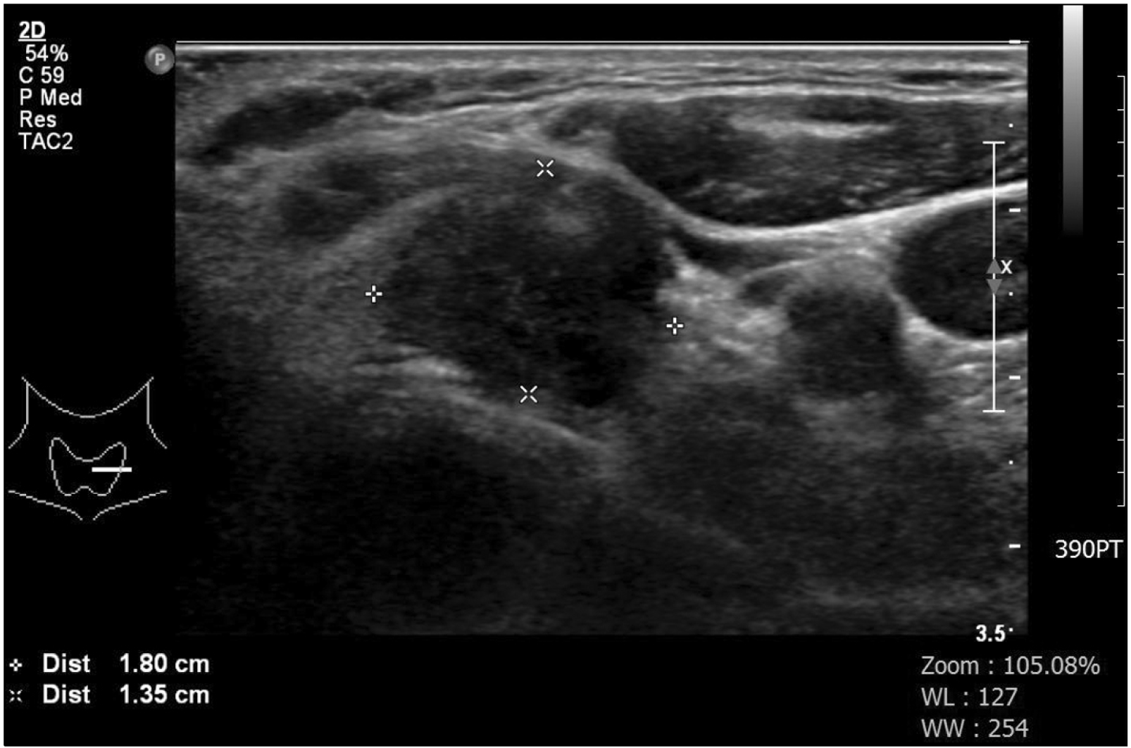
Fig. 4
Ultrasonographic image of N1a. Lymph node without fatty hilum in level VI.
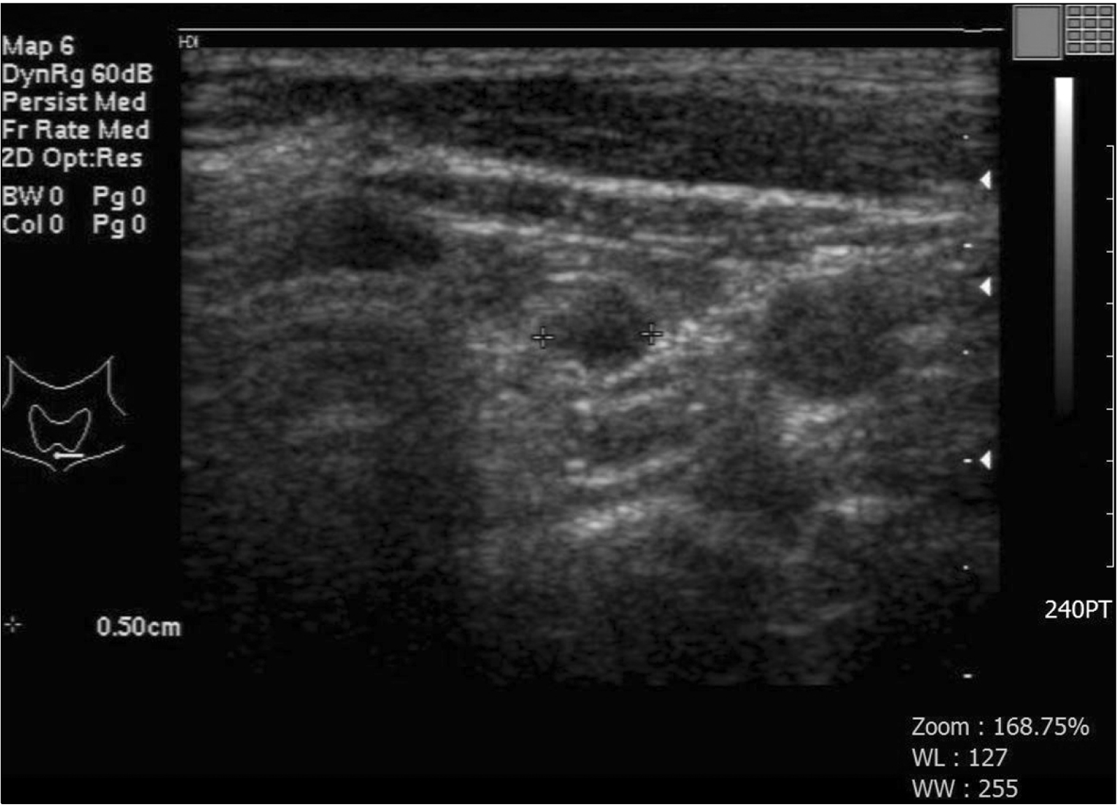
Fig. 5
Ultrasonographic image of N1b. Lymph node with internal microcalcification and without fatty hilum in lateral to left jugular vein.
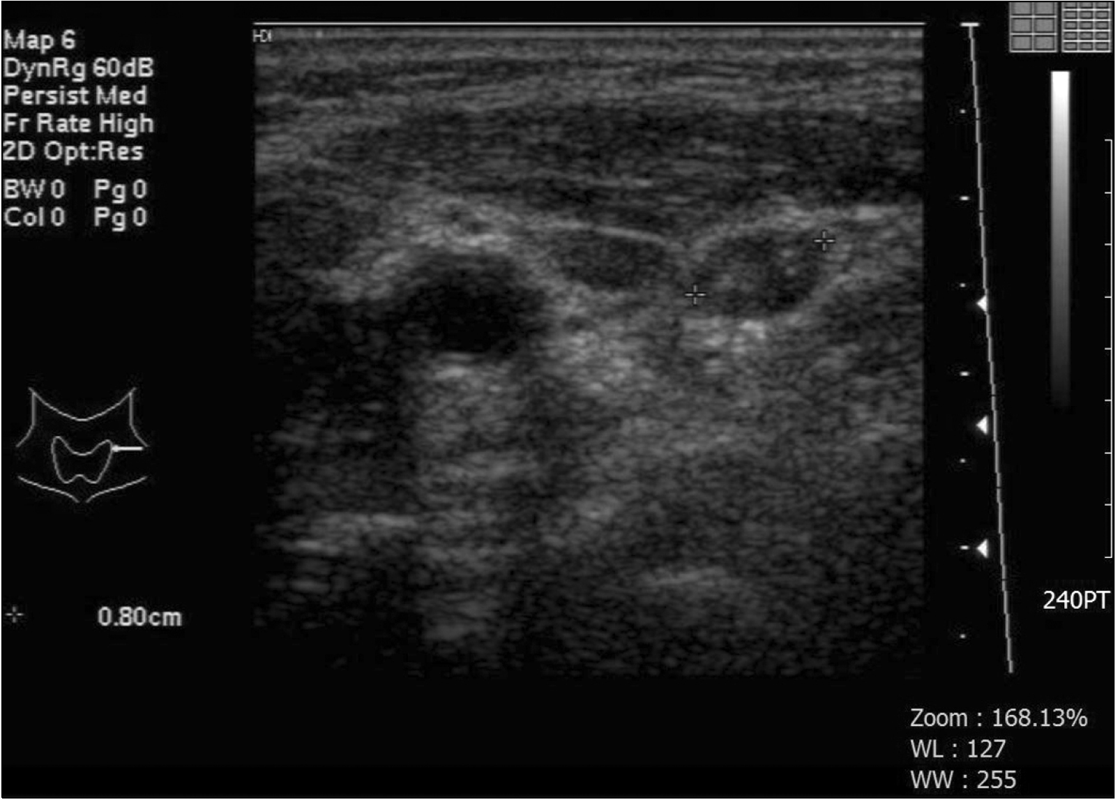
Table 1
Definition of Primary Tumor (T)
|
TX |
Primary tumor cannot be assessed |
|
T0 |
No evidence of primary tumor |
|
T1 |
Tumor ≤2 cm in greatest dimension, limited to the thyroid |
|
T1a |
Tumor ≤1 cm in greatest dimension, limited to the thyroid |
|
T1b |
Tumor >1 cm but ≤2 cm in greatest dimension, limited to the thyroid |
|
T2 |
Tumor >2 cm but ≤4 cm in greatest dimension, limited to the thyroid |
|
T3 |
Tumor >4 cm limited to the thyroid, or gross extrathyroidal extension invading only strap muscles |
|
T3a |
Tumor >4 cm limited to the thyroid |
|
T3b |
Gross extrathyroidal extension invading only strap muscles (sternohyoid, sternothyroid, thyrohyoid, or omohyoid muscles) from a tumor of any size |
|
T4 |
Includes gross extrathyroidal extension |
|
T4a |
Gross extrathyroidal extension invading subcutaneous soft tissue, larynx, trachea, esophagus, or recurrent laryngeal nerve from a tumor of any size |
|
T4b |
Gross extrathyroidal extension invadingprevertebral fascia or encasing the carotid artery or mediastinal vessels from a tumor of any size |
Table 2
Definition of Regional Lymph Node (N)
|
NX |
Regional lymph nodes cannot be assessed |
|
N0 |
No evidence of locoregional lymph node metastasis |
|
N0a |
One or more cytologically or histologically confirmed benign lymph nodes |
|
N0b |
No radiologic or clinical evidence of locoregional lymph node metastasis |
|
N1 |
Metastasis to regional nodes |
|
N1a |
Metastasis to level VI or VII (pretracheal, paratracheal, or prelaryngeal/Delphian, or upper mediastinal) lymph nodes. This can be unilateral or bilateral disease. |
|
N1b |
Metastasis to unilateral, bilateral, or contralateral lateral neck lymph nodes (levels I, II, III, IV, or V) or retropharyngeal lymph nodes |


 PDF
PDF Citation
 Print
Print


 XML Download