

 PDF
PDF Citation
 Print
Print


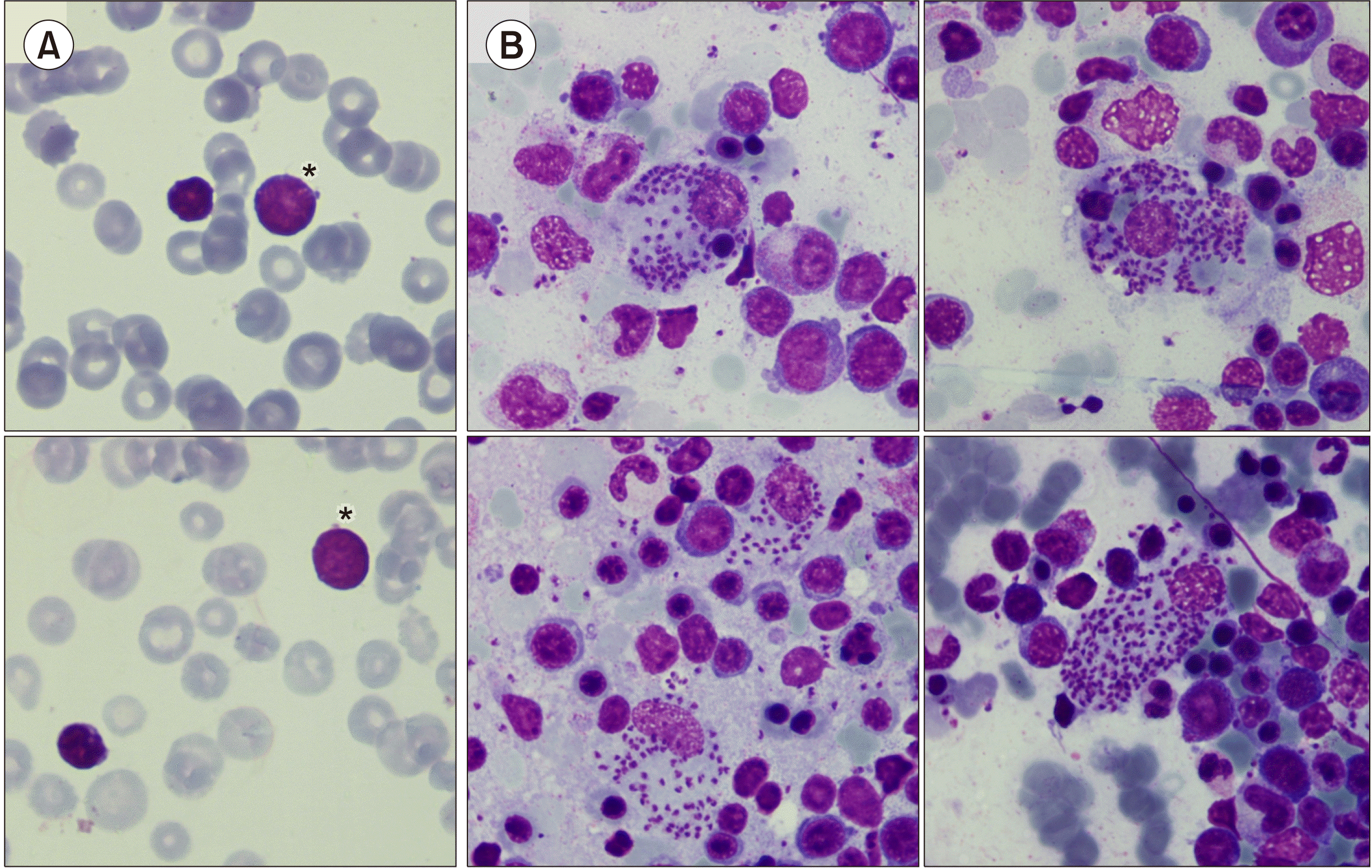
A 21-month-old asymptomatic child was referred to our hospital with leukemia suspicion with pancytopenia and “blasts” in peripheral blood. The blood count showed: hemoglobin 57 g/L, platelets 59×109/L, leukocytes 2.4×10⁹/L and the abdominal ultrasound a spleen of 158 mm. The morphological examination of peripheral blood showed left swift and some medium-sized lymphocytes with a very high nuclear-cytoplasmic ratio and homogenous immature chromatin (A). Manual count: neutrophils 18%, bands 2%, myelocytes 4%, lymphocytes 50%, monocytes 8% and “blasts” 18%. In the bone marrow aspirate multiple images of Leishmania amastigotes were observed both inside and outside the macrophages (B), some of them with erythrophagocytosis. The multiparametric flow cytometry showed a 1.8% of hematogones but no blasts or lymphoid clonality. The study was extended with Leishmania PCR in bone marrow, being positive. The patient received treatment with amphotericin B. After complete treatment the child had hemoglobin 118 g/L, platelets 233×109L, leukocytes 9.73×10⁹/L. Normal peripheral blood lymphocytes from young children may appear immature and can be mistaken for lymphoblasts by physicians who are not used to pediatric smear review. Furthermore, blood may contain hematogones that are indistinguishable from lymphoblasts by morphology. Leishmaniasis should be considered in the differential diagnosis of pancytopenia, especially in endemic areas. The sensitivity of bone marrow examination round 60 to 85%.
 XML Download