

 PDF
PDF Citation
 Print
Print


Coronavirus disease 2019 (COVID-19), caused by severe acute respiratory syndrome coronavirus 2, was first reported in Wuhan, China. Since then, COVID-19 has become a global pandemic and worldwide public health crisis. Over 339 million infections and 5.58 million deaths have been reported by January 20, 2022. To help build protection and reduce the spread of the disease, vaccination is expected to be a safe and effective method.1,2) More than 50 vaccine candidates have reached clinical and some of them including Oxford/AstraZenaca’s ChAdOx1 nCov-19, Moderna’s mRNA-1273, Pfizer/BioNTech’s mRNA BNT162b6, Gameleya’s Sputnik V, Johnson & Johnson’s INJ-7843735/Ad26.COV2.s, Sinopharm’s BBIBP-CorV, and Novavax’s NVX-CoV2373 trials have either been approved or have received emergency use authorization by governing bodies around the world.3) In South Korea, the Ministry of Food and Drug Safety has approved four vaccines against COVID-19: ChAdOx1 nCov-19, mRNA-1273, mRNA BNT162b6, and INJ-7843735/Ad26.COV2.s. As of January 20, 2022, 43,676,631 people (85.1% of the entire population) have received the second dose of the vaccine.
Regardless of the type of vaccine, common side-effects related to COVID-19 vaccination include local (injection site pain, swelling, redness, and itching) and systemic symptoms (fever, headache, vomiting, fatigue, diarrhea, myalgia, and arthralgia).1,3) Recently, a number of case reports have suggested a distinct prothrombotic syndrome associated with ChAdOx1 nCov-19 and INJ-7843735/Ad26.COV2.s vaccines called vaccine-induced immune thrombotic thrombocytopenia (VITT).4-9) Individuals with VITT have a high frequency of spontaneous bleeding or overt and decompensated disseminated intravascular coagulation.
In this report, we describe the case of a patient scheduled for diabetic foot amputation who received the ChAdOx1 nCov-19 vaccine and subsequently developed severe purpura in his genitalia and both of his hands and feet, accompanied by acute renal failure, and we review the relevant literature. We expect that this clinical experience would inform COVID-19 vaccination of patients with diabetic foot and benefit surgeons who treat the diabetic foot concomitant with these rare side effects of COVID-19 vaccination/
CASE REPORT
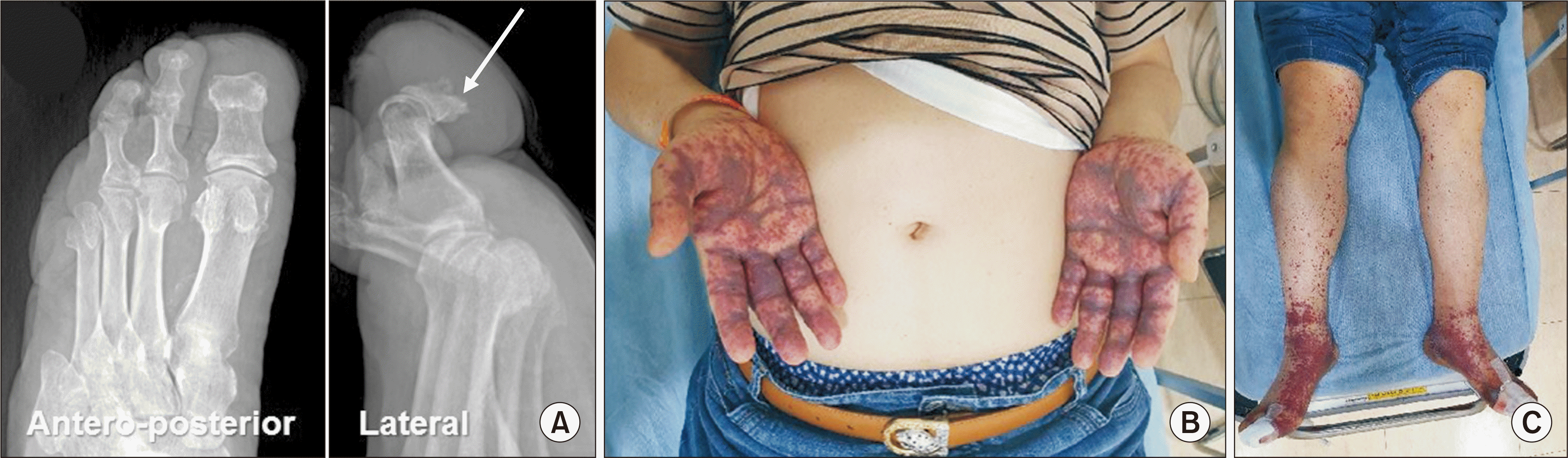
A 73-year-old male who had type 2 diabetes mellitus for 20 years visited the outpatient orthopedic clinic with a 6-month history of swelling and an ulcerative wound with draining sinus on the left great toe. Anteroposterior and lateral toe radiographs showed an osteolytic lesion of the distal phalanx, which suggested chronic osteomyelitis (Fig. 1A). His previous operative history included amputation 3 times on the same foot: second distal phalangeal amputation, fourth metatarsophalangeal (MTP) joint disarticulation, and fifth ray amputation. The patient had numerous medical illnesses, including hypertension, hypothyroidism, dyslipidemia, carotid artery stenosis, chronic renal insufficiency, and more than 30 years of type-2 diabetes mellitus; therefore, an operation (great toe amputation or disarticulation of the first MTP under sciatic nerve block) was planned after 5 days of preoperative risk evaluation. After 3 days, the patient visited the emergency department for limb pain and severe purpura on the genitalia and both of his hands (Fig. 1B) and feet (Fig. 1C). Vaccination of the first dose with ChAdOx1 nCov-19 had been performed a day after the orthopedic clinic visit, and the purpura developed the day after the vaccination. The patient had not mentioned that he would be vaccinated in a few days when he scheduled the operation at the orthopedic clinic.
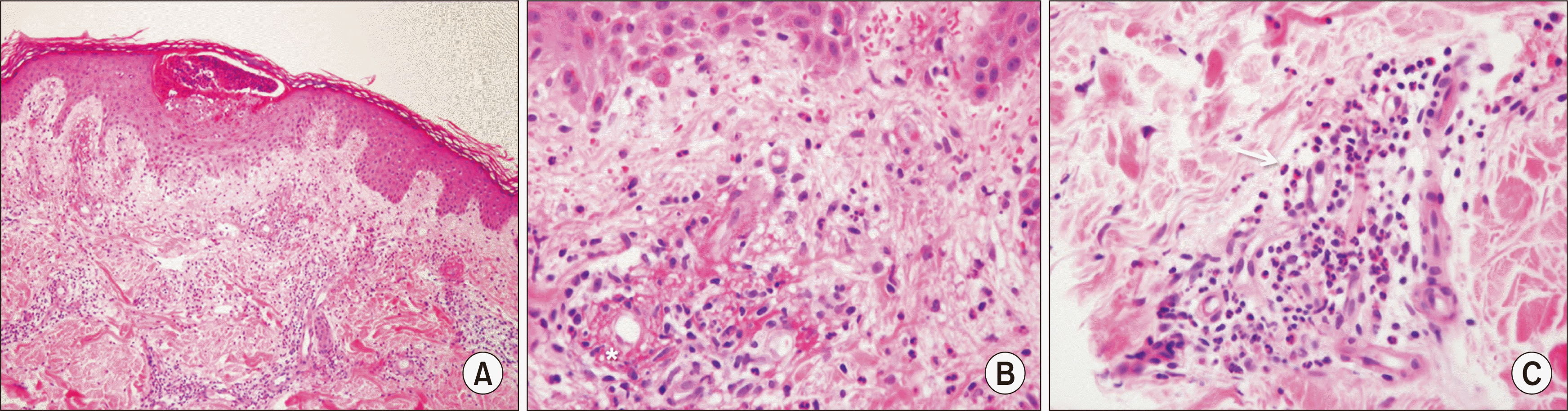
At the emergency department, acute renal failure superimposed on chronic renal insufficiency was suspected as the serum creatinine level was elevated. Laboratory findings revealed elevated C-reactive protein (CRP) and D-dimer levels, while platelet counts, prothrombin time (PT), and activated partial thromboplastin time (aPTT) were within the normal range (Table 1). The patient was then admitted to the nephrology department. After a week of intravenous normal saline hydration, blood transfusion, and furosemide injection, azotemia slowly improved. At the same time, skin biopsy was performed for the hand and foot purpura lesions. Histopathological evaluation confirmed dermal perivascular inflammation and edema with superficial epidermal pustule formation (Fig. 2A). Red blood cell extravasation and variable fibrinoid necrosis of the vessel walls were also observed (Fig. 2B). The blood vessels were surrounded by a dense inflammatory infiltrate composed of eosinophils and lymphocytes (Fig. 2C), which was consistent with vasculitis with unusual eosinophil preponderance.10) Only a topical steroid cream was applied, without systemic high-dose glucocorticoid application, because the patient’s glucose level could become uncontrolled. For the foot wound, intravenous ceftriaxone was administered (2-grams, every 6 hour) with a daily change in wound dressing.
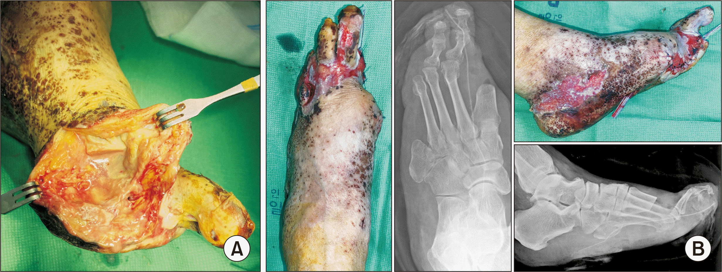
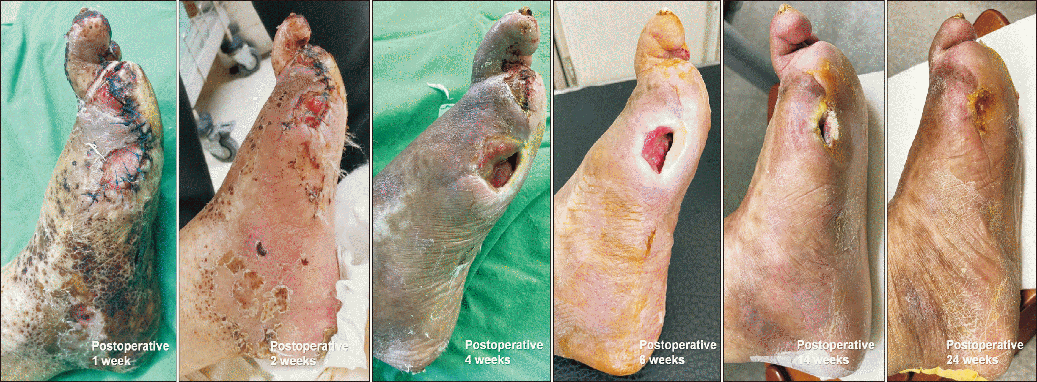
Two weeks after admission, while the purpura slowly improved, the original left great toe lesion remained untreated (Fig. 3). The operation on the left great toe was performed 15 days after hospital admission. Intraoperatively, a pus-like discharge was found around the proximal and distal phalanges, which had sequestrum formation, and it continued proximally along the flexor hallucis longus tendon. It was suspected that the infection had spread proximally due to the delay in the operation; therefore, amputation at a more proximal level (the first ray amputation) was inevitable (Fig. 4).
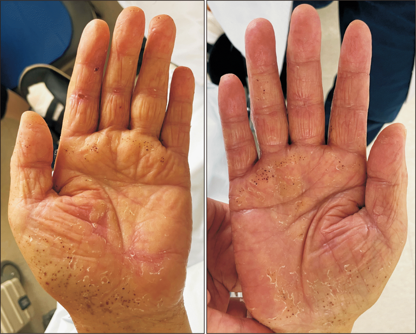
At three weeks of hospital admission, his hand purpura was completely improved (Fig. 5). Intravenous vancomycin injection was started on the fifth postoperative day, since methicillin-resistant Staphylococcus aureus had been cultured from the intraoperative wound specimen. At 2 weeks postoperatively, the CRP level declined to 2.8 mg/dL. However, a persistent, scanty, turbid discharge was drained from the medial side of the amputation wound. Three weeks after surgery, the wound was opened after stitch removal. A cultural study from the deep site through the open wound revealed Acinetobacter baumannii infection; therefore, antibiotics were changed to tigecycline, considering the patient’s impaired renal function. During 24 postoperative weeks, the wound slowly improved with daily dressing, without further deterioration (Fig. 6).
Written informed consent was obtained from the patient, the ethical review committee of our institution approved the study.
DISCUSSION
In this report, we present the case of a patient with diabetic foot complicated with vasculitis-related purpura induced by COVID-19 vaccination. Despite the patient requiring great toe amputation due to intractable chronic wounds and osteomyelitis, the operation had to be postponed as severe limb purpura appeared just before the operation. Patient management was challenging, since no similar case had been previously reported yet.
During our literature search, we found two dermatologic reports of vasculitis-related purpura after COVID-19 vaccination.11,12) While vasculitis in our case followed vaccination with ChAdOx1 nCov-19, Cohen et al.11) reported a case of vasculitis following Pfizer/BioNTech’s BNT162b6 vaccination. In their report, palpable purpuric papules on the bilateral lower legs appeared within two days after the first dose. Histopathological examination confirmed leukocytoclasia and erythrocyte extravasation. The symptoms were successfully treated with a prednisone taper. Within 2 days after the second dose, the purpura was exacerbated again and was distributed bilaterally on the lower and upper extremities and back. The symptoms were successfully treated using the same regimen. Bostan et al.12) presented a similar case following use of an inactivated vaccine. Erythematous macules and palpable papules on the legs, forearms, and right belly started 3 days after the first dose. A histopathological finding with direct immunofluorescence revealed immunoglobulin A deposition within the small vessel walls. The symptoms were controlled successfully using a topical steroid agent.
Several autoimmune diseases such as Guillain–Barre syndrome, systematic lupus erythematosus, rheumatoid arthritis, multiple sclerosis, and vasculitis can be demonstrated as adverse events following vaccination.13) Of these, vasculitis is the most commonly reported autoimmune disease postvaccination. Vasculitis has been widely reported to be secondary to vaccination for influenza, hepatitis B virus, Bacille Calmette–Guerin, and human papilloma virus. Although the incidence for each vaccine varies, it is rare, and the relationship between vaccination and vasculitis has yet to be determined.14) Since vaccines contain antigens from infectious agents, they can induce autoimmunity in genetically susceptible individuals by the same mechanisms as infectious agents.15) In clinical practice, a diagnosis of vaccine-induced vasculitis can be made after histological confirmation of a skin biopsy specimen. However, during the initial examination in an emergency department, without histopathological confirmation, VITT should be ruled out since the initial symptoms can be similar to those of vasculitis. Approximately 40% of VITT patients die, some from ischemic brain injury, superimposed hemorrhage, or both conditions, and often after anticoagulation. The difference between VITT and vasculitis is that VITT likely begins in a narrow window 5 to 10 days postvaccination, leading to identification typically between 5 to 30 days postvaccination;4) the symptoms derived from vasculitis begin within 5 days after vaccination. Thrombosis in VITT can occur in typical sites of venous thromboembolism, including as a pulmonary embolism or deep vein thrombosis in the leg. However, it also includes thromboses at unusual sites, such as cerebral venous sinus thrombosis or thrombosis in the portal, splanchnic, or hepatic veins. Regarding laboratory findings, platelet counts at VITT diagnosis are approximately 20,000 to 30,000/µL without specific abnormalities on a peripheral blood smear; high levels of D-dimer and low levels of fibrinogen are common and suggest systemic activation of coagulation.4) Likewise, PT and aPTT can be increased. With respect to the patient in this report, his symptoms began within 24 hours after being vaccinated. The initial platelet count, PT, and aPTT were within the normal range, although D-dimer levels increased. Furthermore, the patient did not present any thrombosis-induced symptoms, such as severe headache, backache, abdominal pain, chest pain, or altered mental status.
To the best of our knowledge, this is the first report of vasculitis-related purpura following COVID-19 vaccination that is superimposed on a diabetic foot requiring amputation. In this patient’s case, the vasculitis-related purpura induced by COVID-19 vaccination did not aggravate the diabetic wound or change the overall healing progress after amputation. Therefore, although diabetic foot amputation should be delayed due to a patient’s general condition and severe purpura, a successful postoperative outcome could be obtained with adequate management for acute renal failure and topical steroid application on the skin lesions.
In conclusion, despite the patient requiring the great toe amputation due to intractable chronic wounds and osteomyelitis, the operation had to be postponed as vaccination-induced severe limb purpura appeared just before the operation. Therefore, we recommend that COVID-19 vaccination should be carefully administered in patients with a diabetic foot requiring amputation.

 XML Download