

 PDF
PDF Citation
 Print
Print


INTRODUCTION
Low back pain (LBP) is one of the most common reasons why individuals visit hospitals [1]. If pain in the low back lasts for more than three months, this condition is generally defined as chronic LBP. Chronic LBP causes disability, absences from the work force, and economic loss [2]. Despite today’s advanced diagnostic methods, the underlying etiology cannot be elucidated in most patients with chronic LBP, and these cases are identified as nonspecific LBP [3]. Structural changes in one or more of the facet joints, intervertebral discs, ligaments, and paraspinal muscles are thought to be factors underlying the etiology of nonspecific LBP [4]. Thus, clarifying the underlying etiology in nonspecific LBP is very important to create specific treatment protocols [3].
Paraspinal muscles are essential structures for the lumbar region’s stability, balance, and motor control [1,3], because they have been considered dynamic stabilizers, providing stability to the movement of the spinal units. In patients with chronic LBP, their paraspinal muscles could have macroscopic (atrophy) and microscopic (fatty infiltration) changes [5,6]. While atrophy is the decrease of the cross-sectional area (CSA) in the paraspinal muscles, fatty infiltration occurs when muscle tissue is replaced by fatty tissue [7]. Disuse, muscle denervation, and reflex inhibition have been suggested as possible mechanisms for muscle atrophy in patients with LBP [8]. The CSAs of the paraspinal muscles have been shown to correlate with muscle strength [1]. The muscle strength imbalance that develops due to atrophy in the paraspinal muscles can cause the kinetic imbalance of the spine [1]. However, it is still unclear whether structural changes in the paraspinal muscles are the cause or consequence of LBP.
Nonetheless, a limited number of studies have investigated the factors that may affect the CSAs of paraspinal muscles, especially in terms of how the CSA may be decreased in patients with chronic LBP in comparison with individuals in control groups or how it may be decreased on the painful side in comparison to the side without pain [9-11]. Therefore, in this study, we aimed to examine the relationship between the CSA of paraspinal muscle, as well as age, sex, body mass index (BMI), level of physical activity, risk of sarcopenia, posture, and type of disc herniation. This study was also performed to examine the effect of muscle CSAs on the severity of LBP and disability.
MATERIALS AND METHODS
1. Ethical statement
This study was approved by the local institutional ethics committee of University Health of Scienc e Dıiskapi Education and Research Hospital (DERH 2021-106-18), and was conducted in accordance with the Declaration of Helsinki guidelines. Informed consent was obtained from each participant before starting data collection.
2. Study setting and participants
This single-center cross-sectional study was conducted on 164 patients with chronic LBP who were admitted to our physical medicine and rehabilitation outpatient clinic between May 2020 and October 2020. Patients between the ages of 18-65, with LBP for more than 3 months, who were ambulatory with or without a walking stick, and who could speak Turkish, in order to complete the questionnaires, were included. Patients who had received physical therapy or injection for low back in the previous 6 months; patients with inflammatory, severe metabolic, endocrine, cardiovascular, pulmonary, genitourinary, gastrointestinal, and progressive or non-progressive central or peripheral neurological diseases; and patient who had a history of spinal cord compression or radiculopathy, spinal structural deformity, lumbar instability, trauma to the lumbar region, osteoporotic fracture, lumbar surgery, malignancy, pregnancy, or lactation were excluded from the study.
3. Demographic characteristics
Data pertaining to age, gender, BMI (kg/cm2), additional comorbidity, and 25-dihydroxyvitamin D3 (25 [OH] D3) levels were collected. The patients were divided into 3 groups in terms of BMI according to the World Health Organization’s classification: normal, 18.50-24.99; overweight, 25.00-29.99; and obese, ≥ 30 [12]. The patients were divided into two groups according to their 25 (OH) D3 levels: < 20 ng/mL and ≥ 20 ng/mL [13].
4. Radiological evaluation
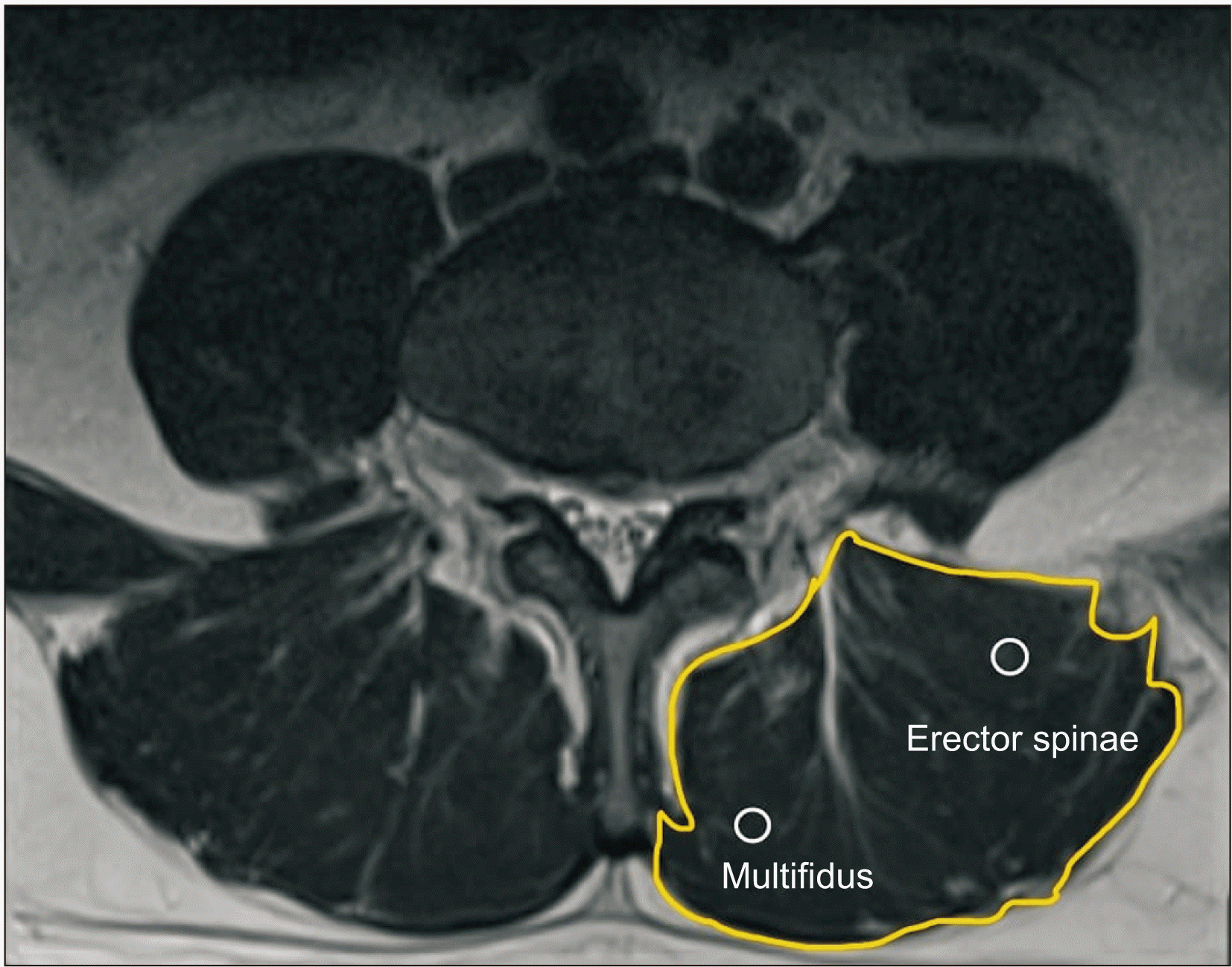
The CSAs of the paraspinal muscles were calculated for each patient at the middle point of the L2-3, L3-4, L4-5, and L5-S1 intervertebral discs by magnetic resonance imaging (MRI) slice as cm2, similar to previous studies [5]. All calculations were done using a 1.5 Tesla MRI scanner (Siemens Medical Systems, Erlangen, Germany) with 5 mm slice thickness and a close polygon region-of-interest tool. The Image J 1.53 software program (produced by Wayne Rasband, United States National Institutes of Health, Bethesda, MD) was used to trace the contour of the muscles of interest on axial T2-weighted MRI images for all CSA measurements. To determine the CSA of the muscle, its location of attachment with the fascia was manually marked using a freehand selection pointer and a touch screen pen on the touch screen monitor. The software automatically measured the area [1,10,14]. We chose to examine muscle CSA on the right side for standardization purposes (Fig. 1). The CSA measurements were made by two different clinicians, who were unaware of the clinical evaluation, and then the two measurements were compared and a value was recorded. Additionally, the type of disc herniation was evaluated as normal, bulging, protrusion, extrusion, or sequestration with MRI views [15].
5. Measurements
Pain intensity, disability, posture, physical activity level, and sarcopenia risk were evaluated in all patients.
6. Pain intensity-disability
Pain intensity was evaluated using the 100 mm visual analog scale. The disability levels of the patients were measured using the Quebec Back Pain Disability Scale, which is a condition-specific questionnaire consisting of 20 questions scored between 0 and 5 points. High scores indicate high disability [16].
7. Posture
The patients’ posture was evaluated using the New York Posture Rating (NYPR). By assigning scores between 0 and 100, the NYPR scale evaluates body segment images; back views of the head, shoulders, spine, hips, and feet; and lateral (left side) views of the neck, upper back, trunk, abdomen, and lower back. Low scores indicate poor posture [17].
8. Physical activity
The International Physical Activity Questionnaire - Short Form was applied to assess the physical activity status of the patients. This scale, which evaluates the intensity of physical activity and sitting time as part of people’s daily lives, is used to estimate the total physical activity in MET-min/week and time spent sitting [18]. According to their physical activity levels, the patients were divided into 3 groups including sedentary, minimally active, and very active individuals [19].
9. Risk of sarcopenia
A simplified screening tool was used to assess and screen for the risk of sarcopenia (SARCF), which can affect muscle mass. SARC-F is a self-administered questionnaire used to determine the level of difficulty experienced along 5 components: strength, assistance in walking, getting up from a chair, climbing stairs, and falling. The total score ranges from 0 to 10. Patients with a SARC-F score of ≥ 4 are considered ‘at risk for sarcopenia’ [20].
10. Comparisons
After the patients’ clinical evaluation and radiological measurements, the relationship between the CSAs of paraspinal muscles at L2-3, L3-4, L4-5, L5-S1 levels were examined. The evaluation parameters and the effect of the CSAs of the paraspinal muscles on pain and disability were also investigated.
11. Statistical analysis
Data were analyzed using the SPSS ver. 22.0 (IBM Co., Armonk, NY). The variables were investigated using visuals (histograms and probability plots) and a Kolmogorov–Smirnov test to determine whether they were normally distributed. In reporting descriptive statistics, the data were expressed as mean ± standard deviation and median (minimum-maximum) for continuous variables and as frequency and percentage (%) for nominal and categorical variables. Spearman and Pearson correlation tests were used to examine the relationship between demographic, clinical, radiologic characteristics, and paraspinal CSAs. One-way analysis of variance tests were used to compare paraspinal CSAs among the patients’ physical activity level and BMI status. When an overall significance was observed, pairwise post-hoc tests were performed using Tukey’s or Tamhane tests. The differences in paraspinal CSAs between genders, between patients with sarcopenia risk and normal patients, and between patients with different 25 (OH) D3 levels were analyzed using independent samples t-tests. Using age as a covariant factor, the differences of paraspinal CSAs between women and men; patients with different physical activity levels: healthy weight, overweight and obese patients; patients with and without the risk of sarcopenia; and patients with different 25-(OH) D3 levels were examined using the analysis of covariance (ANCOVA) tests. BMI was taken as a covariant factor, and the differences between paraspinal the CSAs of the two genders were examined using ANCOVA. The effects of lumbar paravertebral CSAs on pain severity and disability were examined using univariate linear regression analysis. For statistical significance, a value of P < 0.05 was accepted.
RESULTS
The general characteristics of the patients are shown in Table 1, and the correlations between the CSAs of the paraspinal muscles and patient demographics, as well as clinical and radiological characteristics are shown in Table 2. A negative significant correlation was found between age and the CSA at the L2-3, L3-4, L4-5 and L5-S1 levels (r = –0.225, P = 0.004; r = –0.231, P = 0.003; r = –0.256, P = 0.001; and r = –0.335, P = 0.001, respectively) (Table 2). A positive correlation was found between physical activity level and the CSA at the L2-3 and L3-4 levels, and a negative significant correlation was found between the risk of sarcopenia and the CSA at the L4-5 level (r = 0.282, P = 0.001; r = 0.178, P = 0.023; and r = –0.166, P = 0.034, respectively) (Table 2). No significant correlation was found between the type of disc herniation and the CSAs of the paraspinal muscles (PL2-3 = 0.716, PL3-4 = 0.859, PL4-5 = 0.975, PL5-S1 = 0.987, Table 2).
The levels of the paraspinal muscles’ CSAs at L2-3 and L3-4 in male patients were significantly higher than those in female patients (P = 0.001, P = 0.001, respectively, Table 3). No differences were found between genders at the L4-5 and L5-S1 levels (P = 0.534, P = 0.965, respectively). The results revealed that BMI did not affect the statistical results of the paraspinal muscles’ CSAs difference between males and females. A statistically significant difference was found between normal, overweight, and obese patients regarding the CSA at the L2-3 level (P = 0.037). This difference was determined to be due to the fact that the CSA was significantly higher at the L2-3 level in obese patients compared to overweight patients (P = 0.028). When the effect of age was not taken into consideration, CSAs at the L3-4 level in obese patients were found to be significantly higher than those in overweight patients (P = 0.026, Table 3). No differences were found between patients with 25 (OH) D3 levels below and above 20 ng/mL regarding the paraspinal muscle CSAs (PL2-3 = 0.817, PL3-4 = 0.687, PL4-5 = 0.875, PL5-S1 = 0.587) (Table 3). Significant differences were found in the CSAs of paraspinal muscles at the L2-3 and L3-4 levels between sedentary, minimally active, and very active individuals (P = 0.009 and P = 0.046, respectively) (Table 3). These differences were evident between sedentary and very active individuals (P = 0.006). There were no statistical significances in paired group comparisons of sedentary, minimally active, and very active individuals regarding the L3-4 level. When the effect of age was removed, while there was a significant difference in the CSA of the paraspinal muscles at L2-3 according to different physical activity levels (P = 0.038), no significant difference was found the L3-4 level (P = 0.113). When the effect of age was removed, patients who had a risk of sarcopenia were found to have significantly lower CSAs at the levels than those who did not have a risk of sarcopenia (P = 0.046, P = 0.009, P = 0.001, and P = 0.019, respectively) (Table 3). No relationship was found between the CSAs of the paraspinal muscles and pain intensity or disability (PL2-3 = 0.071, PL3-4 = 0.097, PL4-5 = 0.095, PL5-S1 = 0.387) (Table 4).
DISCUSSION
The present study tried to examine factors affecting the CSAs of paraspinal muscles and the relationship between the CSA of the paraspinal muscles and pain intensity and disability in patients with chronic LBP. In patients with chronic LBP, the CSA of the paraspinal muscles was found to decrease with increasing age, and it may increase with an increased level of physical activity. The CSAs of the paraspinal muscles in female patients were lower than those in male patients, and the CSAs were found to be higher in obese patients compared to overweight patients. This study also found that a high degree of atrophy could develop in the paraspinal muscles in patients with sarcopenia risk in comparison with patients without sarcopenia risk. However, posture, 25 (OH) D3 levels, and the type of disc herniation were not found to be influential factors on the CSAs of paraspinal muscles. No relationship was found between the paraspinal muscles CSA and pain intensity or disability. To date, it is unclear whether structural changes in the lumbar musculature are the cause or the result of nonspecific LBP. However, information on whether structural muscle changes occur and how the back muscles specifically change in LBP is essential for preventing and treating nonspecific LBP [6].
Many studies have shown that the quality and CSAs of the paraspinal muscles decrease with age [10,21-23]. In elderly patients, the fat/muscle ratio was found to increase with a decrease in the CSAs of the paraspinal muscles [10]. The decrease in muscle mass, strength, and function that accompanies aging is defined as sarcopenia [20]. Sarcopenia was found to be significantly higher in patients with chronic LBP than in those without chronic LBP [24]. In the present study, we found that the CSAs of the paraspinal muscles decreased with age, and individuals who were at risk for sarcopenia were found to have lower CSAs than normal individuals. Pain physicians should consider that sarcopenia or muscle atrophy may develop in the paraspinal muscles with aging, and patients with chronic LBP should be evaluated for sarcopenia, and precautions should be taken.
In the previous studies, the CSAs of the paraspinal muscles in male patients were significantly higher than those in women [21], which matches our results. This result can be attributed to the fact that men have more muscle mass than women, not only in patients with chronic LBP, but also across the whole population. The greater levels of muscle mass in men are due to hormonal difference between the sexes [25]. A study found that the CSA of paraspinal muscle was significantly higher in obese patients than in overweight and normal patients [10]. In the present study, we found that the CSA of paraspinal muscle was significantly higher at the L2-3 and L3-4 levels in obese patients than in overweight patients. Similar to our study, some previous studies have reported that there may be fat accumulation in the paraspinal muscles as in all the body’s muscles in obese patients; however, the last two vertebral levels are not affected by this fat accumulation [26]. With an increase in BMI, the fat content of the paraspinal muscles was found to increase and the density was found to decrease [6]. Although muscle CSAs increase due to fat accumulation in the attached paraspinal muscles in obese patients, there may be a decrease in muscle strength [27]. The reason that the increase in BMI found in previous studies is strongly associated with the increase in the prevalence of LBP can be attributed to the changes in muscle morphology, and hence muscle strength, due to increased BMI and fat accumulation [27,28]. However, we did not evaluate the density of the muscles in our study, but only the CSA. To further investigate this topic, future studies examining the effect of BMI on paraspinal muscle morphology, pain intensity, and disability in patients with chronic LBP can evaluate the density of the paraspinal muscles together with the CSA.
Our study did not find a relationship between the type of disc herniation and the mass of paraspinal muscle. A few studies have examined whether changes in the paraspinal muscles are associated with structural changes in the spine [26,29]. One study found a significant association between atrophy of the multifidus muscle caused by nerve root compression, herniated nucleus pulposus, and the number of degenerated discs [29]. A computed tomography study found a density change in the paraspinal muscles in patients with lumbar vertebral facet joint osteoarthritis, spondylolisthesis, and disc narrowing [26]. In previous studies, atrophy of the multifidus muscle was found in patients with radiculopathy, due to unilateral disc herniation in comparison with the opposite side [30,31]. Previous studies have produced conflicting results regarding the relationship between the type of disc herniation and the CSA of the paraspinal muscles, and more studies are needed to evaluate this relationship.
Unfortunately, this study did not find any relationship between 25 (OH) D3 levels and the paraspinal muscle’s CSA. However, vitamin D is one of the critical multifunctional mediators in skeletal muscle. In individuals with vitamin D deficiency, the activity of antioxidant enzymes decreases and muscles face more oxidative stress. Vitamin D protects muscles from atrophy [32]. More research is needed to investigate the relationship between vitamin D and the CSAs of the paraspinal muscles. A recent systematic review showed that people with chronic LBP had similar physical activity levels compared to healthy controls [33]. Physical activity is an important mediator of the connective tissue in skeletal muscle. Physical activity increases collagen synthesis in the short term and prevents fibrosis due to age in the long term [34]. In the present study, physical activity level was found to be an influential factor on the CSAs of the paraspinal muscles in patients with nonspecific chronic LBP.
In a recent MRI study, the CSAs of multifidus or erector spine muscles were not found to be associated with the intensity of pain or disability [7]. In a comprehensive review, it was reported that no relationship was found between paraspinal muscles’ CSAs and pain intensity in older adults [35]. While no relationship was found between pain and CSAs in patients with acute LBP, a negative relationship was found between pain and the CSAs in patients with chronic LBP lasting more than 12 months. In the present study, no relationship between the CSAs of the paraspinal muscle and pain intensity or disability could be found. Muscle inhibition and atrophy may directly result from pain, as pain-related nerve inhibition reduces lumbar muscle activity to prevent tissue damage [36]. Moreover, LBP is likely to cause changes in neuromuscular function, which cause changes in muscle histology that appear as atrophy [37]. Fat infiltration replaces muscle with fat, and changing muscle function may not alter muscle CSA [26,38]. The present study did not find a decrease in muscle CSAs. This result may due to its small sample size. Although we did not find any relationship between the CSA and pain or disability, clinicians such as pain physicians who deal with chronic LBP in their daily practice should consider the possibility of structural changes in the paraspinal muscles. Treatment regimens for paraspinal muscles may aid pain physicians in relieving pain, restoring function, and preventing recurrence in patients with chronic LBP. Studies with larger samples are needed to evaluate the relationship between CSAs in the paraspinal muscles and pain and disability.
Liminations of the present study include its cross-sectional design, the lack of a quantitative method to evaluate sarcopenia, as well as the fact that macroscopic or microscopic changes such as fatty infiltration beside the CSAs of the paraspinal muscles were not examined.
In conclusion, this study found that muscle mass decreases in less physically active patients, patients with sarcopenia risk, and older patients, and an association between back pain and CSA was not found. Although no relationship between the CSAs of the paraspinal muscles and pain or disability were found in this study, treatment regimens for preventing paraspinal muscles from atrophy may aid pain physicians in relieving pain, restoring function, and preventing recurrence in patients with chronic LBP. Prospective studies should consider including larger numbers of participants and using more quantitative methods to evaluate the factors affecting the morphology of paraspinal muscles and the relationship between the morphology of the paraspinal muscles and disability resulting from pain in patients with chronic LBP.
 XML Download