

 PDF
PDF Citation
 Print
Print


INTRODUCTION
Discogenic pain (DP) is an important etiology of low back pain (LBP). Diagnosis and management of DP is a debatable topic among physicians who treat LBP [1-4]. Unlike magnetic resonance imaging (MRI), which gives anatomic details, provocative discography (PD) is a physiological test to specifically identify a painful disc. PD is a diagnostic and prognostic test for DP, but, over time, the popularity of PD has decreased, possibly due to a poor correlation between the results of PD and results of spine fixation surgeries (SFS) [5]. The main reason for this discord seems to be non-uniform technique and classification criteria [6] for a painful disc on PD. Most of the publications are ambiguous about the technical aspects of PD [6]. Although the Spine Intervention Society (SIS) has laid down standard criteria for interpreting the results, they are seldom implemented.
In this study, we used PD to identify painful disc in patients who had chronic LBP with a suspicion of DP. To pressurize the posterior part of the disc, we adopted a posterolateral approach for needle placement. The pressure criteria laid down by the SIS were implemented to interpret the results. The aim of this study was to identify the correlation between MRI findings (desiccation, high intensity zone [HIZ], and change in the shape and size of the disc) and the results of PD.
MATERIALS AND METHODS
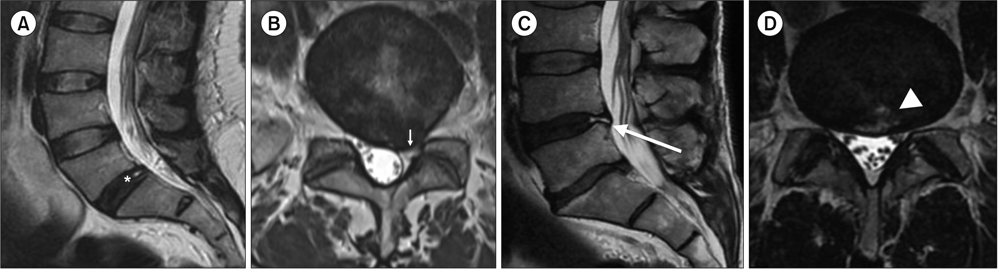
In the current retrospective study, exemption was obtained from the institutional review board (IHEC-LOP/2019/ IM0237). Patients visiting the pain clinic between July 2018 and December 2019 were screened and the records of 576 patients with LBP were analyzed. PD was done in patients who, based on history/examination and radiological findings, had a high index of clinical suspicion for DP; other etiologies of LBP were ruled out. Clinical features considered suggestive of DP were severe episodic axial LBP, increased on prolonged sitting or forward bending and showing centralization on Mckenzie exercises [7,8]. An MRI feature suggestive of DP was a desiccated disc alone (Pfirrman grade 3 or more) [9], or in combination with protrusion/extrusion or a HIZ (Fig. 1).
Patients who had advanced spondylitis changes, spondylolisthesis, multiple level prolapsed intervertebral disc, previous spine surgery, collagen vascular disorders, mental health issues, or any sensori-motor deficit were not subjected to PD. Before subjecting the patients to PD, alternate pain generators like facet joint, sacroiliac joints, and myofascial components were ruled out by means of targeted diagnostic pain relieving interventions.
All the PDs were performed under strict asepsis and fluoroscopic guidance. A single shot of intravenous antibiotic was given 30 minutes before the procedure. Intraoperative monitoring was done using electrocardiogram, pulse-oximetry, and blood pressure monitoring.
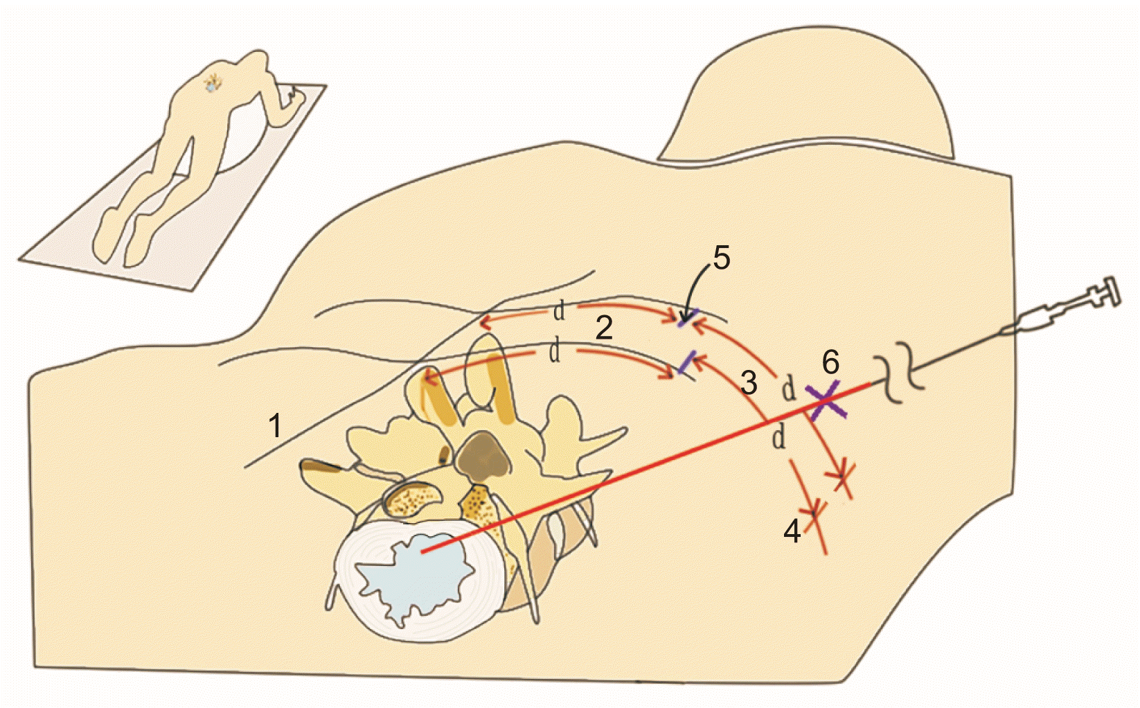
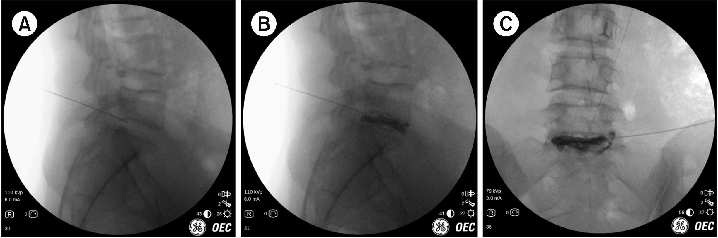
With the patient placed in prone position with a pillow under the belly, PD was performed using the posterolateral approach, as described, for disc access in percutaneous transforaminal endoscopic discectomy (Figs. 2, 3). A 22G, 20 cm Quincke needle was used to access the disc. The suspected disc was pressurized manually by injecting contrast medium in aliquots of 0.5 mL, and the patients’ response to provocation was noticed; categorization of the results was done as per the SIS criteria [10]. The disc was classified as painful if, upon injection of diluted radiocontrast (Omnipaque; GE Healthcare, Bengaluru, India), 1:1 in normal saline, the patient experienced concordant pain that was greater than 6 on the verbal numerical rating scale (NRS) or had more than 70% pain reproduction, at a pressure of less than 50 psi above the opening pressure, with a painless adjacent control disc; no more than 3 mL of contrast medium was injected. Opening pressure was defined as the pressure when the contrast media was first visualized at the tip of the needle by fluoroscopy. The control disc was an adjacent radiologically normal disc. Discography was considered negative if 3 mL of contrast medium was injected and still the pressure in the disc did not reach 50 psi or the elicited pain was discordant in nature. A disc-monitor discography probe (Stryker, Kalamazoo, MI) was used for pressure measurements during PD. A disc-monitor helps to standardize PD by providing and recording the opening pressure, maximum pressure achieved, and volume injected [11].
Based on the formula for sample size calculation by Green [12] (N = 104 + X, where N is the sample size and X is the number of independent variable).We considered age, containment of the disc, disc desiccation, HIZs, and protrusion/ extrusion as the independent variables. Hence, a total of 109 PDs were needed for analysis. The power of study was kept at 80 percent.
1. Statistical analysis
Statistical analysis was done using the SPSS ver. 20.0 (IBM Co., Armonk, NY) software. Quantitative data like age, body mass index (BMI), NRS, and mean disc pressure were expressed as mean ± standard deviation, and were analysed using independent sample Student’s t-test. Categorical variables were expressed as counts (percentage) and were analysed using a chi-square test, Wilcoxon’s U-test, or Fisher’s exact test.
An adjusted multivariate logistic regression model analysis was performed for identifying the association of MRI findings and PD results. Results were presented as odds ratio (OR) with 95% confidence interval (95% CI). All statistical assessments were 2 tailed and significance was set at a P value less than 0.05.
RESULTS
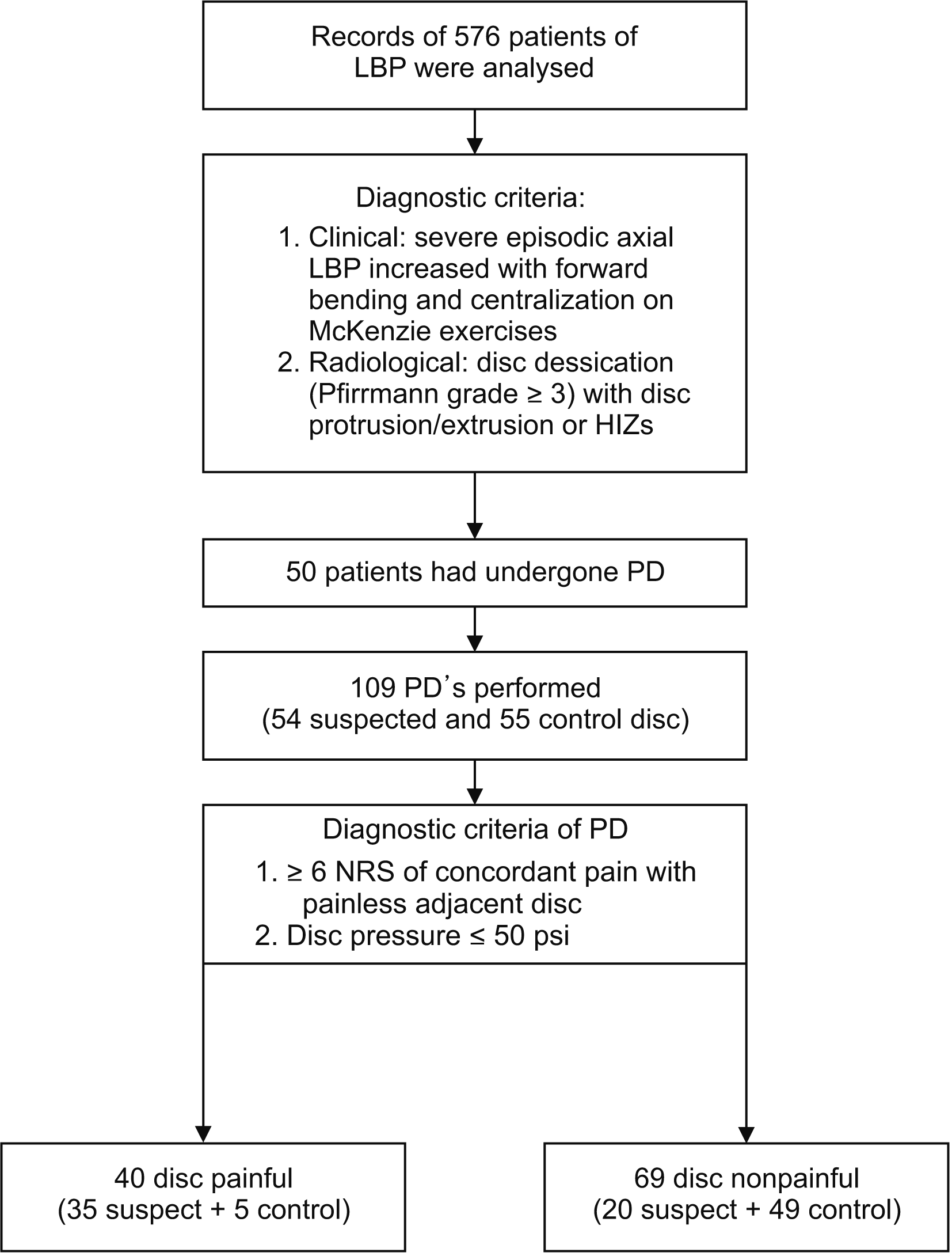
After analyzing records of 576 patients with chronic LBP, 526 were excluded, and 50 patients that had undergone PD were studied; 109 PDs were performed, with 54 suspected discs and 55 control discs (Fig. 4). The mean age 45.3 ± 6.7 years, 54% patients were male, mean BMI was 26.7 ± 2.3, and the mean duration of symptoms was 11.27 ± 4.0 months. The mean pain score before intervention on the NRS was 5.7 ± 1.8. Forty discs were painful on discography, including 35 suspected and 5 control discs. Eighty percent of the suspected discs were at L4-5 level, while the rest were at L5-S1 level. For suspected discs at the L4-5 level, the L3-4 disc acted as the control, while for those at the L5-S1 level, the L4-5 was the control.
The mean disc pressure in the painful discs was 31.9 ± 7.9 psi (range, 15-44 psi), with less than 20 psi in 2, 20-30 psi in 10, 30-40 psi in 15, and 40-50 psi in 8 patients. Out of the 50 patients who underwent PD, 35 had positive MRI findings. Thirty-nine suspect discs were contained (no spillage of contrast in the extra discal space). A significant positive correlation was found only between disc desiccation and the results of PD (Table 1).
An adjusted logistic regression analysis revealed that only disc desiccation successfully predicted the result of discography (P < 0.001; OR, 26.5; 95% CI, 4.5-155.6). Sixty-six percent of discs having HIZs were painful, and 65% discs with protrusion/extrusions were painful, but neither HIZ nor protrusion/extrusion had a statically significant OR (P = 0.330 [OR, 2.3; 95% CI, 0.4-12.5], and P = 0.800 [OR, 1.24; 95% CI, 0.28-6.8], respectively). The regression model had an overall correct prediction rate of 82%.
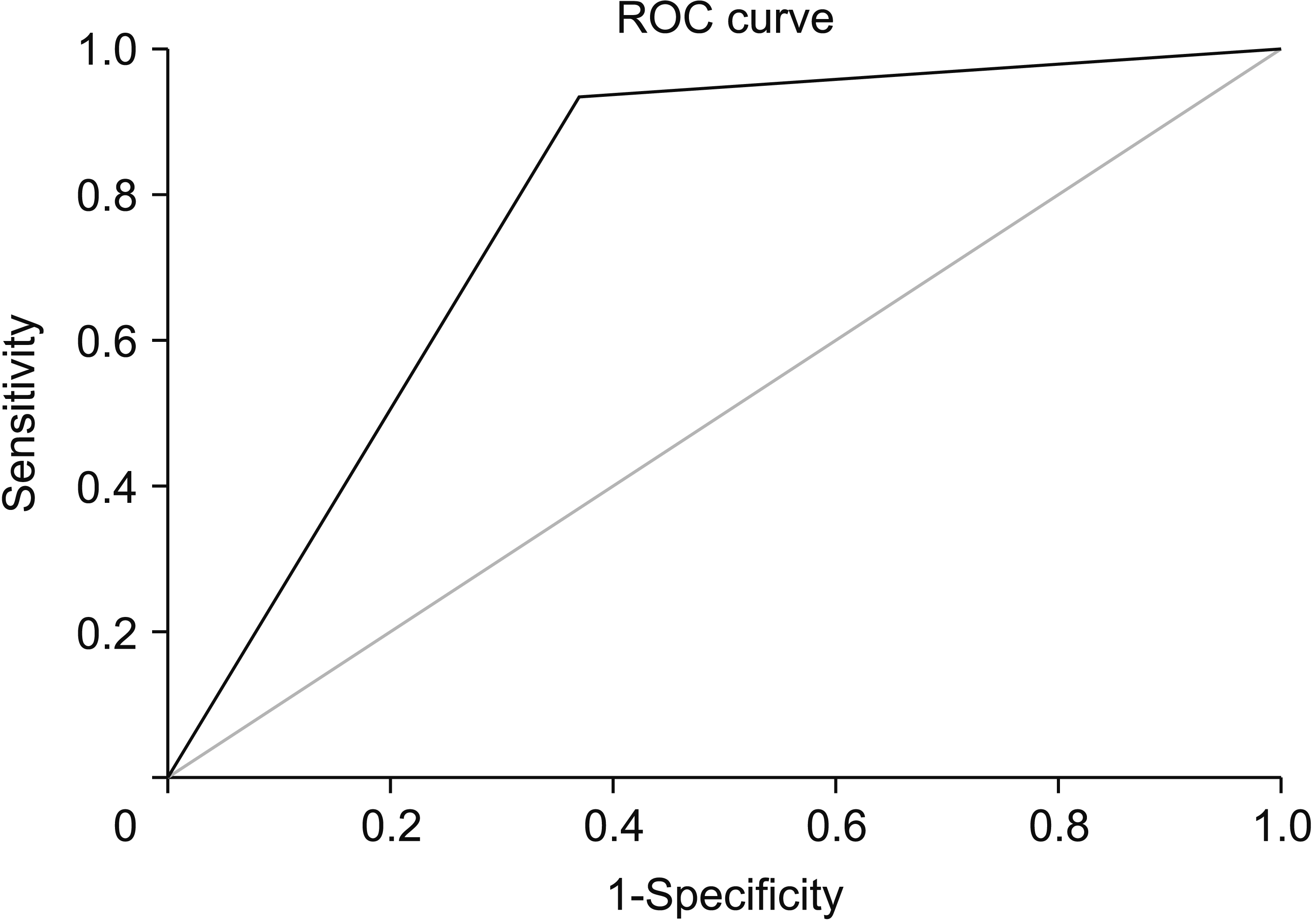
A receiver operating characteristic curve was generated for the result of PD and disc desiccation, the area under the curve was 0.78 (95% CI, 0.64-0.93; P = 0.001), and the sensitivity of disc desiccation in predicting the results of PD was 0.93, while the specificity was 0.64. The positive likelihood ratio (LR+) was 2.58 and the negative likelihood ratio (LR–) was 0.11 (Fig. 5). Apart from a transient increase in the intensity of back ache, no other serious side-effects were observed.
DISCUSSION
PD has been controversial and recently its utility has been questioned. Non-uniform technique and diagnostic criteria are possibly the main reason why the results of PD are difficult to interpret clinically [13]. PD has been demonstrated to have a degenerative effect on intervertebral discs [14]. The study was done in the 1990s, had a small sample size, and did not adhere to the SIS guidelines for PD. They used extremely high pressures (100 psi), and the accelerated degeneration could have been due to the exposure of the annulus to very high intradiscal pressure [15,16]. Another small study (n = 36) of matched control demonstrated no worsening of symptoms or radiological grade of disc degeneration at the 5-year follow-up [17].
With time, PD has also evolved, with the maximum pressure allowed now being much less (50 psi). Needles of a much smaller caliber are also now used, thereby minimizing the damage to the annulus [18]. In this study we implemented a pressure controlled protocol of PD. One important observation of this study is the provocation of concordant pain at a pressure lower than the recommended upper limit. Placement of needle tip in the posterior part rather than the center of disc, and hence nearer to the site of maximum disc damage, seems to be the reason for this observation. This small change in the technique not only increases the clinical relevance of the procedure but also theoretically reduces the chances of disc damage over a period of time.
MRI criteria for diagnosis of a painful disc have been controversial; most of the controversy is due to non-uniformity of patient selection, diagnosis, and procedure related criterias. The sensitivity, specificity, positive predictive value (PPV), and negative predictive value of T2-weighted images in detecting the symptomatic disc have been estimated to be 94%, 71%, 59%, and 97%, respectively [19]. An MRI study on healthy volunteers revealed disc herniation and extrusion in 7% and 13% cases, respectively [20].
High HIZs, disc desiccation, disc enlargement, and reduction in disc height are the most common MRI criteria considered significant by radiologists. Many consider HIZ to be the most relevant feature in a T2W MRI, however the diversity in results in intriguing. A significant proportion of discs having HIZs are painless on PD, and the two have been demonstrated not to have any significant correlation [21-24]. HIZs demonstrated on an axial loaded MRI did not show a significant correlation with a painful disc [25]. On the contrary, HIZs in morphologically abnormal discs (Dallas grades 3, 4, and 5) have been demonstrated to have a significant correlation with concordant pain reproduction (P < 0.001). The sensitivity, specificity, and PPV of HIZs in isolation or in combination with other radiological features ranges from 45.5%-81%, 69%-97.8%, and 39%-87%, respectively [25-27]. HIZ in conjunction with endplate changes can have a sensitivity and specificity of 94 and 77%, respectively [5]. In the current study, we did not find HIZs to predict the results of PD.
A change in the shape and size of the disc has also been considered an important MRI criteria. Disc bulge has been found not to correlate with disc pain [28]. Disc protrusion has been demonstrated to have a sensitivity of 68.2%, specificity of 80.6%, PPV of 53.6% [27], and the PPV of disc extrusion has been found to be 93% [28]. Another criterion considered important in lumbosacral MRI is disc desiccation [29]. Isolated disc desiccation at the L4-5 level may be seen in Bertolotti syndrome [30]. O’Neil et al. [29] demonstrated that a moderate loss of nuclear signal is the most important factor that predicts a positive discography and, along with disc bulge, has the best combination of sensitivity (79.8%) and specificity (79.3%). They concluded that normal or severe loss of signal intensity in the disc is most likely to be painless and, combining other MRI parameters, has no influence on test performance [29]. Although a contrarian view is also available for this parameter [27], our results are in congruence with this observation.
In this study, we carefully selected the patients (based on high probability clinical features and high probability MRI findings), implemented a pressure-controlled protocol of PD, and also stimulated the posterior part of the disc, thereby removing most of the confounding factors that impaired the quality of previous research. In an experimental study in pigs, injection of contrast medium into one disc led to a 16% (3.2-37.0) increase in intradiscal pressure of the adjacent non-injected disc; this observation is important, as it may increase the false positive results of PD [31]. Although this aspect of PD has not been studied in humans, our approach of posterolateral discography can theoretically reduce this problem by utilizing lower intradiscal pressure for diagnosis of DP.
Based on a meta-analysis, if the SIS criteria are followed [10,16], PD has a low false positive rate of 6% per disc [13,32]; however, given the variability of techniques this incidence may be much higher. In order to address this problem of false positive results, a few investigators have suggested an alternative approach, i.e., analgesic discography/discoblock [33,34]. In discoblock, in contrast to PD, a small amount of local anesthetic is injected into the suspected disc and relief in back ache is assessed. If a patient’s pain is relieved after discoblock, then that disc is considered to be the pain generator. An analgesic discography can further solve the problem of false positives. A study with a small study group reported that, at a 3 year follow-up, results of surgery-based discoblock (analgesic discography) were better than those based on PD [35].
Based on the finding in our study, we recommend the use of MRI as a screening tool for chronic LBP and utilization of a pressure-controlled PD in selecting patients for SFS. The findings of this study suggest that over and above clinical features, MRI provides useful information in identifying DP. In MRI, disc desiccation is the only important finding as far as prediction of a painful disc is concerned. In the current study, we adopted a posterolateral approach for needle placement, avoiding puncture of the thecal sac [36], and thereby pressurizing the posterior third of the disc. Stimulation of the posterior part of disc makes the results of this study more relevant from the clinical point of view, as these are the sites where the sinuvertebral nerve is mainly located and sensitized [37,38].
The present study has many limitations. The retrospective nature of the study is the biggest limitation. A larger sample size could have increased the power of study. Although, our data clinically address the usefulness of PD in decision-making for spinal operations, it lacks the corroborative data on the outcome of PD in SFS. Besides, it is data from a single center, so selection bias cannot be ruled out. A prospective randomized study, sequentially using a posterolateral approach for discography along with discoblock can give promising results as far as diagnosing DP is concerned.
In patients with DP, disc desiccation is the most useful MRI feature that predicts a painful disc on PD, with an OR of 26. The sensitivity and specificity of disc desiccation on MRI in predicting a painful disc on PD were 93 and 64, respectively, and the positive likelihood ratio and negative likelihood ratio were 2.58 and 0.11, respectively.
 XML Download