

 PDF
PDF Citation
 Print
Print


Introduction
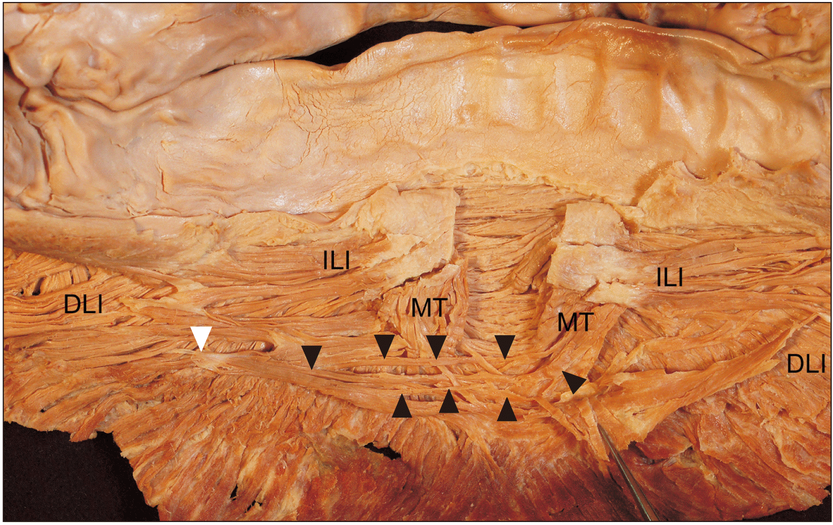
Fibers of the facial muscles occasionally extend, cross the midline, and connect to surrounding structures on the contralateral side, perhaps enabling the mouth to make more delicate movements and generate more facial expressions [1]. In and around the chin area, the mentalis (MT), orbicularis oris, depressor anguli oris, depressor labii inferioris (DLI), and incisivus labii inferioris (ILI) muscles work together to protrude and retract the lower lip [1-4]. The MT, a muscle of the lower lip and chin, consists of upper and lower fibers and courses antero-posteriorly. The upper fibers are generally shorter than the lower fibers [2]. The MT fibers are also classified as medial and lateral. In both the upper and lower portions of the MT, the medial fibers descend and cross the midline to attach to the skin on the contralateral chin while the lateral fibers descend and attach to the skin on the ipsilateral chin. The ILI usually connects between the MT, the periosteum of the mandible, and the buccinator. The ILI fibers inserting into the MT do not cross the midline but terminate into the ipsilateral MT [2]. This case report describes a variant in which the extended fibers of the MT crossed the midline and indicates the relationship of these fibers to the surrounding structures.
Case Report
During routine dissection of a Korean male cadaver whose age at the time of death was 82 years, the MT and related muscles showed anatomical variations (Fig. 1). The lower face with muscle tissues was harvested en bloc with the periosteum. The tissues were then dissected posteriorly. When the course and connection of the MT fibers were examined, some of the deepest fibers were found to descend inferomedially and to cross transversely just below the chin prominence to attach to the periosteum of the mandible on the contralateral side. These crossing fibers of the MT were intermingled with some of the deepest MT fibers on the contralateral side and were extended to the mandible below the corner of the mouth. After crossing the chin, they were continuously arranged below the ILI on the contralateral side. The other fibers of the MT showed the usual morphological patterns of this muscle. The medial fibers of the left and right MT crossed together, attaching to the chin skin of the contralateral side. Their lateral fibers descended inferomedially, intermingling with the DLI to attach to the skin of the ipsilateral side.
This study was conducted in accordance with the Declaration of Helsinki. The cadaver dissected had been legally donated to the Catholic Kwandong University College of Medicine. This study was approved by the Institutional Review Board of Catholic Kwandong University (IRB no. CKU-21-01-0203). A surgical microscope (OPMI-FC; Carl Zeiss, Oberkochen, Germany) was used during detailed dissection on the deep surface of the removed facial muscles.
Discussion
Most human skeletal muscles generally originate from and insert into structures on the ipsilateral sides, but owing to their variability, many exceptions have been reported. Burley et al. (2020) [5] reported that sternal fibers of the pectoralis major crossed the midline. In Mori’s study of Japanese cadavers, different musculatures were reported to have variants that crossed the midline, e.g., the anterior belly of the digastric muscle and the sternalis muscle [6]. Variant muscle fibers crossing the midline have therefore been well documented. A similar variant has also been reported in mimetic muscle [1].
The present study demonstrates a variation of the MT in which its extended fibers attached to the mandible on the contralateral side. These extended fibers passed just below the chin prominence, while the other crossing fibers reached the chin prominence to attach to its skin. These extended crossing fibers of the MT attached to the periosteum of the mandible of the contralateral side below the angle of the mouth instead of the skin of the chin. They might therefore contribute to making a longer sling that helps to support the chin prominence and submental fat.
The MT draws up the skin of the chin and thus indirectly causes the lower lip to protrude. It functions in articulation, in forcing bits of food from between the gums, and in the expression of various emotions such as pride and doubt [7]. Although it is a small muscle, it is located in the center below the lower lip and intermingles with several surrounding muscles such as the ILI, DLI, and platysma [2, 7], implying close anatomical and functional relationships with movements of the lower lip. Therefore, the variation presented in this study was considered in the light of the interactions of the MT with the surrounding muscles.
 XML Download