

 PDF
PDF Citation
 Print
Print


INTRODUCTION
Obesity is a serious medical condition characterized by excessive body fat accumulation which results in significant metabolic complications [1]. The accumulation of body fat greatly affects one’s physical appearance ending up with disfigurement, and obese patients often suffer from negative social consequences [2]. The fat reduction in a certain area is hard to be achieved by general weight loss methods such as exercise and diet. Targeted fat reduction procedures for body and face contouring have gained remarkable global interest, and various noninvasive procedures have been developed worldwide [3]. Injection lipolysis, based on mesotherapy, is one of the easily accepted noninvasive treatment by patients. This procedure is performed by subcutaneous injection of lipolytic substances to induce local fat reduction [4], and several injection formulations have been introduced and utilized in injection lipolysis. Most of the lipid reducing agents used in injection lipolysis formulations are used off-label, and thus their efficacy and safety for the removal of local fat is controversial.
Glycerophosphocholine (GPC), also known as choline alfoscerate, was originally approved for treatment of mental integrity in patients with dementia. GPC shares structural similarity with one of the most widely used ingredient called phosphatidylcholine (PPC) by having choline moiety, and thus GPC was introduced in clinical practice as an alternative lipolytic agent for PPC. GPC is a natural source of choline and the use of GPC as an alternative to enhance lipolysis is based on the hypothesis that GPC would stimulate lipid metabolism as choline plays important role in the lipid metabolism [5]. Despite some anecdotal evidence, the efficacy of GPC onto local fat reduction has not been thoroughly evaluated. Therefore, here we evaluated the lipolytic potential of GPC and compared the effects with two off-label agents for injection lipolysis, PPC and aminophylline (AMPL), using in vitro and in vivo models of obesity.
METHODS
Materials
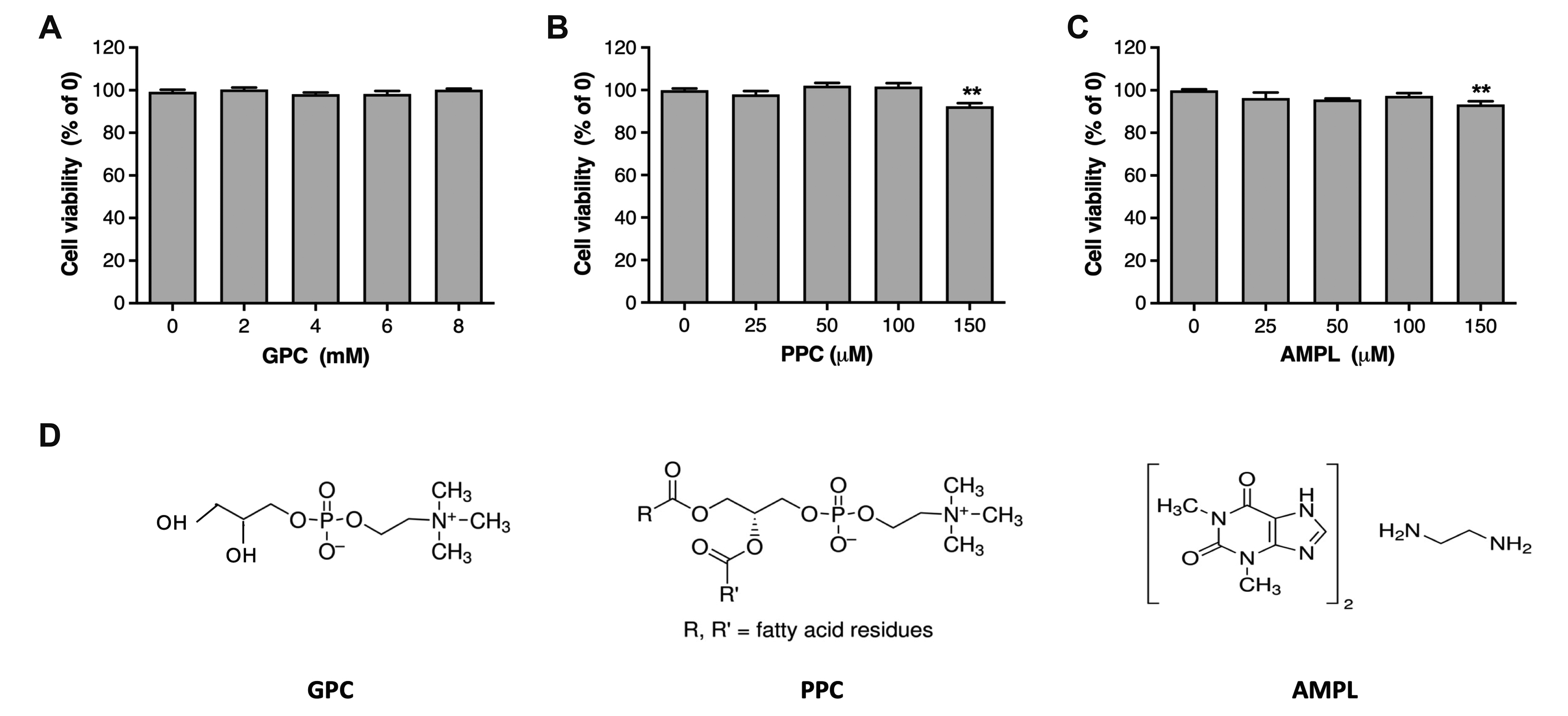
Cell culture reagents including Dulbecco’s modified Eagle medium (DMEM), penicillin-streptomycin antibiotics, and fetal bovine serum (FBS) were purchased from Gibco (Grand Island, NY, USA). The experimental solutions used in this study; Alfocholine, Lipobean, and Aminophylline, were provided from Beauvous Co. (Seoul, Korea), and the active component of each solution is described in Table 1. All other reagents were from Sigma (St. Louis, MO, USA) unless otherwise stated.
Cell culture and cell viability assay
Mouse embryonic fibroblast 3T3-L1 cells were purchased from the American Type Culture Collection (Manassas, VA, USA). 3T3-L1 cells were cultured in DMEM containing 10% bovine serum and 100 units/ml penicillin-streptomycin at 37°C in an atmosphere of 5% CO2. The cytotoxicity of compounds was determined by using tetrazolium compound (MTS)-based CellTiter 96 AQuenous One solution Cell Proliferation Assay Kit (Promega, Madison, WI, USA) following the manufacturer’s protocol. Briefly, 3T3-L1 cells were seeded at 5 × 10³ cells/well in a 96-well plate and incubated with compounds. The experimental concentration of each compound was calculated solely using the content of active component (AMPL, PPC, or GPC) in the stock solution shown in Table 1. After 96 h, 20 μl of assay solution was added to each well and incubated at 37°C for 1 h. The absorbance was then recorded at 490 nm using a microplate reader (ThermoLabSystems, Helsinki, Finland).
Adipocyte differentiation and Oil Red O staining
To induce adipocyte differentiation, 3T3-L1 pre-adipocytes were seeded at 3 × 10⁵ cells/well in a six-well plate. Cells were cultured in differentiation medium (DMEM containing 5% FBS and 1% antibiotics) which was refreshed every other day during differentiation. Two days after confluence (designated day 0), cells were placed in differentiation medium and were induced to differentiate by hormonal cocktail (0.5 mM IBMX, 1 μM dexamethasone, and 10 μg/ml insulin; MDI) for 4 days. After MDI induction, cells were switched to differentiation medium supplemented with 10 μg/ml insulin until day 6, and then maintained in differentiation medium for an additional 2 days. During differentiation, 3T3-L1 cells were treated with the compounds as indicated in the figures from day 0 to day 4. Once the differentiation is complete, cells were washed twice with phosphate buffered saline (PBS), and fixed with 10% neutral buffered formalin (NBF) for 1 h, then stained with Oil Red O solution for 1 h at room temperature. Cells were photographed using a phase-contrast microscope (CKX41; Olympus, Tokyo, Japan) equipped with Olympus DP22 digital camera (Olympus). The Oil Red O dye in adipocytes was eluted with isopropyl alcohol and the absorbance was measured at 540 nm to quantify stained lipid droplets.
Lipolysis assay
Two days after the completion of differentiation, 3T3-L1 adipocytes were washed twice with PBS and incubated with the compounds in low glucose (1 g/L), serum-free and phenol red-free DMEM supplemented with 2% fatty acid free BSA. After a 3 h treatment period, supernatants were collected and analyzed for glycerol content using lipolysis colorimetric assay kit (Sigma) following the manufacturer’s indications.
Animals
All animal experimentation was approved and conducted in accordance with the guidelines from IACUC of Kyung Hee University (approval number KHUASP(SE)17-164). C57BL/6J mice (5 weeks-old, male) were purchased from Daehan Biolink Co. Ltd. (Eumsung, Korea). The mice were randomized and housed in a room under constant temperature (22 ± 2°C) and humidity (50 ± 5%) conditions with 12:12 h light-dark cycle. Food and water were supplied ad libitum. After a week of acclimatization to the environment, mice were given a high fat diet (HFD; 60% fat, #D12762; Research Diets, New Brunswick, NJ, USA) to induce obesity. After 20-weeks on the HFD, the established diet-induced obesity (DIO) mouse models were used for the experiment.
Injection
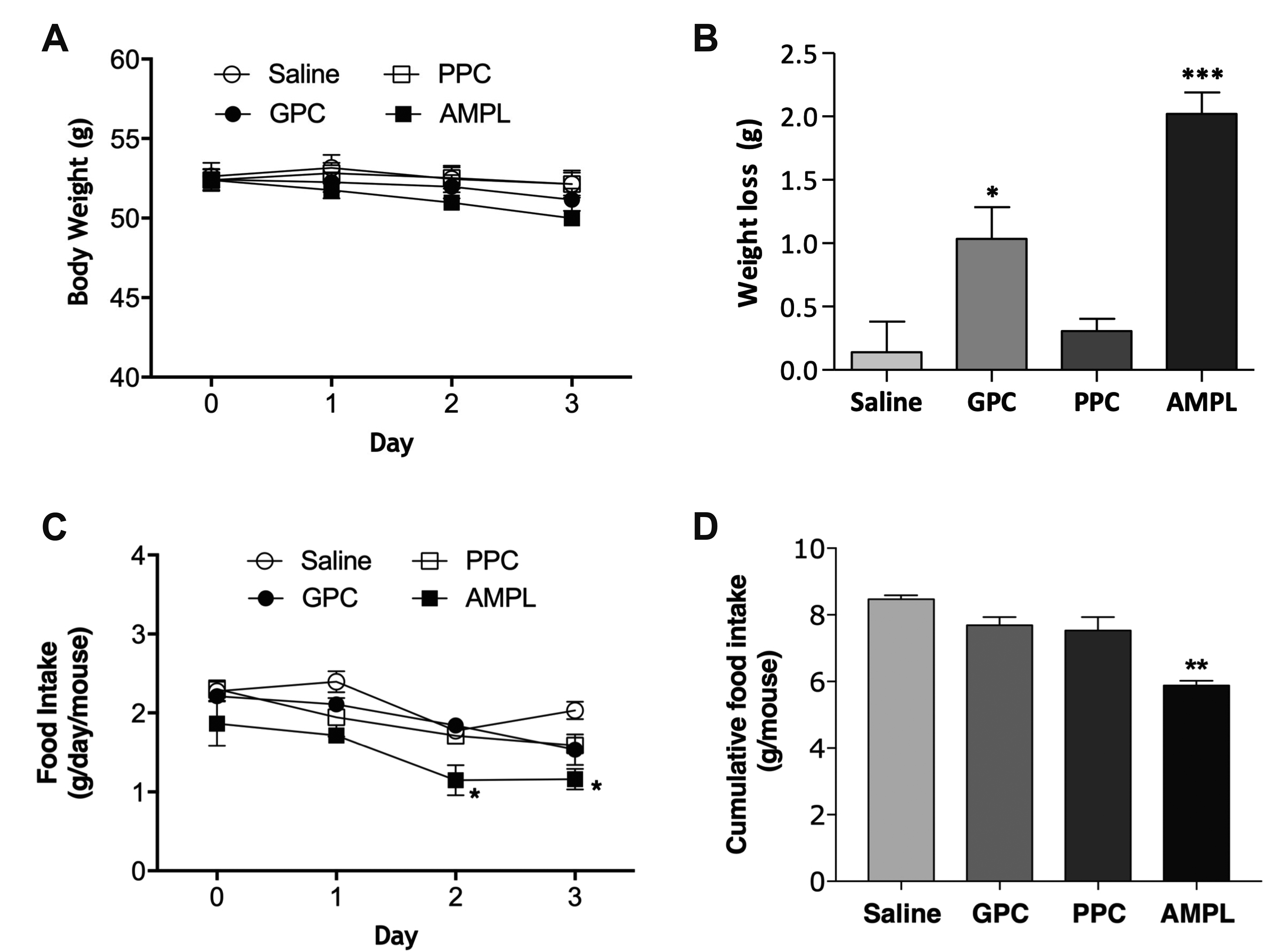
DIO mice were randomly divided into four experimental groups: vehicle (10 ml/kg of normal saline, n = 8), GPC (1,000 mg/kg, n = 8), PPC (40 mg/kg, n = 8) or AMPL (40 mg/kg, n = 8). Mice were given the corresponding treatments by subcutaneous injection onto right inguinal fat pads between 11 AM to 12 PM for three consecutive days by one skilled experimenter. The administered doses for PPC and AMPL were the animal equivalent dose converted from the doses applied to human in the clinic using dose conversion formula [6]. Typical injection protocol for PPC and AMPL is using a single vial solution consist of 250 mg of either PPC or AMPL [7]. For GPC, the most effective non-toxic dose was determined by preliminary experiments. The left inguinal fat pads were injected with equal volume of saline as a negative control. Body weight and food intake were recorded every day during treatment period. Mice were sacrificed 24 h after the last injection.
Tissue preparation and histological analysis
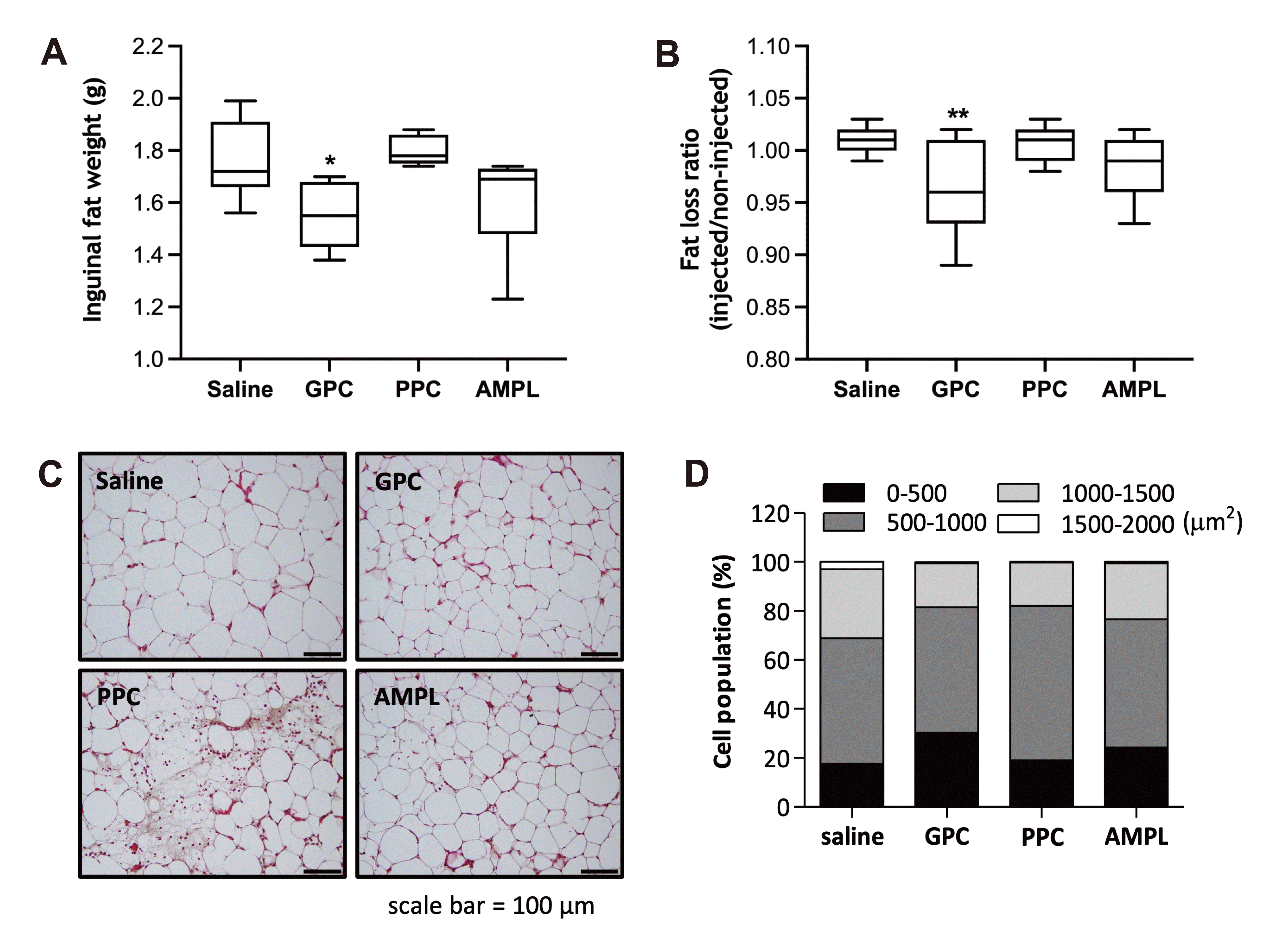
One day after the last injection (on day 4), the mice were euthanized, and inguinal fat pads were removed from both sides and weighed in a blind fashion. Inguinal lymph nodes were carefully removed from the adipose tissue before weighing. The tissues were washed with saline and fixed in 10% NBF for 24 h. Then the tissues were embedded in paraffin wax and sectioned at 5 μm using microtome. Paraffin-sectioned adipose tissues were stained with hematoxylin and eosin (H&E). For image analysis, 3 randomly selected fields per mouse for each treatment group were examined under light microscope (BX51, Olympus) at a final magnification of ×200 and photographed with Olympus DP22 digital camera (Olympus). The size of adipocytes was calculated by using Adiposoft as described previously [8].
Statistical analysis
All values are expressed as mean ± standard error of the mean (SEM). The experiments were statistically analyzed and compared by using one-way ANOVA followed by Dunnett’s post-test in GraphPad Prism 5.0 (La Jolla, CA, USA). p values < 0.05 were considered statistically significant.
RESULTS
Cytotoxicity of GPC on 3T3-L1 cells
To determine non-toxic experimental concentration of GPC in 3T3-L1 cells, we performed MTS-based cell viability assay in 3T3-L1 cells that are treated with varying concentrations of GPC for 96 h. As shown in Fig. 1A, GPC had no significant cytotoxicity on 3T3-L1 cells up to 8 mM. However, slight morphological change was observed at 8 mM of GPC, and therefore 6 mM of GPC was applied as a maximum concentration in the following study with 3T3-L1 cells. Both PPC and AMPL maintained normal cell viability up to 100 μM, and thus 100 μM was set as a maximum concentration of PPC and AMPL for the subsequent experiments (Fig. 1B, C).
GPC suppresses adipogenesis and lipid accumulation in 3T3-L1 adipocytes
To examine the effect of GPC on adipogenesis and lipid accumulation, 3T3-L1 preadipocytes were treated with the indicated concentrations of GPC for 4 days during the course of differentiation. On the completion of adipocyte differentiation, the degree of lipid accumulation was assessed by Oil Red O. Treatment with GPC significantly reduced the numbers of lipid droplet and the lipid contents in the adipocytes in a concentration-dependent manner (Fig. 2A, B). Lipid contents were decreased by 22.3% in the differentiated adipocytes treated with 4 mM GPC when compare to non-treated control (Fig. 2B). Similarly, 100 μM of PPC markedly reduced lipid contents in 3T3-L1 adipocytes during differentiation by 42.4% compared to that of non-treated control, and no significant differences in lipid contents were observed in AMPL-treated cells with three different concentrations.
GPC enhances basal lipolysis in 3T3-L1 adipocytes
Next, to determine whether GPC exerts any positive effect on lipolysis, we evaluated basal lipolysis by measuring glycerol release from matured 3T3-L1 adipocytes incubated with GPC. As shown in Fig. 2C, GPC significantly enhanced basal lipolysis in a concentration-dependent manner (2 mM; 1.14-fold, 4 mM; 1.8-fold, and 6 mM; 2.0-fold vs. non-treated control), to the similar degree with isoproterenol-induced glycerol release (2.1-fold vs. non-treated control) in which isoproterenol was used as a positive control in this experiment. AMPL also increased glycerol content in media by 1.2-fold, but no significant release of glycerol was detected in adipocytes treated with PPC (Fig. 2C).
GPC reduces body weight and food intake in DIO mice
In order to investigate potential effect of GPC in fat reducing, GPC was injected to DIO mice for three consecutive days. When the changes in body weight was monitored during the injection period, no significant changes were observed between GPC and saline control groups (Fig. 3A). Total weight losses during 3 days of injection period in saline-, GPC-, PPC- or AMPL-treated groups were 0.14 ± 0.24 g, 1.04 ± 0.25 g, 0.31 ± 0.10 g and 2.02 ± 0.17 g, respectively (Fig. 3B). Interestingly, AMPL-injected mice showed a significant decrease in food intake from day 2 of injection compared to saline-injected mice (Fig. 3C). Total food intake during the injection period was decreased in all experimental groups by 9.17%, 11.0% and 30.5% in GPC, PPC and AMPL groups, respectively, although the decreases in food intake by GPC and PPC were not statistically significant (Fig. 3D).
GPC reduces inguinal fat mass and adipocyte size
Inguinal fat mass of injected side was significantly decreased by 11.5% in GPC-injected mice when compared to the saline-injected mice (Fig. 4A). Reduction in inguinal fat weight was also observed in AMPL-injected mice by 8.9% although the difference was not statistically significant. The fat loss ratio was calculated by injected tissue mass divided by non-injected tissue mass. The calculated ratios for saline, GPC, PPC and AMPL groups were 1.01 ± 0.01, 0.96 ± 0.02, 1.01 ± 0.01 and 0.98 ± 0.01, respectively (Fig. 4B). Only the difference between saline- and GPC-injected mice showed statistical significance at p < 0.01, suggesting that GPC reduced fat accumulation in the injected area. Next, histological analysis was performed using adipose tissue sections prepared from the injection site to compare morphological changes of inguinal adipose tissue between experimental groups. GPC- and AMPL-injected mice exhibited reduced adipocytes size compared to the saline-injected mice while PPC-injected mice show marked adipocyte destruction (Fig. 4C). The result of histological analysis was also confirmed by distribution of cell population which demonstrates the adipocytes of GPC-injected mice were distributed over smaller size ranges compared to saline-injected mice (Fig. 4D). In addition, mean sizes of GPC, PPC and AMPL-treated adipocytes were 17.9%, 10.7% and 18.7% smaller than those of saline-injected mice (data not shown).
DISCUSSION
Mesotherapy and injection lipolysis have gained popularity for reduction of local fat and the appearance of cellulite. The use of lipolytic solutions in injection lipolysis is primarily based on anecdotal evidence and empirical observations in clinics. Here, we evaluated the potential efficacy of GPC on local fat dissolve and compared the effect with commonly used lipolytic agents (PPC and AMPL) using in vitro and in vivo models of obesity. In 3T3-L1 adipocytes, GPC inhibited fat accumulation during adipocyte differentiation while enhancing basal lipolysis in mature adipocytes (Fig. 2). The results from in vitro studies were in parallel with the reduction of adipocyte size and inguinal fat mass in GPC-injected mice (Fig. 4). These effects of GPC were more pronounced when compared to PPC and AMPL. The lipolytic effect of GPC was previously demonstrated in an experiment performed with obese-diabetic KK-Ay mice. Ten weeks of GPC administration in KK-Ay mice significantly reduced total cholesterol and triglyceride levels in serum [9]. These effects of GPC may be ascribed to choline-induced lipid metabolism. GPC is a useful source of choline, an essential nutrient required for cell membrane integrity and signaling and lipid transport [10,11]. This hypothesis can be supported by results reported by Kawamura et al. [12] that a single oral ingestion of 1,000 mg GPC significantly elevated serum level of choline after 60 min of ingestion and subsequent increase in serum glycerol level after 120 min of ingestion in humans.
Lipolysis is the hydrolysis of triglyceride stored in intracellular lipid droplets for generation of energy in the form of fatty acids and glycerol. In adipose tissue, adipose triglyceride lipase (ATGL) is highly expressed and functions in hydrolyzing triglyceride and ATGL overexpression in 3T3-L1 adipocytes by adenovirus-infection resulted in an elevation of glycerol release [13]. ATGL is modulated by Pparα, a transcription factor that regulates fatty acid oxidation. Choline is reported to reduce hepatic triglyceride accumulation by upregulating Pparα, and genes involved in fatty acid oxidation [14]. The increase in mRNA expression level of Pparα has been reported in the liver of GPC-fed mice suggesting that GPC may regulate lipid metabolism by activating downstream targets of Pparα such as ATGL [15].
One of the major findings of the present study is the histological changes in subcutaneous adipose tissue of mice injected with experimental solutions. In DIO mice, GPC and AMPL markedly reduced the size of adipocytes. However, PPC caused disrupted structure of adipocyte with noticeable signs of inflammation and tissue necrosis (Fig. 4C). Similarly, Noh and Heo [16] have reported significant inflammation, necrosis, and fibrosis in response to injection of PPC preparation in rat inguinal fat pad. Subcutaneous fat reduction by PPC injection has been suggested as a result of inflammation-mediated adipocyte apoptosis and resorption [17,18]. The results from histological analysis strongly support the different action mechanism of GPC and PPC on local fat dissolve, in which GPC reduces adipose tissue mass by stimulating lipolysis while PPC induces apoptosis in adipocytes. Moreover, PPC significantly inhibited 3T3-L1 adipocyte differentiation, however lipolytic activity was not observed in DIO mice injected with PPC (Fig. 2). Adipogenesis is a process of new fat cell formation and it can be regulated by apoptosis [19,20]. PPC has been reported to inhibit adipogenesis via inducing apoptosis in several studies [21,22], in parallel to our in vitro results showing decreased adipogenesis and in vivo results displaying adipose tissue necrosis by PPC (Fig. 4C). Surprisingly, we could not observe lipid reducing effect of PPC in mice (Fig. 4). This contradictory result could be explained by a previous study which reported that PPC injection reduced subcutaneous fat mass at day 7 post-injection but did not affect the tissue mass in a shorter period of injection [23]. It is worth noting that the lipolytic effect of AMPL was not significant when injected in inguinal fat pad in this study. Although AMPL is recognized as a stimulator of lipolysis, several studies reported that AMPL injection is not effective in reducing local fat [24,25]. The body weight loss and decrease in adipocyte size by AMPL are likely to be attributed to significant decrease in food intake.
In summary, we found that GPC inhibited adipogenesis and enhanced basal lipolysis, leading to a reduction of adipocyte size and subsequent loss of local fat. GPC also exhibited relatively low cytotoxicity. Together, these observations support the potential use of GPC as an injection lipolytic agent. However, further studies are needed to investigate the molecular mechanism of GPC in reducing local fat and the safety of using GPC for the purpose of injection lipolysis.

 XML Download