

 PDF
PDF ePub
ePub Citation
 Print
Print


INTRODUCTION
Coronary artery aneurysm (CAA) is uncommon, exhibiting an incidence of 0.3% to 5.3% (1). Most CAAs occur in the epicardial space, along the course of major coronary arteries. We encountered a patient with a giant aneurysm embedded in the basal posterior wall of the left ventricle (LV), with a direct fistulous connection to a dilated, tortuous left circumflex artery (LCX). Additionally, the LCX directly communicated with the right coronary artery (RCA) at the crux cordis. To our knowledge, this phenomenon has not been reported; herein, we describe this rare and incidental finding by focusing on its presentation in echocardiogram (ECG)-gated multidetector computed tomography (MDCT).
CASE REPORT
A 43-year-old woman was referred to our institution for the evaluation of palpitation that had been present for 2 months. Notably, the palpitation occurred in the resting condition and exhibited a duration of 20 minutes. The patient had no remarkable medical history such as hypertension, diabetes, or trauma; however, she had received hormone replacement therapy for premature menopause throughout the prior 18 months. Her blood pressure was 122/72 mm Hg and her pulse rate was 103 beats/min. Chest radiography showed normal heart configuration and size.
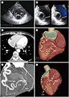
ECG showed normal sinus rhythm and 24 hours Holter monitoring showed ventricular premature contraction. On ECG (Vivid 7 ultrasound, GE Healthcare, Chicago, IL, USA), a 2.3 × 2.0-cm-sized encapsulated, homogeneous, and anechoic round mass was identified at the basal posterior wall of the LV (Fig. 1A, B). Subsequently, further evaluation comprised cardiac CT, which was performed by using a 64-slice MDCT (Lightspeed VCT, GE Healthcare); this examination revealed a 2.1 × 2.4-cm-sized intramyocardial coronary aneurysm (IMCA) with thin cap at the basal posterior wall of the LV (Fig. 1C). Additionally, a diffuse, dilated, and tortuous LCX was observed to communicate with the IMCA (Fig. 1D, E); there was direct intercoronary communication (ICC) between LCX and RCA at crux cordis (Fig. 1F). The RCA was straight without evidence of abnormal dilation; MDCT did not demonstrate any myocardial thickening or stenosis in the coronary arteries. The coronary sinus was normal without any abnormal dilation; thus, the patient was discharged with medication, but without any surgical or interventional treatment, because there was no symptom related to these findings.
DISCUSSION
CAA constitutes localized dilation of a coronary artery segment by more than 1.5-fold, compared with adjacent normal segments (12); Atherosclerosis and kawasaki disease are the most common causes of CAA. Notably, CAA may also develop secondary to arteritis, such as in Behçet's disease, syphilis, and takayasu arteritis, as well as after various coronary interventions; with these causes, most CAAs occur along major coronary arteries in the epicardial space (1234). IMCA as exhibited in our patient is very rare with an unknown incidence; extensive review of the literature revealed a case report that introduced IMCA arising from the septal branch of the left anterior descending artery (LAD), secondary to primary percutaneous transluminal coronary angioplasty (5). The authors of that report suggested that intramyocardial localization of the aneurysm resulted in different angiographic, pathophysiological, and clinical features of the disease, compared with epicardial coronary aneurysm (5). Most CAAs are asymptomatic; in those that are symptomatic, thrombus is frequently found within the aneurysm (1). Rupture of the CAA can be a life-threatening condition (2). Therefore, early diagnosis of CAA is critical.
Another remarkable finding of our case is the ICC between LCX and RCA at the crux cordis; this comprises a very rare subset of coronary artery anomalies, involving unidirectional or bidirectional blood flow between two or more coronary arteries (67). The true prevalence in the general population is not known; however, coronary angiographic findings have shown incidences of 0.002% in 126595 patients and 0.02% in 9726 patients (89).
Two types of ICC have been reported thus far: 1) between LAD and posterior descending arteries in the distal interventricular groove, and 2) between the LCX and RCA in the posterior atrioventricular groove (6), as in our patient. The practical significance of ICCs and their consequences remain unknown. Some authors speculate that these connections may play a protective role for the myocardium upon the development of significant coronary artery obstruction in one of the connecting vessels. Importantly, myocardial ischemia can result from the coronary steal phenomenon by unidirectional flow (6). Collateral vessels and ICC are quite different: collaterals develop in obstructive coronary artery disease, are typically less than 1 mm in diameter, and appear tortuous and twisted with a corkscrew shape, whereas intercoronary anastomosis in the absence of obstructive lesions tends to be straight or gently curved (7). Histologically, collaterals that develop in the presence of obstructive coronary artery disease are composed of endothelium supported by poorly organized collagen, muscle, and elastic fibers; in contrast, ICCs are similar to an epicardial vessel with a well-defined muscular layer. (10). Persistence of fetal coronary circulation has been suggested as the underlying mechanism for the development of ICC. A true intercommunication in the coronary system is benign, and may serve as a collateral source if a coronary artery obstruction develops (6).
We suspect either of two possibilities for intramyocardial CAA with this finding. First, spontaneous closure of a preexisting fistula between the LAD artery and LV may have resulted in aneurysmal dilation at the distal intramyocardial fistulous segment; second, the straight RCA, with narrower caliber than the LCX, has higher pressure relative to the LCX, such that persistent unidirectional or bidirectional flow with dominant flow from RCA to LCX may cause dilation and tortuosity of LCX, resulting in a giant intramyocardial CAA.
In conclusion, we have reported a rare instance of an IMCA in a patient with ICC between a dilated tortuous LCX and straight RCA.
 XML Download