Journal List > Yonsei Med J > v.61(2) > 1141543

Lee, Kang, Kim, Lee, Lee, Cho, Yoon, Jung, Park, Oh, Hong, and Hong: NOTCH1 Pathway is Involved in Polyhexamethylene Guanidine-Induced Humidifier Disinfectant Lung Injuries

Abstract

An outbreak of fatal humidifier disinfectant lung injuries (HDLI) occurred in Korea. Human studies on mechanisms underlying HDLI have yet to be conducted. This study aimed to investigate methylation changes and their potential role in HDLI after exposure to HDs containing polyhexamethylene guanidine-phosphate. DNA methylation analysis was performed in blood samples from 10 children with HDLI and 10 healthy children using Infinium Human MethylationEPIC BeadChip. Transcriptome analysis was performed using lung tissues from 5 children with HDLI and 5 controls. Compared to healthy controls, 92 hypo-methylated and 79 hyper-methylated CpG sites were identified in children with HDLI at the statistical significance level of |Δβ|>0.2 and p<0.05. NOTCH1 was identified as a candidate network hub gene in cases. NOTCH1 transcripts significantly increased in lung tissues from HDLI cases compared to unexposed controls (p=0.05). NOTCH1 may play an important role in pulmonary fibrosis of HDLI.

An outbreak of fatal lung injuries occurred in Korea between early 2000 and 2011, characterized by rapidly progressing respiratory failure with lung fibrosis, extensive air leak syndrome in many cases, a lack of responsiveness to any treatment, and high mortality rate.1,2,3,4,5 This fatal interstitial lung disease (ILD) was distinct from previously identified ILDs in terms of clinical course as well as radiologic and pathologic findings; therefore, it was considered to be idiopathic.1,2 Toxic chemicals, including polyhexamethylene guanidine (PHMG), in humidifier disinfectants (HDs) were subsequently identified as the cause.1,2 The unique features of this fatal lung disease raised questions regarding the distinct mechanisms underlying the disorder.6 However, there has been no report on the mechanisms underlying HD-associated lung injuries (HDLI) in humans. As altered DNA methylation is associated with development of idiopathic pulmonary fibrosis,6,7 we investigated whether DNA methylation plays a role in HDLI using human samples.
Blood samples from 10 children with HDLI and 10 healthy control children with no exposure to HDs were used to analyze methylation profiles. Clinical characteristics of the study population are summarized in Table 1. The mean age at diagnosis of HDLI was 35.4 months (range, 12–81 months) and blood samples for methylation analysis were obtained at a mean age of 11.4 years (range, 7–15 years). Male-to-female rate was 7:3. None of the children in sex-matched control group had any respiratory diseases and their mean age was 7 years. DNA extracted from the peripheral blood mononuclear cells of each subject was analyzed using Infinium Human MethylationEPIC BeadChip (Illumina, San Diego, CA, USA). For quality check (QC) of the methylation data, beta-mixture quantile normalization, and Pearson's correlation (range: −1≤r≤1) for reproducibility between samples were performed. For QC of the transcriptome data, all data were normalized with the robust multi-average method implemented in in Affymetrix® Power Tools (Thermo Fisher Scientific, Waltham, MA, USA). Statistical significance for differentially methylated CpG sites was set at |Δβ|>0.2 and p<0.05 using a t-test. Ingenuity® Pathway Analysis (IPA, Ingenuity Systems, Redwood City, CA, USA) was used to represent the functional networks of genes containing differentially methylated CpG sites. Transcriptome analysis was performed using lung tissues from five pediatric patients with HDLI and five control children. Lung tissue was obtained from children with no abnormal lung lesions from Bio-Resource Center at Asan Medical Center to form a control group. The Institutional Review Board of Asan Medical Center reviewed and approved the study protocol (IRB No. 2016-0885).
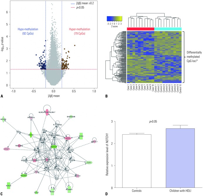
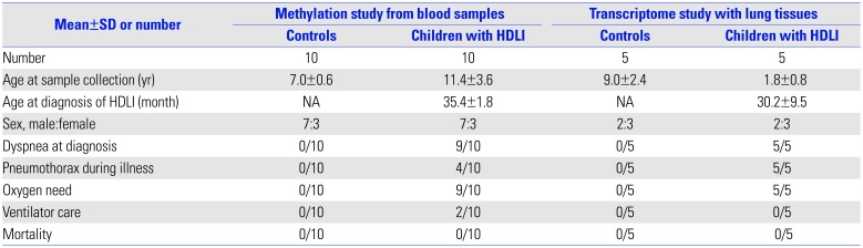
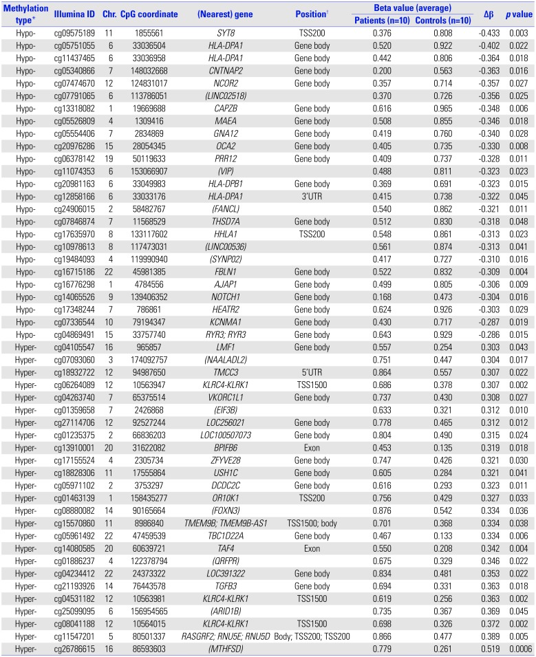
A total of 171 CpG loci (79 hypermethylated, 92 hypomethylated) showed significantly differential methylation patterns in children with HDLI compared to the controls (Fig. 1A), with a distinctive clustering observed between the two groups (Fig. 1B) (Table 2). The top 25 hypomethylated and 25 hypermethylated CpG loci are listed in Table 2. SYT8 cg09575189 showed the highest hypomethylation level (|Δβ|=0.433, p=0.003), whereas cg26786615 (chr16: 86593603) had the highest hypermethylation level (|Δβ|=0.519, p=0.0006). However, there are a few functional studies of these two genes and no reports in existing literature that provide any clues to the associations between them and fibrosis and/or lung diseases. Potential upstream and downstream regulators of NOTCH1 based on IPA network analysis and its signaling (https://www.rndsystems.com/pathwyas/notch-signaling-pathway) are described in Table 3.
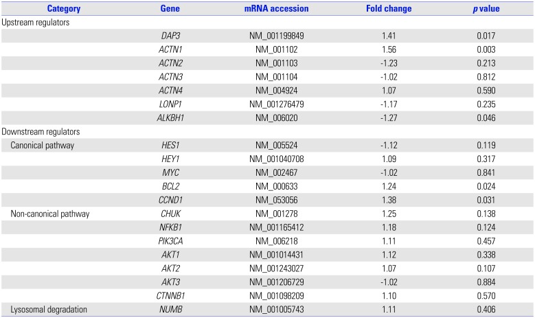
NOTCH1 cg14065526 showed a significant degree of hypomethylation (|Δβ|=0.304, p=0.016). In further network analysis of the genes containing differently methylated CpG sites, “cancer, organismal injury and abnormalities, reproductive system disease (score=41)” was identified as the top network for HDLI, indicating NOTCH1 as a hub gene (Fig. 1C). The cg14065526 (chr9: 139406352) of NOTCH1 showed a significantly hypomethylated level (|Δβ|=0.304, p=0.016). NOTCH1 transcripts from lung tissues were significantly elevated in HDLI cases compared to unexposed controls (p=0.05, each group n=5) (Fig. 1D).
Our present findings from methylation and transcriptome analysis of human blood and lung tissues have identified that NOTCH1 is involved in the pathogenesis of HDLI. This is the first study to investigate DNA methylation changes and network analyses combined with transcriptomics in pediatric patients with HDLI, which may partially explain the underlying mechanisms of HDLI.
Although NOTCH1 may be common to the mechanisms of other types of ILDs,8 the results of our current analysis suggest that it also plays a central role in the mechanism of HDLI. Notch1 is involved in angiogenesis, abnormal remodeling of vessels, and mucus hypersecretion, and thereby is associated with pathogenesis of diverse lung diseases.9 The apoptosis of bronchial epithelial cells following exposure to toxic chemicals affects the clearance of apoptotic debris combined with lung fibrosis.10 The overexpression of NOTCH1, which is related to its gene hypomethylation, as shown in this study, promotes the differentiation of myofibroblasts, which is a critical step in pulmonary fibrosis.3 NOTCH1 has been identified to be involved in bleomycin-induced lung diseases and paraquat poisoning, for which the main mechanism is pulmonary fibrosis.11,12 The results of previous reports and our present findings provide strong evidence for the involvement of NOTCH1 in the pathogenesis of fatal fibrotic lung diseases and give new insights into the possible mechanisms of lung injuries caused by inhalation of unidentified but harmful chemicals that are commonly used.
The inhalation of toxic chemicals damages the epithelial lining in the airway, initiating a series of processes including disruption of epithelial lining, alterations of diverse mediators and chemokine levels, and induction of epithelium-to-mesenchymal transition (EMT).13 NOTCH1 regulates EMT through various signaling factors, such as TGF-β, NF-κB, and β-catenin.10 It has been reported that exposure to PHMG phosphate can induce EMT in a dose-dependent manner.14 A previous study identified that PHMG could induce EMT through the Akt/Notch signaling pathway.15 This prior evidence, in combination with our current data, further supports the notion that NOTCH1 plays a role in the pathogenesis of HDLI via EMT following exposure to HDs that contain PHMG.
Our study had some limitations, including its small sample size. However, the results of the current study are significant in that HDLI is an exceptional disease, and the acquisition of blood and lung tissue in our patients was not easy. In our present cohort, there were time lags with a mean of 9 years between diagnosis of HDLI and blood sampling. The methylation patterns in the blood obtained after a time lag of 9 years may have been affected by diverse factors.16 A previous study showed that less than 30% of individuals showed methylation changes in epigenome- wide DNA methylation analysis on average 11 years apart, even with intra-individual variations.16 We could not perform methylation analysis in human lung tissues in the current study, as these samples were not available. In spite of the limitations, methylation changes observed in the present study could be helpful to elucidate the mechanisms underlying HDLI with stable disease state.
In conclusion, we have identified NOTCH1 pathways as one of the possible main fibrogenetic mechanisms of HDLI in children following exposure to PHMG phosphate. Further identification and elucidation of the mechanisms underlying this fatal lung disease are essential for the future development of therapeutics and prevention of lung diseases after exposure to harmful domestic chemicals.

ACKNOWLEDGEMENTS

This study was funded by the Korea Ministry of Environment (MOE) as “the Environmental Health Action Program (2016001360006)” and partially supported by the Environmental Health Center for Hazardous Chemical Exposure funded by the Ministry of Environment Republic of Korea (2019).

Notes

The authors have no potential conflicts of interest to disclose.

AUTHOR CONTRIBUTIONS:

  • Conceptualization: Eun Lee, Mi Jin Kang, Jeong-Hyun Kim, and Soo-Jong Hong.

  • Data curation: Seung-Hwa Lee, So-Yeon Lee, Hyun-Ju Cho, Jisun Yoon, Sungsu Jung, Yangsoon Park, Dong Kyu Oh, and Sang-Bum Hong.

  • Formal analysis: Mi Jin Kang and Jeong-Hyun Kim.

  • Funding acquisition: Eun Lee and Soo-Jong Hong.

  • Investigation: Eun Lee, Mi Jin Kang, Jeong-Hyun Kim, Dong Kyu Oh, Sang-Bum Hong, and Soo-Jong Hong.

  • Methodology: Mi Jin Kang, Jeong-Hyun Kim, Seung-Hwa Lee, and Yangsoon Park.

  • Project administration: Eun Lee, Mi Jin Kang, Jeong-Hyun Kim, and Soo-Jong Hong.

  • Resources: Eun Lee and Soo-Jong Hong.

  • Software: Mi Jin Kang and Jeong-Hyun Kim.

  • Supervision: Soo-Jong Hong.

  • Validation: Eun Lee, Mi Jin Kang, Jeong-Hyun Kim, Seung-Hwa Lee, and Yangsoon Park.

  • Writing—original draft: Eun Lee.

  • Writing—review & editing: Eun Lee, Jeong-Hyun Kim, and Soo-Jong Hong.

  • Approval of final manuscript: all authors.

References

1. Kim KW, Ahn K, Yang HJ, Lee S, Park JD, Kim WK, et al. Humidifier disinfectant-associated children's interstitial lung disease. Am J Respir Crit Care Med. 2014; 189:48–56. PMID: 24199596.
2. Hong SB, Kim HJ, Huh JW, Do KH, Jang SJ, Song JS, et al. A cluster of lung injury associated with home humidifier use: clinical, radiological and pathological description of a new syndrome. Thorax. 2014; 69:694–702. PMID: 24473332.
crossref
3. Ryu SH, Park DU, Lee E, Park S, Lee SY, Jung S, et al. Humidifier disinfectant and use characteristics associated with lung injury in Korea. Indoor Air. 2019; 29:735–747. PMID: 31278778.
crossref
4. Park DU, Ryu SH, Lim HK, Kim SK, Choi YY, Ahn JJ, et al. Types of household humidifier disinfectant and associated risk of lung injury (HDLI) in South Korea. Sci Total Environ. 2017; 596-597:53–60. PMID: 28415004.
crossref
5. Yoon J, Cho HJ, Lee E, Choi YJ, Kim YH, Lee JL, et al. Rate of humidifier and humidifier disinfectant usage in Korean children: a nationwide epidemiologic study. Environ Res. 2017; 155:60–63. PMID: 28189074.
crossref
6. Yang IV, Pedersen BS, Rabinovich E, Hennessy CE, Davidson EJ, Murphy E, et al. Relationship of DNA methylation and gene expression in idiopathic pulmonary fibrosis. Am J Respir Crit Care Med. 2014; 190:1263–1272. PMID: 25333685.
crossref
7. Sanders YY, Ambalavanan N, Halloran B, Zhang X, Liu H, Crossman DK, et al. Altered DNA methylation profile in idiopathic pulmonary fibrosis. Am J Respir Crit Care Med. 2012; 186:525–535. PMID: 22700861.
crossref
8. Zong D, Ouyang R, Li J, Chen Y, Chen P. Notch signaling in lung diseases: focus on Notch1 and Notch3. Ther Adv Respir Dis. 2016; 10:468–484. PMID: 27378579.
crossref
9. Jiang J, Xiao K, Chen P. NOTCH signaling in lung diseases. Exp Lung Res. 2017; 43:217–228. PMID: 28636457.
crossref
10. Kage H, Borok Z. EMT and interstitial lung disease: a mysterious relationship. Curr Opin Pulm Med. 2012; 18:517–523. PMID: 22854509.
11. Yin Q, Wang W, Cui G, Yan L, Zhang S. Potential role of the Jagged1/Notch1 signaling pathway in the endothelial-myofibroblast transition during BLM-induced pulmonary fibrosis. J Cell Physiol. 2018; 233:2451–2463. PMID: 28776666.
crossref
12. Li T, Yang X, Xin S, Cao Y, Wang N. Paraquat poisoning induced pulmonary epithelial mesenchymal transition through Notch1 pathway. Sci Rep. 2017; 7:924. PMID: 28424456.
crossref
13. Liu T, Hu B, Choi YY, Chung M, Ullenbruch M, Yu H, et al. Notch1 signaling in FIZZ1 induction of myofibroblast differentiation. Am J Pathol. 2009; 174:1745–1755. PMID: 19349363.
crossref
14. Shin DY, Jeong MH, Bang IJ, Kim HR, Chung KH. MicroRNA regulatory networks reflective of polyhexamethylene guanidine phosphate-induced fibrosis in A549 human alveolar adenocarcinoma cells. Toxicol Lett. 2018; 287:49–58. PMID: 29337256.
crossref
15. Jeong MH, Kim HR, Park YJ, Chung KH. Akt and Notch pathways mediate polyhexamethylene guanidine phosphate-induced epithelial-mesenchymal transition via ZEB2. Toxicol Appl Pharmacol. 2019; 380:114691. PMID: 31348943.
crossref
16. Gervin K, Andreassen BK, Hjorthaug HS, Carlsen KCL, Carlsen KH, Undlien DE, et al. Intra-individual changes in DNA methylation not mediated by cell-type composition are correlated with aging during childhood. Clin Epigenetics. 2016; 8:110. PMID: 27785156.
crossref
Fig. 1

Results of methylation, network, and NOTCH1 expression analysis in pediatric HDLI cases. (A) Volcano plot of differentially methylated CpG sites. (B) Heatmap of differentially methylated CpG sites between children with humidifier disinfectant associated lung injuries and unexposed healthy controls. Differentially methylated CpG loci indicated by asterisk. (C) The top network of differentially methylated CpG sites was found to be “cancer, organismal injury and abnormalities, reproductive system disease” and was derived from genes containing hyper-/hypo-methylated CpG sites associated with HDLI. (D) The transcriptional expression of NOTCH1 between HDLI cases and the control group (p=0.05, t-test, nonparametric methods were applied, and no correction for multiple testing was done due to the small sample size of each group, n=5 for each group). HDLI, humidifier disinfectant lung injuries.

ymj-61-186-g001
Table 1

Clinical Characteristics of the Study Population

ymj-61-186-i001
Mean±SD or number Methylation study from blood samples Transcriptome study with lung tissues
Controls Children with HDLI Controls Children with HDLI
Number 10 10 5 5
Age at sample collection (yr) 7.0±0.6 11.4±3.6 9.0±2.4 1.8±0.8
Age at diagnosis of HDLI (month) NA 35.4±1.8 NA 30.2±9.5
Sex, male:female 7:3 7:3 2:3 2:3
Dyspnea at diagnosis 0/10 9/10 0/5 5/5
Pneumothorax during illness 0/10 4/10 0/5 5/5
Oxygen need 0/10 9/10 0/5 5/5
Ventilator care 0/10 2/10 0/5 0/5
Mortality 0/10 0/10 0/5 0/5

HDLI, humidifier disinfectant lung injuries; NA, not applicable.

Table 2

Top 25 Hypomethylated and Top 25 Hypermethylated Sites Showing Significantly Different Levels in Pediatric Patients with HDLI Compared to Unexposed Healthy Control Children

ymj-61-186-i002
Methylation type* Illumina ID Chr. CpG coordinate (Nearest) gene Position Beta value (average) Δβ p value
Patients (n=10) Controls (n=10)
Hypo- cg09575189 11 1855561 SYT8 TSS200 0.376 0.808 -0.433 0.003
Hypo- cg05751055 6 33036504 HLA-DPA1 Gene body 0.520 0.922 -0.402 0.022
Hypo- cg11437465 6 33036958 HLA-DPA1 Gene body 0.442 0.806 -0.364 0.018
Hypo- cg05340866 7 148032668 CNTNAP2 Gene body 0.200 0.563 -0.363 0.016
Hypo- cg07474670 12 124831017 NCOR2 Gene body 0.357 0.714 -0.357 0.027
Hypo- cg07791065 6 113786051 (LINC02518) 0.370 0.726 -0.356 0.025
Hypo- cg13318082 1 19669688 CAPZB Gene body 0.616 0.965 -0.348 0.006
Hypo- cg05526809 4 1309416 MAEA Gene body 0.508 0.855 -0.346 0.018
Hypo- cg05554406 7 2834869 GNA12 Gene body 0.419 0.760 -0.340 0.028
Hypo- cg20976286 15 28054345 OCA2 Gene body 0.405 0.735 -0.330 0.008
Hypo- cg06378142 19 50119633 PRR12 Gene body 0.409 0.737 -0.328 0.011
Hypo- cg11074353 6 153066907 (VIP) 0.488 0.811 -0.323 0.023
Hypo- cg20981163 6 33049983 HLA-DPB1 Gene body 0.369 0.691 -0.323 0.015
Hypo- cg12858166 6 33033176 HLA-DPA1 3′UTR 0.415 0.738 -0.322 0.045
Hypo- cg24906015 2 58482767 (FANCL) 0.540 0.862 -0.321 0.011
Hypo- cg07846874 7 11568529 THSD7A Gene body 0.512 0.830 -0.318 0.048
Hypo- cg17635970 8 133117602 HHLA1 TSS200 0.548 0.861 -0.313 0.023
Hypo- cg10978613 8 117473031 (LINC00536) 0.561 0.874 -0.313 0.041
Hypo- cg19484093 4 119990940 (SYNP02) 0.417 0.727 -0.310 0.016
Hypo- cg16715186 22 45981385 FBLN1 Gene body 0.522 0.832 -0.309 0.004
Hypo- cg16776298 1 4784556 AJAP1 Gene body 0.499 0.805 -0.306 0.009
Hypo- cg14065526 9 139406352 NOTCH1 Gene body 0.168 0.473 -0.304 0.016
Hypo- cg17348244 7 786861 HEATR2 Gene body 0.624 0.926 -0.303 0.029
Hypo- cg07336544 10 79194347 KCNMA1 Gene body 0.430 0.717 -0.287 0.019
Hypo- cg04869491 15 33757740 RYR3; RYR3 Gene body 0.643 0.929 -0.286 0.015
Hyper- cg04105547 16 965857 LMF1 Gene body 0.557 0.254 0.303 0.043
Hyper- cg07093060 3 174092757 (NAALADL2) 0.751 0.447 0.304 0.017
Hyper- cg18932722 12 94987650 TMCC3 5′UTR 0.864 0.557 0.307 0.022
Hyper- cg06264089 12 10563947 KLRC4-KLRK1 TSS1500 0.686 0.378 0.307 0.002
Hyper- cg04263740 7 65375514 VKORC1L1 Gene body 0.737 0.430 0.308 0.027
Hyper- cg01359658 7 2426868 (EIF3B) 0.633 0.321 0.312 0.010
Hyper- cg27114706 12 92527244 LOC256021 Gene body 0.778 0.465 0.312 0.012
Hyper- cg01235375 2 66836203 LOC100507073 Gene body 0.804 0.490 0.315 0.024
Hyper- cg13910001 20 31622082 BPIFB6 Exon 0.453 0.135 0.319 0.018
Hyper- cg17155524 4 2305734 ZFYVE28 Gene body 0.747 0.426 0.321 0.030
Hyper- cg18828306 11 17555864 USH1C Gene body 0.605 0.284 0.321 0.041
Hyper- cg05971102 2 3753297 DCDC2C Gene body 0.616 0.293 0.323 0.011
Hyper- cg01463139 1 158435277 OR10K1 TSS200 0.756 0.429 0.327 0.033
Hyper- cg08880082 14 90165664 (FOXN3) 0.876 0.542 0.334 0.036
Hyper- cg15570860 11 8986840 TMEM9B; TMEM9B-AS1 TSS1500; body 0.701 0.368 0.334 0.038
Hyper- cg05961492 22 47459539 TBC1D22A Gene body 0.467 0.133 0.334 0.006
Hyper- cg14080585 20 60639721 TAF4 Exon 0.550 0.208 0.342 0.004
Hyper- cg01886237 4 122378794 (QRFPR) 0.675 0.329 0.346 0.022
Hyper- cg04234412 22 24373322 LOC391322 Gene body 0.834 0.481 0.353 0.022
Hyper- cg21193926 14 76443578 TGFB3 Gene body 0.694 0.331 0.363 0.018
Hyper- cg04531182 12 10563981 KLRC4-KLRK1 TSS1500 0.619 0.256 0.363 0.002
Hyper- cg25099095 6 156954565 (ARID1B) 0.735 0.367 0.369 0.045
Hyper- cg08041188 12 10564015 KLRC4-KLRK1 TSS1500 0.698 0.326 0.372 0.002
Hyper- cg11547201 5 80501337 RASGRF2; RNU5E; RNU5D Body; TSS200; TSS200 0.866 0.477 0.389 0.005
Hyper- cg26786615 16 86593603 (MTHFSD) 0.779 0.261 0.519 0.0006

HDLI, humidifier disinfectant lung injuries; TSS, transcription start site; UTR, untranslated region.

*Hyper- and hypo- indicate the methylation levels of patients compared to controls, TSS200 and TSS1500 indicate the distance within 200 bp and 1500 bp from TSS, respectively.

Table 3

Gene Expression of Potential Upstream and Downstream Regulators of NOTCH1 in Formalin-Fixed, Paraffin-Embedded Lung Tissue Specimens from Children with HDLI and the Control Group

ymj-61-186-i003
Category Gene mRNA accession Fold change p value
Upstream regulators
DAP3 NM_001199849 1.41 0.017
ACTN1 NM_001102 1.56 0.003
ACTN2 NM_001103 -1.23 0.213
ACTN3 NM_001104 -1.02 0.812
ACTN4 NM_004924 1.07 0.590
LONP1 NM_001276479 -1.17 0.235
ALKBH1 NM_006020 -1.27 0.046
Downstream regulators
 Canonical pathway HES1 NM_005524 -1.12 0.119
HEY1 NM_001040708 1.09 0.317
MYC NM_002467 -1.02 0.841
BCL2 NM_000633 1.24 0.024
CCND1 NM_053056 1.38 0.031
 Non-canonical pathway CHUK NM_001278 1.25 0.138
NFKB1 NM_001165412 1.18 0.124
PIK3CA NM_006218 1.11 0.457
AKT1 NM_001014431 1.12 0.338
AKT2 NM_001243027 1.07 0.107
AKT3 NM_001206729 -1.02 0.884
CTNNB1 NM_001098209 1.10 0.570
 Lysosomal degradation NUMB NM_001005743 1.11 0.406

HDLI, humidifier disinfectant lung injuries.

Upstream regulators are predicted using Ingenuity Pathway Analysis. Downstream regulators of NOTCH1 are notified from (https://www.rndsystems.com/pathways/notch-signaling-pathway). Formalin-Fixed Paraffin-Embedded lung tissue specimens from HDLI cases (n=5) and controls (n=5).

TOOLS
Similar articles