

 PDF
PDF Citation
 Print
Print


INTRODUCTION
Syncope is a common problem in clinical practice.1 Although reflex syncope is frequently identified, approximately 30% of conventional diagnostic workups return a result of unexplained syncope (US).23 US can be a clinical manifestation of rare arrhythmia, and prolonged electrocardiogram (ECG) monitoring can detect intermittent or transient arrhythmia-related syncope if a symptom-correlated ECG is documented.
An implantable loop recorder (ILR) is an effective tool for detecting arrhythmia-related syncope.45 The European Society of Cardiology (ESC) guidelines recommend ILR in the early phases of evaluation for recurrent US.6 In pooled data from nine studies,27891011121314 176 of 506 patients (35%) with US had a syncope-correlated ECG at the end of a complete workup; of these, 56% had asystole, 11% had tachycardia, and 33% had no arrhythmia. A significant asymptomatic arrhythmia, such as a prolonged pause (≥ 3 seconds) or supraventricular tachycardia (SVT) (i.e., > 160 beats/min for > 32 beats), and ventricular arrhythmia have been considered possible causes of US.1516
Previous ILR studies have shown that more than half of all cases of US are caused by asystole,1718 and pacemaker (PM) implantation is highly effective in the case of syncope caused by brady-arrhythmia. Several studies have reported clinical predictors for later PM implantation in US patients.192021
We examined the usefulness of ILR-guided diagnosis of US (syncope-correlated arrhythmia, significant arrhythmia) and also analyzed the characteristics of patients likely to undergo PM implantation.
METHODS
Study population
Data for this multicenter, retrospective, observational study of 173 patients who received an ILR at 11 hospitals in Korea were gathered from February 2006 to April 2018. Enrolled patients with recurrent US and negative conventional diagnostic examinations received an ILR (Reveal LINQ ICM, Reveal Plus, or Reveal DX-XT; Medtronic Inc., Minneapolis, MN, USA or ConfirmTM; St. Jude Medical, St. Paul, MN, USA). The initial examinations for syncope evaluation included careful history-taking (total number and duration of syncope, prodromal symptoms, syncope-related injury, underlying disease), physical examination, baseline 12-lead ECG, two-dimensional echocardiography, 24-hour Holter, treadmill test, head-up tilt test (HUT), carotid sinus massage, and orthostatic blood pressure measurement. Invasive diagnostic tools, such as an electrophysiological study and coronary angiography were performed if clinically indicated. When a neurologic cause was suspected, brain computed tomography or magnetic resonance imaging was performed. The conventional diagnostic tests performed for syncope evaluation before ILR implantation are shown in Supplementary Table 1.
Any hypertensive medication that could cause symptomatic bradycardia or hypotension, such as a beta-blocker or Ca blocker, was withheld before the conventional evaluation or ILR implantation at the physician's discretion to determine whether they were causing the syncope. If the conventional examination did not reveal the cause of syncope, an ILR was implanted. Some physicians implanted an ILR when the clinical syncope was atypical for reflex syncope or orthostatic hypotension, even when the HUT or orthostatic BP measurement was positive.
Prodromal symptoms were defined as autonomic activation that occurred before unconsciousness, such as dizziness, nausea, vomiting, sweating, palpitation, or chest or abdominal discomfort. Major syncope-related injuries were defined as a major bone segment fracture, such as a skull fracture causing intracranial hemorrhage, and tooth subluxation. Minor syncope-related injuries were defined as swelling, ecchymosis or bruising, abrasion or laceration, and external superficial hematoma or minor bleeding.
Any bundle branch blocks (BBBs) were defined as left bundle branch block (LBBB) or right bundle branch block (RBBB) by a baseline 12-lead ECG, following the recent American Heart Association/American College of Cardiology/Heart Rhythm Society recommendations.22 Structural heart disease was defined as coronary artery disease (CAD), heart failure (HF), cardiomyopathy (CMP), or moderate to severe valvular heart disease (VHD).
ILR implantation
Each ILR was positioned subcutaneously in the left parasternal area at the level of the 4th–5th intercostal space under local anesthesia. The ILR stores rhythm strips when the patient presses a button in response to symptom occurrence (patient activation) or when the heart rate meets a preset limit (auto activation). The auto activation setting varied among patients, but it was generally programmed for a ventricular pause of > 3 seconds, a ventricular rate of < 40 beats/min, or a ventricular rate > 180 beats/min for more than 16 beats. Patients were taught to use the device prior to discharge after ILR implantation. Since small, insertable ILR devices (Reveal LINQTM ICM; Medtronic Inc., Minneapolis, MN, USA and Confirm RxTM; St. Jude Medical, St. Paul, MN, USA) came to the market in 2014 and 2017, respectively, ILR procedures have been simple and rapid and rarely entail major complications.23
Follow-up after ILR implantation
Regular ILR follow-up at an outpatient clinic was performed at 3- to 6-month intervals at the physician's discretion. Immediate ILR interrogation was performed in the case of recurrent syncope. The ILR remained implanted until a diagnostic event was recorded or until the end of the battery life. If no diagnosis of syncope was made before the end of the battery life, removal or reinsertion of the ILR was performed at the physician's discretion.
Study endpoint
The study endpoint was a symptom-correlated ECG diagnosis by ILR interrogation during the first recurrent syncope after implantation of the ILR. The secondary endpoint was significant arrhythmia irrespective of syncope, defined as a ventricular pause of > 3 seconds, a ventricular rate of < 40 beats/min, or a ventricular rate > 180 beats/min for more than 16 beats.
Statistical analysis
Continuous variables are expressed as the mean ± standard deviation. A χ2 analysis was used for categorical data, independent t-tests for continuous data, and Mann-Whitney tests for nonparametric cases. Cumulative incidence and event-free curves were derived using Kaplan-Meier analyses, stratified by study groups, and compared using the log-rank test. To identify independent predictors for PM implantation, we first conducted univariate analyses and then included predictors with a significance level < 10% in a multivariable Cox proportional hazards model with a 95% confidence interval (CI). All P-values were two-sided, and statistical significance was accepted at P < 0.05. Data were analyzed using Statistical Package for the Social Sciences, version 11.0 (SPSS, Inc., Chicago, IL, USA) with Windows 2000 (Microsoft, Redmond, WA, USA).
RESULTS
Patient characteristics
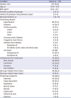
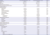
The study population is summarized in Fig. 1 and baseline characteristics of the study population are shown in Table 1. A total of 173 recurrent US patients (mean age, 67.6 ± 16.5 years; 62% men) were analyzed. The median number of previous cases of syncope was 3 (interquartile range, 2–5). Hypertension was present in 89 patients (51.4%), and a history of paroxysmal atrial fibrillation (AF) was noted in 41 (24%). Structural heart disease was present in 30 patients (17.3%): CMP in 13 (7.5%), CAD in 9 (5.2%), HF in 5 (2.9%), and VHD in 3 (1.7%). Significant CAD was re-vascularized. The VHD patients were 2 cases with moderate aortic valve stenosis and 1 with a well-functioning prosthetic valve. Those conditions were not directly associated with syncope.
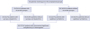
Table 1
Baseline characteristics of the study population
First syncope recurrences after ILR implantation
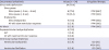
Symptom-correlated arrhythmia and ILR-guided therapy are shown in Table 2. A total of 52 patients (30.1%) experienced a first recurrence of syncope after ILR implantation during the mean follow-up period of 9.1 ± 13.7 months. The cumulative incidence of recurrent syncope was 34.3% and 47.2% at 1 and 2 years, respectively (Fig. 2A). Among those patients, a symptom-correlated ECG diagnosis during syncope was made by the ILR in 34 (65.3%) cases. Bradycardia related arrhythmia diagnoses were as follows: 24 sinus node dysfunction and 2 sudden atrioventricular (AV) blocks. Tachy-arrhythmia syncope diagnoses were as follows: 4 SVT, 2 ventricular tachycardia (VT), and 2 ventricular fibrillation (VF). As ILR-guided therapy, 25 (73.5%) patients underwent PM implantation, and 3 patients underwent implantable cardioverter defibrillator (ICD) implantation.
Table 2
Documented arrhythmia during first syncope and ILR-guided therapy
Significant arrhythmia documented by ILR interrogation irrespective of syncope
Significant arrhythmia documented by IRL irrespective of syncope and the corresponding ILR-guided therapies are shown in Supplementary Table 2. Significant arrhythmia was documented by ILR irrespective of syncope in 99 (57.2%) patients after the mean follow-up of 5.5 ± 7.9 months. The cumulative incidence of ILR-guided therapy was 47.8% and 57.1% at 1 and 2 years, respectively. (Fig. 2B). A device (PM or ICD) was implanted if the significant arrhythmia found by ILR interrogation was deemed a possible cause of the clinical syncope by the physician, following the ESC guidelines.24 A PM was implanted in 60 patients (34.7%), 5 (2.9%) received an ICD, and 4 (2.3%) underwent radiofrequency catheter ablation (RFCA).
Subgroup analysis between PM groups
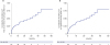
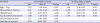
A total of 60 patients (34.7%) underwent PM implantation during a mean follow-up period of 6.0 ± 9.3 months. Comparisons between patients who received a PM and those who did not are shown in Table 3. The PM group was older, took more antihypertensive medication, had a higher proportion of paroxysmal AF history and relatively short duration of syncope, and were more likely to have had any BBB on the baseline 12-lead ECG. The univariable analyses showed that age > 75 years (hazard ratio [HR], 1.97; 95% CI, 1.18–3.30; P < 0.01), a history of paroxysmal AF (HR, 2.73; 95% CI, 1.62–4.58; P < 0.01), and any BBB on the baseline 12-lead ECG (HR, 2.98; 95% CI, 1.35–6.58; P < 0.01) were significantly associated with later PM implantation (Fig. 3). The multivariable analysis showed that a history of paroxysmal AF (HR, 2.34; 95% CI, 1.33–4.12; P = 0.01) and any BBB on the baseline 12-lead ECG (HR, 2.52, 95% CI, 1.09–5.85; P = 0.03) were significantly associated with PM implantation (Table 4).
Table 3
Clinical characteristics of patients receiving pacemaker implantation
Data are presented as number (median), or mean ± standard deviation, and number (%).
BMI = body mass index, AF = atrial fibrillation, ACE = angiotensin-converting enzyme, ECG = electrocardiogram, AV = atrioventricular, PVC = premature ventricular complex, IVCD = intraventricular conduction block, RBBB = right bundle branch block, LBBB = left bundle branch block, LAFB = left anterior fascicular block.
Fig. 3
Cumulative incidence of pacemaker implantation by risk factors. (A) Cumulative incidence of pacemaker implantation by older age (≥ 75 years). (B) Cumulative incidence of pacemaker implantation by history of paroxysmal AF. (C) Cumulative incidence of pacemaker implantation by any bundle branch block.
ILR = implantable loop recorder, PAF = paroxysmal atrial fibrillation, AF = atrial fibrillation.
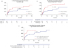
Table 4
Clinical predictors of pacemaker implantation in patients with implantable loop recorder
DISCUSSION
The main finding of our study is that ILR for US diagnosis detected recurrent syncope in 52 patients (30%), symptom-correlated ECG in 34 (19.6%), and significant arrhythmia irrespective of syncope in 99 (57.2%). A total of 69 (39.8%) patients were effectively treated with a PM, ICD, or RFCA as a result of ILR-guided diagnosis. These results are comparable to those of previous studies.15232526 Elderly US patients with a history of paroxysmal AF or any BBB on the baseline ECG could have a higher risk of later PM implantation. To our knowledge, this is the first study to evaluate the utility of ILR and clinical predictors of PM implantation in a large number of Asian patients.
The detection rate for significant arrhythmia irrespective of syncope was 57%, which is also comparable with a previous study23 that reported that 60% of ILR patients were diagnosed as having had an arrhythmic event. Significant arrhythmia regardless of syncope was diagnosed more frequently than syncope-related ECG. Krahn et al.15 reported that pre-specified significant asymptomatic arrhythmia was automatically detected by the ILR in 9 US patients (15%), with 7 of those patients later undergoing PM implantation. We did not classify the pre-specified significant vs. borderline asymptomatic arrhythmia as they did; we speculated that physicians might implant a PM under the assumption that a pre-specified significant arrhythmia is likely to cause syncope.24
Some previous studies have reported finding symptom-correlated ECG in 36%–64% of US patients, depending on the study population and incidence of structural heart disease,132526 which is known to be an important predictor of cardiac syncope.27 Although previous studies differ in their reporting, structural heart disease is present in 33%–48% of US patients.252628 In our study, the ILR established a symptom-correlated ECG in 19.6% (34/173) of the whole study population, which is lower than reported in other studies,2526 possibly because our study population included fewer patients with structural heart disease than previous studies and our follow-up period was relatively short.
The PM subgroup analysis showed that age > 75 years, a history of paroxysmal AF, and any BBB on the baseline 12-lead ECG were also associated with PM implantation. Several previous studies reported clinical predictors for PM implantation in US patients.19202126 Old age is the most common predictor, as expected.192026 Intermittent sinus node dysfunction (SND) from degenerative fibrosis or a paroxysmal AV block with aging are associated with US.2930 SND at an older age could manifest as tachycardia-bradycardia syndrome or AF with slow ventricular response (SVR).3132 In 40% to 70% patients, AF is present at the time of initial diagnosis with SND.3334
Brignole et al.9 reported that a paroxysmal AV block was a frequent cause of syncope in recurrent syncope patients with a BBB and a negative electrophysiological test, suggesting that BBB carry a high risk of pathological conduction-system abnormalities. In our study population, 70% (7/10) of patients with any BBB received a PM. If we initially evaluated US patients with a history of paroxysmal AF or any BBB on the baseline ECG, arrhythmic cause might be more likely than reflex syncope.
Our study has several limitations. First, we could not accurately classify the PM-indicated group because a detailed clinical history of syncope and meticulous ILR electrocardiogram data were not acquired in every hospital. If all patients with significant arrhythmia were classified as the PM-indicated group, it could have biased the results because that group might have included patients with bradycardia from reflex syncope or those with asymptomatic bradycardia. Second, sinus bradycardia in young patients, increased vagal tone during sleep or pain, or reflex syncope might not be distinguishable from SND. Some paroxysmal AV blocks can occur in association with increased vagal tone. Although each physician carefully evaluated clinical histories, we cannot exclude the possibility that a reflex mechanism was responsible for the arrhythmia. Third, a fine F wave in AF with SVR and a small P wave with a paroxysmal AV block could not be differentiated by ILR interrogation because of the low amplitude of the ILR-detected P waves or undersensing of the P wave. Fourth, because the sensing value of R or P waves was not acquired at every hospital, false bradycardia from undersensing and false tachycardia from oversensing caused by artifacts or poor R-wave sensing are possible. Fifth, the number of patients with any BBB (RBBB, LBBB, or a bi-fascicular block) is relatively small. Lastly, a longer follow-up period might have revealed further syncope-related ECG among patients with significant arrhythmia.
In conclusion, the diagnostic utility of ILR for detecting symptom-correlated ECG and significant arrhythmia in a large number of Asian US patients was comparable to previous Western results. Bradycardia was the most common etiology of US. US patients with a history of paroxysmal AF or any BBB on the baseline ECG were at higher risk of PM implantation.
 XML Download