

 PDF
PDF ePub
ePub Citation
 Print
Print


Introduction
Digital workflow has increased recently in dentistry and contributed to the improvement of all dental treatments. Digital techniques strengthen diagnostic visualization and predictability of the prosthetic treatments, and improve the information exchange between patients, doctors and technicians.12 The fundamental steps of a digital workflow are data acquisition, data processing, computer-aided design (CAD), and computer-aided manufacturing (CAM).3456 Three-dimensional (3D) digital image data in dentistry can be obtained from a range of different sources including computed tomography, magnetic resonance imaging, extra-oral optical scanning, and intraoral optical scanning. The use of digitalized scanners minimized human manipulation, reducing inaccuracy and time for the fabrication of restorations.78 Furthermore, previous studies have reported high dimensional accuracy of digital casts compared with stone casts.91011 Dental restorations fabricated from digital scans exhibited similar or even better marginal and internal discrepancies to those fabricated from conventional impressions.1213
A fundamental issue with restorative treatments is the unpredictability of esthetic outcomes due to the incapacity to have direct preceding clinical method.14 Therefore, the definitive restorations are modified in clinic or remade additionally, which increases overall treatment time and charge. Advancements in stereo-photogrammetric facial scanning technology have led to the development of 3D visualization of the orofacial region.15 These systems have been successfully employed to create a virtual patient replica for comprehensive diagnosis and treatment planning in orthodontics and orthognathic surgery.1617 More recently, the concept of applying facial scanning for esthetic analysis during the design phase of dental restorations has been introduced in prosthodontics.18 The main idea is to integrate a virtual teeth setup with a replica of the patient face and to digitally evaluate the impact of changing teeth positions, forms, and colors on facial appearance. The 3D design information obtained from this virtual clinical evaluation phase can then be used to fabricate mockups or provisional restorations. A virtual setup phase might be a good option to provide the patient and the treatment provider with a tool regarding the facial appearance of the prospective definitive restoration. The purpose of this clinical case is to propose a digital approach integrating 3D facial scanning, a virtual mockup and direct conversion of design to definitive restoration in the workflow of a restorative treatment.
Case report
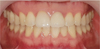
A 39-years-old male patient came for esthetic treatment for maxillary central incisors. The extraoral examination showed no deviation in the facial and dental midlines. The patient had a medium lip line, concave smile line, and 1 mm of tooth visibility at rest. The intraoral examination presented acceptable oral health with periodontal probing depths of no more than 3 mm but a generalized moderate periodontitis at posterior regions at both maxilla and mandibular with probing depths varying from 3 to 4 mm and localized gingival recession. The two traumatized central maxillary incisors had been restored with composite, and showed restorative materials with defective margins and poor color match to the adjacent teeth. Some of the occlusion surfaces of posterior teeth had also been filled with amalgam material with adequate marginal integrity without color infiltration. The occlusal plane was not altered. The two central maxillary incisors were indicated to be restored with a laminate veneer on #11 tooth and a monolithic zirconia crown on #21 tooth and chosen using the stepwise procedure with a virtual mock-up on 3D face scan to increase the esthetic results.
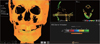
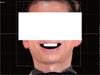
First, 3D facial scanning was taken to create a virtual patient replica for the clinical evaluation phase (Fig. 1A). Using a come-beam computed tomography (CBCT)-scan device (R2 STUDIO, Mega- Gen, Daegu, Korea), maximum smile facial scans were obtained at 3 different angulations (frontal, 80-degree right side, 80-degree left side) with the Frankfort horizontal plane of head to be parallel to the floor. The obtained data was saved in the form of object (OBJ) format. The underlying bone morphology was obtained by using the CBCT-scan device with a field of view of 120 × 85 mm, voxel size of 0.2 mm, exposure conditions of 80 kVp, 8 mA, and a 24-s pulsed scan (Fig. 1B). Data were saved in the digital imaging and communications in medicine (DICOM) format. While taking the CBCT, a face scan was taken simultaneously (Scout image).
The teeth #11 was prepared to receive a laminate veneer on the labial surface and #21 was prepared for a crown, then the maxillary and mandibular dental impression were taken with an additional type silicone impression material (Virtual, Ivoclar Vivadent, Schaan, Liechtenstein). The cast was made with type IV dental stone. Then, the cast models were submitted to a 3D scanning procedure using a desktop scanner (IDC S1, Amann Girrbach, Koblach, Germany) (Fig. 1C). The scanned models were subsequently saved as standard tessellation language (STL) file.
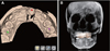
Afterward, all digital files were imported into a dental design software program (R2GATE v2.0, MegaGen, Daegu, Korea) The DICOM data of CBCT was converted to 3D reconstructed mesh format (Fig. 2), and the facial scan and mesh data were aligned to each other using the outlines of Scout image (Fig. 3). This was necessary in order to bring all facial scans in a single corresponding coordinate system. To combine the intraoral scan image to CBCT, remaining teeth were used as fixed anatomical reference (Fig. 4). Using this stepwise method, OBJ, DICOM and STL files were linked.
Based on the merged image, the restorations of laminate and complete crown were designed under consideration of the patient smile and esthetics lines using Digital Oral Design module in R2Gate (MegaGen, Daegue, Korea) (Fig. 5). The designed restoration was shown to the patient in clinic as a virtual mock-up on a 3D face scan of the patient to get the patient's feedback. The designed virtual restorations were transfer to CAD software (R2CAD, MegaGen, Daegu, Korea), and revised based on the patient's feedback in collaboration with the dentist professional considerations. The confirmed design was exported to a 5-axis milling machine (Ceramill Motion 2, Amann Girrbach, Koblach, Germany) to produce the definitive restorations using a hybrid ceramic block (VITA Enamic, Amann Girrbach, Koblach, Germany) for the laminate veneer and a zirconia block (Zolid fx white, Amann Girrbach, Koblach, Germany) for the complete crown. The definitive restorations were delivered to the patient, and the patient was satisfied with the predictability of treatment workflow and esthetics of the restorations (Fig. 6).
Discussion
Scientific revolution of the clinical procedures and technical methods used to rehabilitate oral defects of patients are currently ongoing.19 It could be challenging to meet individual esthetics and functions of patient, which led to digital advances in prosthodontic treatments. Currently, various CAD/CAM technologies are available during treatment plan, design, fabrication treatments phases of fixed and removable implant prostheses. The soft tissue analyses obtained by 3D facial scanning are certainly an optimal supplement to the existing scanning technologies.2021 All pictorial information obtained by intra- and extra-oral scanners with the assistance of facial scanner facilitates the restoration design to reflect the esthetic demands of the patient.22 That would be beneficial to both the patient and clinician.
A virtual teeth setup allows several rapid adjustments of tooth size/position and generalized modifications of the occlusal scheme and compensation curves of the dental arch.23 This procedure would require a lot of manual labor, time, and cost in a traditional wax-up method. Additionally, the virtual setup is digitally stored and thus can be consecutively used to fabricate a new prosthesis, thereby reducing treatment time. Integrating virtual teeth setup with the facial scan permits immediate inspection of the prosthesis considering facial esthetics.24 Similarly, tooth position and morphology can be altered while visualizing the impact on the patient's facial appearance. This would definitely improve on the current clinical procedure of multiple esthetic teeth try-ins that typically required for comprehensive reconstructions and treatments at anterior region. Moreover, the use of this digital approach improves communication between clinicians and lab technicians. A technician, who is usually limited to the waxup on the articulator and selected photographs of the patient, would benefit from this virtual setup, in which changes in the restoration design can immediately be visualized on the patient's face.
Nowadays, two-dimensional (2D) virtual smile design approach is still gaining a reputation as a conceptual tool to improve communication within related parts and to enhance treatment predictability.20 However, perspective distortion may cause inaccuracies or errors in the conversion process from 2D design to 3D diagnostic waxing.2025 In 3D methods, definitive restorations can be directly fabricated from an approved virtual diagnostic wax-up without the need for further conversion after the confirmation of simulated esthetic outcome in the 3D virtual patient. Accordingly, the use of a 3D virtual patient can overcome the limitation of 2D methods.
There are several considerations for the clinical use of the present workflow. First, the alignment of the dental cast models and the facial scans depends on the visibility of the labial surfaces of the teeth as a common fixed reference. Because the intraoral scan needs to be merged with the virtual patient using the remaining teeth, the visibility of the labial surface of the remaining dentition at a full smile is important.2627
Furthermore, some inaccuracies in image margining might be generated in case the remaining teeth are insufficient. Alternatively, an additional radiopaque tray can be applied during face scan and CBCT taking. The extraoral portion of the tray in deed can be used as a congruent area for the image superimposition. Additionally, alignment of the neutral smile and full smile scans rely on the stability of the forehead as a reference. In patients with deep facial corrugations, this region can deform when the patient is smiling thereby hampering an accurate registration procedure. The clinician should also verify that the patient's facial expression and head positioning are consistent when collecting facial scans to improve the accuracy of the registration procedure for the virtual patient.2627
The digital technologies of face scan and image merging for 3D diagnosis and esthetic treatment are promising, but are still at inceptive stages for the practical applications in prosthodontics. More scientific trials are needed for more scientific evidence to explore the possibilities.
Conclusion
This clinical report describes a digital treatment workflow based on the combination of face and oral cavity scans to create a 3D virtual patient for the clinical and laboratory phases of an esthetic treatment with a laminate veneer and a monolithic zirconia crown in the anterior region. Within the limit of this report, the digital workflow has the potential to enhance the predictability of esthetic outcomes.


 XML Download