

 PDF
PDF ePub
ePub Citation
 Print
Print


INTRODUCTION
Classical swine fever (CSF), on the list of diseases notifiable to the World Organisation for Animal Health (OIE), is one of the most serious contagious viral diseases of pigs, with the potential to cause enormous economic losses in the pig industry worldwide. A typical clinical sign of acute CSF is high fever (> 41 ℃), conjunctivitis, anorexia, ataxia, and purple discoloration of the skin, notably at the ears, lower abdomen, and extremities (123). Highly virulent virus strains cause an acute form of the disease with high morbidity and mortality in pigs (particularly in young animals). The causative agent of the disease, CSF virus (CSFV), is a small-enveloped, positive-sense single-stranded RNA virus that belongs to the genus Pestivirus in the Flaviviridae family. The 12.3 kb CSFV genome encodes a polyprotein of 3,898 amino acids (4). A single long open reading frame (ORF) encodes four structural (core, Erns, E1, and E2) and eight non-structural proteins (Npro, p7, and NS2-NS5B) (5). The E2 protein is the most immunogenic structural glycoprotein of CSFV, inducing neutralizing antibodies that provide protection against lethal CSFV challenge (6).
Two main strategies for controlling CSF, depending on the epidemiological conditions of the affected geographical area, are systemic prophylactic vaccination with a live attenuated vaccine and a non-vaccination stamping-out policy (7). Routine vaccination is the most widely used method for controlling CSF in endemically infected countries (including Asia, Eastern Europe, the Americas, and some African nations). Several types of efficacious live attenuated CSF vaccines have been developed over recent decades. Currently, modified live vaccines (MLVs), mainly containing Chinese (C) strain (8), low-temperature-adapted Japanese guinea pig exaltation-negative (GPE-) strain (9), or the French cell culture-adapted Thiverval strain (10) and its derivatives, are being used worldwide. These vaccines are inexpensive and can induce complete protection against virulent CSFV.
In South Korea, the LOM-850 strain has been the MLV strain to eradicate CSFV since 1974. Previous study reported that CSF outbreaks happening in naive swine herds on Jeju Island, South Korea, after the vaccination of the LOM-850 strain (11). In addition, eradication of CSF based on the MLV vaccination alone difficult because differentiation between infected and vaccinated hosts is impossible based on the antibodies induced. Therefore, marker subunit vaccine based on expressed E2 protein has been developed (121314).
Meanwhile, transgenic plants are one of the most cost effective and safe systems for large scale production of proteins without pathogenic animal contaminants (151617). Clinical trials and approval of plant-based vaccine products demonstrated that the use of transgenic plants is becoming a useful tool to control diseases (1819). Tobacco (Nicotiana bentamiana) has become the primary vehicle for proof of concept work in recombinant protein production (20). It is a promising alternative expression system for production of recombinant subunit vaccines due to its high biomass yield and high soluble protein levels compared with many other model and crop species (21).
Our previous report showed that plant-produced E2 fusion protein was expressed at a high level, and determined an immunogenicity (22). This study was conducted to evaluating histopathological data of ppE2 vaccine against CSFV, which is one of the most economically and industrially important in pig industry.
MATERIALS AND METHODS
Experimental design
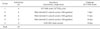
Challenge with YC11WB (106.0 TCID50/ml), a virulent CSFV strain was performed through intramuscular routes. Vaccination of experimental groups was performed only one time. The piglets were randomly divided into six, five groups of four animals each with 100 ug of ppE2 combined with adjuvant IMS1313 or LOM-850 strain vaccine as a positive control (Table 1). Four unvaccinated piglets were used as a positive control group. Group I and group II piglets were challenged at days 7 post vaccination. Group II and III piglets were challenged at days 11 post vaccination (plant-derived green marker vaccine candidate, 100 ug/dose). Group IV piglets were challenged at days 14 post vaccination. Group IV piglets were challenged at days 14 post vaccination (LOM-850 strain vaccine) (22). Histopathological scores applied for each lesion were graded as (−) no lesion, (+) mild, (++) moderate, or (+++) severe. All animal experiment was performed in the Animal and Plant Quarantine Agency Animal Care and Use Committee (QIA-ACUC) with permit number 2017-369.
Histopathological findings
All experimental piglets were sacrificed through the intramuscular inoculation of 10% succinylcholine. The pathologically important organs (tonsil, lung, heart, liver, spleen, kidney, inguinal lymph nodes, mesenteric lymph node, ileum, and cecum) for the diagnosis of CSF were aseptically collected from the sacrificed piglets. The collected organs were fixed in 10% neutral buffered formalin for 24 hr at room temperature. The organs were fixed in paraffin-embedded histological cassettes. The paraffin-embedded organ blocks were cut into 5-µm sections and then were stained with hematoxylin and eosin (H&E). To detect histopathological characteristics, such as inflammation, bleeding, and tissue damage, all tissue sections were observed by light microscopic examination.
RESULTS
Histopathological examination of tissues
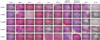
Microscopic lesions were observed in the examined organs (tonsil, lung, heart, liver, spleen, kidney, inguinal lymph nodes, mesenteric lymph nodes, ileum, and cecum) of piglets in all the experimental groups (Fig. 1). Histopathological studies revealed that piglets in the unvaccinated group (group I) displayed lesions after challenge such as moderate hemorrhage in the tonsil and heart, severe hemorrhage in the spleen, liver, lung, kidney, and mesenteric lymph node, loss of germinal center in the tonsil, spleen, and inguinal lymph node, inflammation in the tonsil, heart, lung, and kidney, lymph node enlargement in the inguinal and mesenteric nodes, and depletion of the follicles in the mesenteric lymph node. The scores for the histopathological examination in each group are shown in Table 2.
The histopathological examination of the tonsil showed congestion of the blood vessels, multifocal hemorrhages, lymphoid necrosis with ectatic crypts, and accumulation of cellular debris in groups I and II. Mild hemorrhages were observed in group III. Tonsils in group II especially showed congestion of the blood vessels along with focal to diffuse areas of hemorrhage. The spleen in groups I and II showed extensive hemorrhages in red pulp with depletion and loss of germinal centers. In contrast, mild depletion and atrophy of the lymph follicles were observed in groups IV and V. Furthermore, the heart revealed congestion of the capillaries in groups I, II, and III. Histopathological changes in the liver were characterized by congestion of the central veins, loss of hepatic lobules, and connective tissue proliferation, especially in groups I and II. Severe alveolar hemorrhages were noticed in groups I, II, and III. The inter-alveolar septa were especially thickened with infiltration by mononuclear cells and congestion of the alveolar capillaries was observed. The lung section displayed extensive hemorrhages in the alveoli and interstitium, and diffuse severe infiltration of mononuclear cells in the alveoli, bronchi, and bronchiolar lumen in almost all the experimental groups, except group IV. In groups I, II, and III, the kidneys showed congestion of the blood vessels and severe multifocal interstitial nephritis characterized by infiltration of mononuclear cells in the interstitial spaces. In particular, focal areas of glomerulonephritis were observed, characterized by swelling of the glomerular tuft expanding with cellular infiltration. The inguinal and mesenteric lymph nodes showed a depletion/atrophy of the lymphoid follicles and congestion of the blood vessels along with focal to diffuse areas of hemorrhage in groups I and II. Notably, necrotic cellular debris and severe hemorrhages were observed in groups II and III. In the ileum, depletion of lymphocytes and infiltration of eosinophils in the lymphoid follicles and submucosal layer were observed in groups I and II. Development of blood vessels were observed in the cecum of all the examination groups.
DISCUSSION
General clinical symptoms of CSFV-infected pigs vary for several reasons, including the age and lineage of the pigs and the differences in the immunogenicity and pathogenicity of the infecting viruses. Symptoms are affected by the condition of the pigs even when the same virus is being inoculated (23). If a CSFV-infected pig recovers, long-term defense is possible; if the pig does not recover, it usually dies as a consequence of the inhibition of its immune function (24). Most CSFV-infected pigs 12 weeks or younger develop an acute form of CSF, the symptoms of which tend to weaken over time as the immune response develops (23).
After widespread vaccination under the national eradication policy of South Korea, the virulence of CSFV began to subside. This pattern was also observed in the United States. Before 1950, most CSFV strains had been highly virulent; but after the eradication policy was implemented, less virulent strains appeared (25).
In South Korea, the genotypes of group 2 strains isolated since 2002 were found to be 92% to 100% identical to the viruses circulating in China (26). In addition, when the CSF symptoms that appeared after 2002 were compared with those that appeared before 2000, a decrease in atrophy and constipation and an increase in conjunctivitis were observed. The gross findings showed a marked decrease in epiretinal membrane, infarction, and laryngeal flare (27). Swine fever caused by inoculating viruses that were isolated in Europe after 1990 was found to be difficult to diagnose solely on the basis of clinical symptoms appearing within two weeks (28).
In South Korea, a mandatory vaccination policy has been imposed since the short-term nationwide outbreak of CSF early in 2003. This outbreak was caused by the sale of pigs infected with swine fever that had been raised in a few pig-breeding farms in the affected areas. Since then, CSF outbreaks have occurred regularly according to 2017 disease statistics and livestock epidemic statistics by Animal and Plant Quarantine Agency: two outbreaks in 2006, five in 2007, seven in 2008, two in 2009, one in 2013, and two in 2016.
In recent years, in order to avoid the side effects that may occur in vaccinated piglets because the period of vaccination for swine fever overlaps with the period during which chronic wasting diseases like porcine reproductive and respiratory syndrome (PRRS) and postweaning multisystemic wasting syndrome (PMWS) are likely to develop, this vaccination has been either avoided or deferred until piglets reaches seven to nine weeks old. Given the fact that the incidence of swine fever is lower than that of wasting diseases such as PRRS and PMWS, in the present study a single vaccination test was conducted with 90-day-old piglets.
The histopathologic changes in the organs of CSFV-infected pigs include hemorrhage of the spleen and loss of lymph follicle; pulmonary hemorrhage and purulent exudation; enlargement of the capillary blood vessels; eosinophil infiltration; and thickening of the myenteric and serosal layers of the colon (29). Reduction in lymphocytes in the tonsil and the lymph nodes and hemorrhage in the kidney have also been reported as critical pathologic symptoms of CSF (30). Histopathologic findings have indicated that pigs infected with a highly virulent strain begin to die on the seventh day after inoculation, exhibiting the main symptoms of vasculitis and necrosis of B lymphocytes (31). Consistent with these findings, piglets in the negative control group of the present study also began to die on the seventh day while exhibiting similar symptoms, including necrosis of lymphocytes in the lung. When the pathologic findings of ten organs were compared, group II was also found to exhibit the symptoms reported in earlier studies, including hemorrhage in the tonsil and the kidney.
In this study, the most common pathological detections in the PPE2-treated groups (groups II, III, and IV), especially group II, were mild to moderate hemorrhage, inflammation, and loss of germinal center. These data showed that the histopathological observations seen in the piglets in the virulent CSFV challenge after 7 days of one-shot vaccination were more severe than those in the virulent CSFV challenge after 11 or 14 days of one-shot vaccination and 14 days of LOM-850 strain vaccine.
Collectively, it was found that after the single vaccination test with the ppE2, sufficient protection against the wild-type strain was possible after neutralizing antibodies began to form. On the 14th day after the vaccination, full protection against the wild-type strain was possible. The results indicate that although two vaccinations are required for the formation of sufficient defense ability, a single vaccination can also provide satisfactory protection at least 11 days after the inoculation for the purpose of emergency protection.
 XML Download