

 PDF
PDF Citation
 Print
Print


Cor triatriatum is a relatively rare congenital heart disease found in humans, dogs, and cats. Cor triatriatum sinister [1] and cor triatriatum dexter [2] have been reported in humans, whereas cor triatriatum dexter [3] and cor triatriatum sinister [4] have been reported in dogs and cats, respectively. Cor triatriatum dexter occurs through a different mechanism from cor triatriatum sinister, and is believed to be caused by a persistent right sinus venosus valve [5]. Cor triatriatum dexter requires treatment in the juvenile stage because it presents with clinical symptoms, such as ascites from right heart failure at an early stage. This paper reports the successful treatment of cor triatriatum dexter in a small puppy with hybrid balloon dilation.
The case was a 4-month-old, female Shiba dog weighing 4.0 kg that presented with abdominal distension due to accumulation of ascites in the previous 2 months. The dog was brought to the author's hospital for further examination and treatment with the chief complaints of ascites and dyspnoea during sleep.
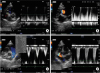
A physical examination revealed no distention of the jugular vein, dilation of the superficial epigastric vein, or abnormal lung sounds. A Levine 2/6 systolic murmur was heard on auscultation. The blood tests were within the normal reference values. The electrocardiogram (ECG) revealed a normal sinus rhythm. The chest X-ray did not show an enlarged heart size, with a Vertebral Heart Score of 8.5, but a dilation of the caudal vena cava was evident. An abdominal X-ray showed reduced abdominal serosal details that were indicative of ascites. Transthoracic echocardiography (TTE) revealed an abnormal membrane separating the right atrium into a cranial chamber (RA1) and caudal chamber (RA2) of the right atrium, with fenestration, 2 mm in size in the membrane (Fig. 1A). The maximum stenotic blood flow velocity from RA1 to RA2 was more than 200 cm/s (Fig. 1B). The right ventricular outflow tract (RVOT) was 88.3 cm/s (Fig. 1C) and the tricuspid regurgitation velocity was 216.6 cm/s (Fig. 1D).
Based on these findings, ascites caused by cor triatriatum dexter was diagnosed, and hybrid balloon dilation was planned on day 6.
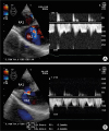
On day 6, anaesthesia was induced with atropine (0.05 mg/kg, subcutaneous injection), ampicillin (Ampicillin sodium injection 1 g; Kyoritsuseiyaku Corporation, Tokyo, Japan) (30 mg/kg, intravenous injection [iv]), butorphanol (Vetorphale®; Meiji Seika Pharma Co. Ltd., Tokyo, Japan) (0.2 mg/kg, iv), midazolam (Dormicum Injection 10 mg; Astellas Pharma Inc., Tokyo, Japan) (0.2 mg/kg, iv), and propofol (Mylan Injection 1%; Mylan Inc., Tokyo, Japan) (to effect, iv). Anaesthesia was maintained with isoflurane (isoflurane for animals; Intervet K.K., Tokyo, Japan) (1.5%–2.6%). A thoracotomy was performed through the sixth right intercostal space and an incision was made in the pericardium from the right ventricle to the right atrium. A pericardium tent was then made using 3-0 silk sutures. The right atrium observed with transesophageal echocardiography (TEE) showed no pulsatile blood flow from RA1 into RA2 through the small fenestration in the abnormal membrane (Fig. 2A). A purse-string suture was placed with 4-0 polypropylene suture thread (PROLENE; Johnson & Johnson K.K., Tokyo, Japan) on the right atrium. Through the purse-string suture site, an 18G indwelling needle followed by a 0.035-inch guidewire was inserted into the RA2. An attempt was made to insert a paediatric balloon catheter (balloon diameter: 10 mm, length: 2.0 cm; Tokai Medical Products Inc, Aichi, Japan). The small hole in the abnormal membrane was expanded by ballooning approximately 10 sec under TEE guidance. After the procedure, TEE confirmed that the small hole had expanded and the blood flow through the hole had become pulsatile with no arrhythmia (Fig. 2B). The right atrial puncture site was ligated with the purse-string suture. Chest drainage (Phycon tube SH No.3: 2.5 mm inner diameter, 4.0 mm outer diameter; Fuji Systems Corporation, Tokyo, Japan) was set up, and the dog's chest was closed using the standard method.
Surgery was followed by respiratory management in a 40% oxygen chamber. Urination occurred at 2 h and 30 min after surgery, and she showed an appetite at 7 h after surgery. No substantial pleural effusion was present 10 h after surgery (0.03 mL/kg/h).
The day after surgery (day 7), her respiratory condition was good. TTE showed that the small hole opened in the abnormal membrane in the right atrium had expanded to 5.2 mm in diameter. The abdominal X-ray showed that the ascites had disappeared. The dog's appetite and level of activity were also normal, and she was discharged on the day after surgery. ECG revealed a normal sinus rhythm in the perioperative period up to discharge.
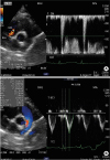
TTE on day 36 (30 days after surgery) showed that the fenestration in the separating membrane between RA1 and RA2 had contracted slightly to 4.9 mm. The stenotic blood flow velocity from RA1 to RA2 was 117 cm/s with an assumed pressure gradient of 5.7 mmHg (Fig. 3A). The RVOT was 124.5 cm/s (Fig. 3B) and tricuspid regurgitation was not observed. The dog's general condition was good, including a healthy appetite, and there were no problems with breathing during sleep at home. Her body weight had also increased to 6.1 kg. The abdominal X-ray also showed no ascites.
Methods for treating cor triatriatum dexter in dogs include open-heart surgery to remove the separating membrane [6] or endovascular therapy, which would be a technique for using a percutaneous balloon to expand the fenestration in the separating membrane [7] or a method for placing a percutaneous stent in the fenestration [8].
Removal of the separating membrane with a cardiopulmonary bypass is an effective curative treatment, but treatment with extracorporeal circulation poses a very high risk for small breed puppies with a low body weight and low circulating blood volume.
Percutaneous endovascular therapy is far less invasive than open-heart surgery, but advanced catheter manipulation techniques are required. This is because it is very difficult to pass the guidewire and balloon catheter through the small hole, which is opened in the abnormal membrane by remote control under X-ray guidance. Another possible limitation is the small size of the blood vessels in cases with low body weight, which limit the size of the balloon catheter that can be used. Previously cases of cor triatriatum dexter treated by percutaneous balloon have often been on large breed puppies [37]. Success with a percutaneous balloon in small breeds has been reported [9]. On the other hand, the blood vessel size/diameter is sometimes limitations to interventional radiology for small breed dogs. Therefore, an increase in the number of options for this disease would be excellent news for patients and owners alike. A new treatment option that is less invasive and does not depend on the physique will benefit small breed puppies with this disease.
Hybrid balloon dilation is a method that is not open-heart and involves the insertion of a device directly into the heart with a thoracotomy; fenestration in the separating membrane is expanded with a balloon. This technique is not as invasive as open-heart surgery, and it is also not dependent on the blood vessel diameter. A hybrid balloon has been used to treat cor triatriatum sinister in humans [10] and cats [4]. On the other hand, cor triatriatum differs considerably between whether the right or left atrium is affected, ranging from occurrence to pathology, and even the animal species involved.
Another benefit of hybrid balloon treatment is that X-ray guidance is not required. This method, which does not require protective clothing, is also advantageous because of its ease of use by animal healthcare professionals. The combined use of TEE with a hybrid balloon reduces the invasiveness and improves the reliability and ease of use.
In the present case, hybrid balloon dilation was performed on a small breed puppy with cor triatriatum dexter, and the clinical symptoms were eliminated. This method appears to be a useful, new, less-invasive treatment option for cor triatriatum dexter.
 XML Download