

 PDF
PDF Citation
 Print
Print


INTRODUCTION
Advancements in multiple detector computed tomography (MDCT) and the development of nonionic contrast material have had an important influence on computed tomography (CT) imaging. The use of MDCT has significantly reduced the scanning time involved in the imaging procedure. Nonionic contrast material is the optimized choice in CT scanning but its high cost and disadvantages for patients with renal insufficiency have compelled clinicians and radiologists to investigate alternative methods that can reduce the dose of contrast material used [14].
Saline chaser, a technique that involves immediate flushing out of the contrast material by injecting a saline solution after contrast material administration is complete, is commonly used in CT applications, particularly in CT angiography in human medicine. The procedure pushes the bolus further into the blood circulation, decreasing the amount of contrast material in the anatomic locations where it is not required. Therefore, accumulation of contrast material in the peripheral vein and injection tubing, which can occur with the use of MDCT and the reduced scanning time in CT imaging, is prevented by applying the saline chaser technique [56789]. Studies conducted on this topic report that reducing the amount of contrast material does not decrease vascular enhancement. Therefore, the use of a saline chaser in CT examination can be a cost-effective option and can decrease the risk of contrast material-related side-effects [101112].
However, while there are numerous reports on the saline chaser protocol in human medicine, there are few studies on the use of saline chaser in small animal CT. In particular, studies focused on reducing the dose of contrast material in CT imaging of dogs have not been reported.
The purpose of this study is to assess the extent to which a saline chaser could reduce the dose of intravascularly injected contrast material in the abdominal CT of dogs without decreasing vascular and hepatic parenchymal enhancement.
MATERIALS AND METHODS
Animals
The study subjects were 5 adult beagle dogs weighing 12.6–16.3 kg (mean weight: 14.2 kg). None of the dogs exhibited abnormal signs related to the cardiovascular system. In addition, all dogs had no abnormal blood analysis, abdominal sonography, or echocardiography results. Details of the anesthesia used during CT scanning are as follows: sedated with an intravenous injection of 0.020 mg/kg body weight acepromazine (Sedaject; Samu Median, South Korea), induced with an intravenous injection of 1.0–1.5 mg/kg of body weight of etomidate (Etomidate-Lipuro; B. Braun Melsungen AG, Germany), and maintained with isoflurane (Ifran; Hana Pharm, South Korea) in 100% oxygen administered via an endotracheal tube. For individual scans, a breath-hold protocol with a peak inspiratory pressure of 10 cm H2O was employed during image acquisition to prevent respiratory motion artifacts. Electrocardiography, oxygen saturation, and breathing were monitored during all procedures. The animal care and experimental procedures were approved by the Seoul National University Institutional Animal Care and Use Committee (IACUC No. SNU-160812-1).
Contrast material injection and CT scan protocol
The contrast material used in this study was iohexol (Omnipaque 300; GE healthcare, Ireland) with an iodine concentration of 300 mg I/mL. Three injection protocols, based on related studies and preliminary experiments were used; 600 mg I/kg contrast material injection at a rate of 3 mL/sec without a saline chaser (protocol 1), 420 mg I/kg contrast material (30% dose reduction) at the same rate as protocol 1 followed by a 10 mL saline chaser (protocol 2), and 360 mg I/kg contrast material (40% reduction) at the same rate followed by a 10 mL saline chaser (protocol 3). In protocols 2 and 3, the saline chaser was injected at a rate of 6 mL/sec. The contrast material and saline chaser were injected through a 22-gauge IV catheter into the cephalic vein. Two long tubes of the injectors were connected with a non-return valve and a Y-shaped tube leading to the IV catheter. A dual-head power injector (Stellant; MedRAD, USA) was used. Scanning was typically started 10 sec after the initiation of the contrast material injection to achieve adequate enhancement in the arteries and veins.
Study experiments were repeated 6 times with the 3 protocols being applied twice for each dog. A minimum 3-day interval was included between CT scanning sessions of each dog.
The study involved 30 dynamic CT scanning sessions using a 64-row MDCT scanner (Aquillion 64; Toshiba, Japan) at a fixed level of the porta hepatis for 40 sec. The scanning parameters included 4 × 1.0 mm detector collimation, 4.0 mm slice thickness, 120 kVp, 50 mAs, and 0.75 sec rotation time.
Analysis of CT data
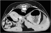
The CT images were reconstructed for analysis by using image analysis software (INFINITT; Infinitt Healthcare, South Korea). Circular regions of interest (ROIs) were placed at the aorta, portal vein, and liver parenchyma with areas ranging from 15 to 30 mm2 at the level of the porta hepatis (Fig. 1). Artifacts were carefully excluded from the measurements. In the liver parenchyma, ROIs were measured in 3 distinct areas including both the left and right lobes, and their average was used as the result. Attenuation values were measured at soft tissue windows (window width +400, window level +40) in Hounsfield units (HU) using 2 parameters: the maximum enhancement value (MEV) and the time to MEV (tMEV); time-intensity curves were obtained from the attenuation values recorded in each ROI.
Statistical analysis
Statistical analyses were carried out by using a statistical software program (SPSS version 23.0 for Windows; SPSS, USA). The difference between the attenuation values obtained in the first and second scans was analyzed by using the Wilcoxon signed-rank test. Further, Kruskal-Wallis and Mann-Whitney U tests were used to compare attenuation parameters of MEV and tMEV obtained from the aorta, portal vein, and liver parenchyma. A p-value of 0.05 was considered to be statistically significant.
RESULTS
Attenuation of the aorta
Among the 5 dogs, there was no significant difference in the mean attenuation values obtained from the aorta between the first and second scans. The MEVs recorded using protocol 2 were significantly higher than those obtained using protocols 1 and 3 (p < 0.05); no significant difference was detected between protocols 1 and 3 (Fig. 2, Table 1). Further, tMEV was not significantly different among the protocols (Table 2).
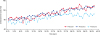
Fig. 2
Mean time-intensity curves for the aorta (n = 5). The maximum enhancement values recorded for protocol 2 are significantly higher than those noted for protocols 1 and 3 (p < 0.05). There is no significant difference between protocols 1 and 3.
HU, Hounsfield units.
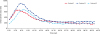
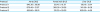
Table 1
Mean maximum enhancement values in HU according to protocol at each region of interest in 5 beagle dogs

Attenuation of the portal vein
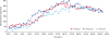
There was no significant difference in the mean attenuation values obtained from the portal vein between the first and second scans. Further, there was no significant difference in the MEVs of the portal vein among the protocols (Fig. 3, Table 1). The tMEV values were not significantly different among the protocols (Table 2).
Attenuation of the liver parenchyma
There was no significant difference in the mean attenuation values obtained from the liver parenchyma between the first and second scans. The MEVs obtained from protocol 3 were significantly lower than those from protocols 1 and 2, and there was no significant difference between protocols 1 and 2 (p < 0.05) (Fig. 4, Table 1). Further tMEV was not significantly different among the 3 protocols (Table 2).
DISCUSSION
Various effects of the saline chaser technique have been reported since its introduction in medical imaging. The method increases the peak attenuation, reduces the artifacts produced by contrast materials [4111314], and decreases the amount of the contrast material used during the imaging procedure [38915].
Previous human medicine-based research using a canine experimental model conducted experiments with various modified protocols involving a saline chaser. According to the report, peak enhancement increased upon increasing the flow rate and volume but revealed no significant difference with the use of different chaser fluids [16]. Based on these results, it was estimated that the dose required for appropriate contrast enhancement can be reduced by the saline chaser. For practical clinical application purposes, the focus can be on reducing the dose of the contrast material used in abdominal CT. Previous studies have shown the possibility of 15% to 40% reductions in the dose of contrast material when saline chasers were used in helical CT of the thorax and abdomen in humans [381115]. A veterinary study suggested that the use of a saline chaser allows a 30% reduction in the dose of the contrast material without significantly decreasing vascular enhancement during CT examination of the heads of cattle [6]. Since there are no previous reports that focused on reducing the contrast material dose in dogs, and based on preliminary experimental results, the enhancement values obtained after reducing the contrast material dose by 30% and 40% and applying saline chaser were investigated. Further, as there was no significant difference noted in enhancement values with the use of different chaser fluids in previous research, in this study, the most commonly used fluid, 0.9% sterile normal saline, was applied as the chaser fluid type.
In this study, there was no significant difference in MEVs of the aorta between protocols 1 and 3. In fact, protocol 2 (a 30% reduction in the applied contrast material and a saline chaser) showed significantly higher MEVs in the aorta than those obtained via other protocols. This result is due to the “pushing effect” of the saline chaser propelling the remaining contrast material into the peripheral veins and the injection tubing into the heart [101112131718]. There was no significant difference among the protocols in the MEV values recorded for the portal vein. This implies that there was no significant difference in contrast enhancement by decreasing the amount of contrast material in the major vessels by 40%.
However, in the liver parenchyma, the HU values recorded while using protocol 3 (a 40% reduction in the contrast material dose and a saline chaser) were significantly lower than those recorded with the other 2 protocols. This is because visualization of the parenchymal organ is likely to have occurred after that of the blood vessels, implying limitations in assessing liver parenchyma when reducing the contrast material dose by 40% from that used in a conventional contrast material protocol without a chaser. Nevertheless, the liver parenchymal enhancement results indicate that a saline chaser allows a 30% reduction in the dose of contrast material without decreasing vascular and hepatic parenchymal enhancement.
Similar to the results of previous studies, pushing the contrast material with a saline fluid would cause an effect that is similar to that achieved by increasing the amount of contrast material. As a result, the use of a saline chaser increases peak attenuation and results in a significant delay in the tMEV [131920]. In this study, the peak time results displayed a pattern indicating a slight increase after the application of the saline chaser. However, there was no statistical difference in tMEV for the aorta and portal vein among the protocols. This result may due to the differences in the total dose of the contrast material used in each study, as opposed to the results in previous studies that injected similar doses of contrast material [21].
There are several limitations to this study. The study included relatively few animals and failed to monitor their blood pressure. Further, since the breath-hold protocol was used to prevent motion artifacts, each dynamic scan was performed for 40 sec; as a result, the peak time for the hepatic parenchyma assessment could not be evaluated. Also, since each scan was performed at a fixed level, assessment of the complete liver parenchyma and other parenchymal organs was not possible. Furthermore, evaluation of the saline chaser technique in other organs and its application in the clinical patients with decreased renal functions would be relevant for increased understanding of the topic. In addition, determining the effect of a saline chaser on tumor conspicuity and attenuation would be useful.
In conclusion, the saline chaser technique allows a 30% reduction in the dose of the contrast material used in abdominal CT without significantly decreasing vascular and hepatic parenchymal enhancement in clinically healthy dogs.
 XML Download