

 PDF
PDF ePub
ePub Citation
 Print
Print


I. Introduction
Bone morphogenetic proteins (BMPs) are natural molecules that were first identified in dentin and demineralized bone matrix in ectopic sites of rats and are capable of bone formation1. In 1968, Urist suggested that these BMPs are released from native dentinal tubules and act as osteoinductive proteins2. Since then, several members of the BMP family have been isolated, and BMP-2 has been produced and refined for therapeutic purposes (recombinant human BMP, rhBMP-2)34.
In 2007, the United States (US) Food and Drug Administration (FDA) approved rhBMP-2 INFUSE BONE Graft (Medtronics, Memphis, TN, USA) at a concentration of 1.5 mg/mL along with an absorbable collagen sponge (ACS) as an alternative to autogenous bone grafting for maxillary sinus grafting and localized alveolar ridge augmentation56. Collagen is a natural polymer that can be processed into powder, membrane film, liquid, gel, and nanofiber forms and is the most widely used carrier for delivery of rhBMP-27. In general, proteins can be inserted into the pores of ACS immediately before grafting by soaking the ACS in rhBMP-2 protein solution (this method is used in commercially available INFUSE)8.
In an in vivo physiological environment, the proteins from the ACS can be released rapidly by diffusion, compression during handling, and/or degradation of the ACS, despite its biocompatibility, biodegradability, and solubility in a physiological environment. Therefore, placing a graft into an environment in which sponges are compressed by the surrounding muscles and tissues might result in local release of unnecessarily large amounts of rhBMP910.
Several adverse events, including those related to wound healing (hematoma, wound dehiscence, and infection), inflammation (cervical spine swelling), ectopic bone formation (bone cyst formation), osteoclast activity (vertebral bone resorption and osteolysis), and urogenital events (bladder retention), have been reported with the use of BMPs11. A common side effect of rhBMP-2/ACS reported in a meta-analysis is ectopic bone formation associated with leakage of rhBMP-2 outside the graft site due to mishandling during manual manipulation of the ACS11. This ectopic bone formation occurs primarily due to the type of rhBMP-2/ACS used in rehabilitation of spine-related disorders. These side effects are known to occur because the above-mentioned dosage is higher than that of osteogenic proteins naturally acting in the human body, and the carrier delivering the protein lacks the ability to maintain the concentration exclusively at the graft site.
The US FDA investigated the safety and effectiveness of localized alveolar ridge and sinus augmentation by comparing 120 oral and maxillofacial surgery patients treated with 1.5 mg/mL rhBMP-2/ACS and 91 patients treated with autologous bone graft. According to their results, the most common adverse events were oral pain, oral edema, facial edema, and oral erythema. No statistically significant adverse events were reported in patients exposed to rhBMP-2/ACS, but the incidence of facial edema was relatively higher in the rhBMP-2/ACS group12. Thus, although the ACSs have been approved and used in humans, the optimal scaffold for delivery of rhBMP-2 in human has not yet been established.
In dentistry, Ike and Urist13 hypothesized that demineralized dentin matrix (DDM) collagen of humans can act as an effective carrier of rhBMP-2 to overcome the reported side effects in association with the supraphysiologic concentration of rhBMP-2 (1.5 mg/mL) and properties of the carrier material1314151617. Therefore, we describe and propose the release profile of rhBMP-2 from DDM collagen (DDM/rhBMP-2) based on related studies.
II. Demineralized Dentin Matrix as an rhBMP-2 Carrier
1. Human dentin
The dentin consists of collagen (18%), hydroxyapatite (70%), non-collagenous protein (2%), and body fluids (10%). It is composed of an avascular, acellular, dense collagen matrix that contains non-collagenous growth factors, such as BMPs. These dentin-matrix-derived BMPs are similar to BMPs derived from the bone matrix, and they have been shown to have identical functions in vivo1819.
The inorganic component of dentin is physiologic calcium phosphate with various degrees of resorbability, which promotes degradation of dentin collagen20 and leads to predictable and controlled biodegradation of the dentin matrix21.
The dentin matrix contains nano-sized dentinal tubules (1–3 µm in diameter) that are important for release of endogenous growth factors present in the matrix and of hydroxyapatite-binding proteins. The number of dentinal tubules is 18,000–21,000 (tubules/mm2)22. The volume porosity of the dentinal tubule is 3.47%±1.46% on average, which is lower than that of natural bone in humans (6.2%)2324.(Fig. 1) In vitro, in vivo, and clinical studies have shown that dentin collagen has superior cell adhesion capacity, excellent tissue compatibility and absorptivity, and weak antigenicity1925.
2. Demineralized dentin matrix (AutoBT, Korea Tooth Bank, Seoul, Korea)
The DDM powder is produced from extracted human teeth according to the protocol of the Korea Tooth Bank (Korea Tooth Bank). Briefly, the teeth are divided separated into groups based upon whether they are crowns or tooth roots, and then the tooth roots are crushed to obtain particles 300–800 µm in size. These particles are demineralized with 0.6 N HCl for 30 minutes. Through demineralization, the mineral phase and major immunogenic components of the dentin are removed, while maintaining the collagen that plays a structural role as an osteoconductive scaffold and contains soluble protein fractions (such as BMPs and growth factors)161726.(Fig. 1)
Any residual hydroxyapatite that remains after demineralization is maintained at a 10%–30% concentration to retain the mechanical properties of the DDM27. Demineralization widens the two types of pores in dentin: tubules and interfibrillar spaces (collagen meshwork).(Fig. 1. B, 1. C) The total porosity of dentin increased to approximately 70% after demineralization and freeze-drying, and the degree of increase varied depending on the processing method. The porosity of dentinal tubules increased from 3%–6% to 20% on average (Fig. 1. B)2829, whereas that of the uncollapsed, freeze-dried interfibrillar space increased to approximately 50%2829.(Fig. 1. C)
III. Incorporation of rhBMP-2 with DDM (DDM/rhBMP-2)
1. Physical adsorption
According to Luginbuehl et al.30, physical adsorption on a surface could be regarded as the simplest method for delivering proteins. A pre-fabricated DDM scaffold is dipped in a protein solution and left to dry. In DDM collagen without specific protein affinity, rhBMP-2 is physically adsorbed into the interfibrillar space pores on the DDM surface31.(Fig. 2; red circle) The amount of interfibrillar space porosity not deformed by freeze-drying, with a pore diameter less than 0.2 µm, is almost 37%32. Given that increased porosity is a key parameter influencing adsorption, the unique geometrical shape of dentin collagen can provide a significant advantage for physical adsorption.
The dentinal tubule on the surface of DDM is also enlarged during demineralization and becomes a pore with an average diameter of 3–3.5 µm, which is similar to the pore size of natural human bone. Therefore, rhBMP-2 is also physically adsorbed into the dentinal tubule pores located on the surface of DDM24.(Fig. 1. A, 1. B and Fig. 2; green circle)
2. Modified physical entrapment
Physical entrapment is achieved by mixing rhBMP-2 with a scaffold material in liquid form, causing a phase change such as gelation; this phase change induces physical entrapment of proteins within the carrier33. Mixing proteins with the ACS is a modified method where the proteins are inserted into the pores of a scaffold by dipping the ACS in a protein solution immediately before implantation (this method is used in commercially available INFUSE).
Like the ACS, denser and highly cross-linked DDM collagen allows modified entrapment of rhBMP-2, which is loaded in the pores in deeper areas of the dentinal tubule.(Fig. 1. B and Fig. 2; green circle) It has been reported that the diameter of the dentinal tubule increases to 3–3.5 µm through demineralization34, and its porosity increases to 20%29. As freeze-drying reduces shrinkage and decreases collagen fiber melting, rhBMP-2 is more densely entrapped in the deeper nanoporous area of the dentinal tubule and interfibrillar space pores. This structural characteristic has the advantages of preventing the initial burst release when the ACS is used172324.(Fig. 2; green circle)
3. Endogenous BMP in the DDM
Binding of non-collagenous endogenous proteins to the dentin matrix and minerals is well established1418. Urist suggested that, if the dentin matrix undergoes biodegradation or resorption through a remodeling process after grafting, disintegration of covalent bonding occurs and releases osteoinductive proteins, which play a key role in stimulating stem cells in the surrounding tissues and causing phenotypic changes to produce osteoblasts2. This mechanism has been determined by several in vitro and in vivo studies1617183536.
Overall, DDM/rhBMP-2 contains physically adsorbed and then modified, physically entrapped rhBMP-2 and endogenous BMP that remains in the DDM as matrix and/or mineral-binding proteins.
IV. The Postulated Release Profile of rhBMP-2 from the DDM (DDM/rhBMP-2)
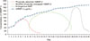
The original release profile of DDM/rhBMP-2 showed sustained, slow release of rhBMP-2 over the experimental period of 36 days (Fig. 3; blue line), and a significantly larger amount of rhBMP-2 was released from the DDM powder than from any other materials16. The postulated release profile of DDM/rhBMP-2 involves sequential delivery of exogenous rhBMP-2 and endogenous BMP from the DDM in a physiological environment116. This sequential release profile occurs in the reverse order of topographical characteristics of the DDM and may be dependent on the type of incorporation of exogenous rhBMP-237.
In the first stage after implantation, the physically adsorbed proteins are released from the surface of the DDM collagen to induce phenotypic changes in the fibroblasts to produce osteoblasts35.(Fig. 3; red line) Because DDM collagen is mechanically strong and the proteins are lyophilized on the collagen surface, the exogenous proteins cannot be released rapidly and resist squeezing and muscle strength, unlike the simple wetting required for ACS. Therefore, the early release profiles were slightly modified from the burst release mode assumed by El Bialy et al.37.
In the second stage, deeply entrapped proteins are released by collagen degradation38.(Fig. 3; green line) This process is accelerated via both dentinal tubule and interfibrillar space pores and is induced by collagenolytic degradation or osteoclastic resorption, which might start at the beginning of implantation but before the actual release of proteins11736.
In the later stages, when osteoclastic resorption of the mineralized core of DDM proceeds as a remodeling process, release of endogenous BMP from the DDM is promoted17.(Fig. 3; black line) Thus, we adopted the release profile of covalent bonding postulated by El Bialy et al.37 for release of endogenous BMPs present as mineral and matrix-binding proteins in the DDM.
V. Concluding Remarks
Herein, we postulated the release profile of rhBMP-2 from the DDM according to the type of incorporation of exogenous rhBMP-2 and endogenous BMPs in the DDM as follows: 1) physically adsorbed rhBMP-2 is released during the early stage of implantation; 2) modified physically entrapped rhBMP-2 is released during the second stage; and 3) the endogenous BMPs in the DDM are released in the later stage. Considering that several side effects have been reported in association with a supraphysiologic concentration (1.5 mg/mL) of rhBMP-2 and uncontrolled release of proteins, if the postulated sequential release profile can reduce localized complications and be effective even at a low concentration (0.2 mg/mL) of rhBMP-2, then DDM can be regarded as a potential carrier of rhBMP-2. However, there are several fundamental questions regarding the effectiveness of rhBMP at a concentration of 0.2 mg/mL instead of 1.5 mg/mL as well as the relationship between the type of incorporation and release kinetics. Further studies should be conducted to obtain more precise release kinetics and to identify a safer and more effective rhBMP-2 concentration for delivery via DDM.


 XML Download