

 PDF
PDF ePub
ePub Citation
 Print
Print


Introduction
Congenital scalp hemangioma is a very rare disease. The sonographic appearance and Doppler characteristics of hemangioma are variable and sometimes difficult to differentiate from fetal teratoma or encephalocele.12 We report a case of fetal scalp hemangioma found by prenatal ultrasonography and evaluated by magnetic resonance image (MRI) and 4D power Doppler ultrasonography. It is important to suspect and diagnose fetal scalp hemangioma as early as possible, because such an early diagnosis can help decide the delivery mode and timing of delivery so as to reduce the risk of fetal injury or bleeding caused by labor or vaginal delivery.
Case
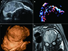
A 43-year-old multigravida woman was referred to Seoul St. Mary's Hospital at 32+4 weeks of gestation with a fetal scalp mass. The patient's antenatal examination was unremarkable until the scalp mass was found by third-trimester screening ultrasonography performed at a local clinic. An integrated test performed at the second-trimester showed low risks for the detection of Down syndrome, Edward syndrome, and neural tube defect. No other anomalies were observed on level II ultrasonography performed at 25+4 weeks of gestation. The ultrasonography at our hospital, performed with a Voluson E10 (GE healthcare, Tiefenbach, Austria), showed a single female fetus in cephalic presentation with fetal biometry appropriate for the gestational age. Ultrasonography showed a 6.3×3.7 cm homogeneously echogenic solid mass covering the fronto-parieto-occipital area of the fetal scalp (Fig. 1A, 1C). Color/power Doppler images identified prominent vascular flow at the periphery of the tumor (Fig. 1B). Neither skull defects nor intracranial extension were visible on ultrasonography. For further evaluation, fetal MRI was performed using the Ingenia 3.0 Tesla MR imaging system (Philips Healthcare, Eindhoven, the Netherlands) to obtain T2-weighted images (TR 1614.2 ms/TE 80.0 ms), and these showed a 3.0×6.0×5.0 cm mass with mixed low and high signal intensities and internal signal void, suggesting hemorrhage or calcification in the tumor (Fig. 1D). The mass had a broad base on the scalp extending to the left periorbital region. No skull defects or abnormal intracranial lesions were noted. Considering the mixed signal intensity in the MRI and prominent flow signals in the ultrasonography Doppler study, prenatal diagnosis considered fetal scalp hemangioma.
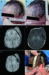
Until 36 weeks of gestation, follow-up ultrasonography examinations showed no significant change in mass size. An early delivery was planned for 36+6 weeks of gestation, because spontaneous labor may cause massive compression that could result in traumatic bleeding or damage to the fetal scalp mass; thus cesarean delivery was performed in order to avoid traumatic hemorrhage of the mass and the risk of dystocia. A live baby girl weighing 2,970 g was delivered with Apgar scores of 6 and 8 at one and five minutes, respectively. A 9.0×7.0 cm well-defined mass was present in the left fronto-parietal area (Fig. 2A, 2B).
Postnatal MRI with Achieva 1.5 Tesla MR imaging system (Philips Healthcare) (TR 5235.9 ms/TE 100.0 ms) showed that the fetal scalp mass had a homogenous appearance and intense enhancement. The lesion was located between the orbicularis occuli and the temporalis muscle and extended to the soft tissue of the left lateral orbit. Compressive contour deformity and mild erosive thinning of adjacent skull were observed as well. The tumor showed intermediate signal intensity on T1-weighted images (Fig. 2D) and high signal intensity on T2-weighted images (Fig. 2C). MRI also demonstrated a signal void, suggesting the tumor's vascular structure. There was multifocal low signal intensity on the susceptibility weighted imaging, suggesting hemangioma with petechial hemorrhage or thrombus (Fig. 2E). Therefore, the postnatal diagnosis of the fetal scalp mass was hemangioma. After consulting with a neurosurgery team and general pediatric surgery team, the baby was treated with beta blockers without excision in order to decrease the size of the mass. Following seven days of treatment, the mass size had decreased from 9.0×7.0 cm to 7.3×6.8 cm (Fig. 2F). The dose of atenolol was gradually increased, and the neonate is currently taking 9 mgs of atenolol twice a day. Computed tomography (CT) will be performed after four to five months of medical treatment, and surgical excision may be performed based on those CT results.
Discussion
Congenital hemangiomas are rare benign vascular tumors that are fully developed at birth.345 They have two major subtypes: rapidly involuting congenital hemangiomas (RICHs) and non-involuting congenital hemangiomas (NICHs), based on their clinical prognosis after birth.345 RICHs are more common and usually involute spontaneously within the first 12–14 months of life.56 In contrast, NICHs typically require surgical excision.345 Clinically, RICHs and NICHs have overlapping characteristics and are difficult to differentiate at the prenatal stage.7 The most common locations of congenital hemangioma are the neck, head, and limbs;3 scalp hemangiomas are relatively rare.
The ultrasonography features of congenital hemangioma vary depending on the sizes and types of vessels present, the amount of arteriovenous shunting, and the degree of endothelial proliferation.8 Sonographic characteristics range from solid masses with homogeneous echogenic patterns to mixed solid-cystic lesions with heterogeneous appearance.5910 Frequently, fetal hemangiomas demonstrate calcification on ultrasonography.311 Doppler ultrasonography depicts vascular blood flow or microshunts within the tumor.311
Recently, fetal MRI has been used more frequently as an adjunctive diagnostic tool for the differential diagnosis of fetal scalp masses. On MRI, congenital hemangiomas present as well-defined masses with mixed signal intensities and a reticulated appearance on T1- and T2-weighted images.3
Once a fetal scalp mass has been found prenatally, several diseases should be considered in differential diagnosis, including teratoma, hemangioma, encephalocele, cystic hygroma, lymphangioma, and mesenchymal sarcoma.311 On ultrasonography, teratomas appear as masses with calcifications and solid or cystic components.12 Encephalocele is the most common posterior midline extracranial mass, and shows skull defects and associated cerebral abnormalities such as hydrocephalus.1 Lymphangiomas and cystic hygromas appear on prenatal ultrasonography as fetal neck masses of various sizes with cystic components and multiple septa.3 The differential diagnosis of those tumors is important because they differ in prognosis and treatment. In the case of congenital hemangioma, spontaneous labor or vaginal delivery may cause traumatic hemorrhage in a tumor, so an elective cesarean section should be considered prior to labor.
In our case, a fetal scalp hemangioma on the left fronto-parieto-occipital area was found at 32 weeks of gestation, and there were no associated anomalies or other complications such as fetal cardiac dysfunction. On ultrasonography, the fetal scalp mass was a homogeneously echogenic solid mass with prominent vascular flow. Prenatal MRI indicated that the extracranial mass was heterogeneous in appearance with an internal signal void that suggested hemorrhage or calcification in the tumor. Early cesarean section was performed at 36+6 weeks of gestation in order to avoid traumatic hemorrhage of the mass as well as the risk of dystocia. Postnatal MRI showed intermediate signal intensity on T1-weighted images and high signal intensity on T2-weighted images, which are characteristic features of infantile hemangioma.210 Following consultation, the baby was treated with beta blockers. The exact mechanism of the benefits of using beta blockers for congenital hemangioma is yet to be elucidated, although it has been suggested that vasoconstriction, the inhibition of angiogenesis, and the induction of apoptosis of capillary endothelial cells inhibit tumor growth.13 As has been reported in previous studies, fetal scalp hemangioma may have spontaneous resolution after birth.36 However, in this case, considering the size and position, it could affect the visual axis when the size has increased. Moreover, there was a risk of complications such as bleeding. Therefore, after consultation with the parents, the neonate was treated with medication.
In conclusion, the proper diagnosis of hemangioma with antenatal ultrasonography and MRI is crucial once a scalp mass has been found in utero. This can help to decide the timing and method of delivery and thus reduce the risk of traumatic hemorrhage during vaginal delivery.
 XML Download