

 PDF
PDF ePub
ePub Citation
 Print
Print


INTRODUCTION
Bowel ischemia, a rare condition in children, is a life-threatening emergency owing to the high rates of mortality and morbidity associated with this disease entity [12]. For this reason, an early diagnosis is challenging because patients usually demonstrate a nonspecific clinical presentation, particularly in young children who cannot communicate well their symptoms or specific experiences related to this condition [1]. Unfortunately, however, delayed diagnosis can result in subsequent bowel necrosis followed by an overwhelming inflammatory response and even result in the death of the patient, whereby time is of the essence in these cases [12]. Imaging studies such as abdominal ultrasonography and computed tomography (CT) are helpful for establishing the diagnosis; however, the reliability of these tests varies and tends to be significantly affected by the skills of the interpreter of the results at the time of the review [3].
Here we report a case of rapidly progressive small bowel necrosis in a previously healthy child without proven mechanical obstruction. The purpose of this case report is to emphasize the clinical importance of careful observation of the patients with suspected strangulation of a small bowel obstruction (SBO), especially in the situation whereby image findings are determined to be inconclusive in this case.
CASE REPORT
A previously healthy 23-month-old boy presented to our hospital with a 2-day history of fever, postprandial diffuse abdominal pain, and non-bilious vomiting (>5 episodes/day) that began 1 day before admission. After admission, the patient developed a watery diarrhea. In addition to food refusal, he was observed to produce lesser urine than usual upon void review. His medical and birth history were unremarkable (he was not administered any medications, he had no known allergies, and his immunizations were up to date). His family medical history was unremarkable, and he had no travel history at that time.
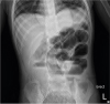
Upon examination, the patient looked acutely ill and dehydrated. He was 90.2 cm (in the 75–85th percentile) tall and weighed 11.6 kg (in the 15–25th percentile). His abdomen was soft and flat with normoactive bowel sounds. Upon palpation of the abdomen the examination revealed the existence of a mild tenderness over the entire abdomen, without rebound tenderness or muscle guarding or rigidity. The rest of his physical examination was noted to be normal. The patient's vital signs showed a heart rate of 141 bpm, respiratory rate of 22 with a normal respiratory pattern, temperature of 38.6°C, and oxygen saturation at 98% on room air. He was observed to be mentally alert at the time of the examination. The initial laboratory tests revealed the following: white blood cells (WBCs) 6,500 cells/μL, hemoglobin (Hb) 13.2 g/dL, platelets 423,000/μL, C-reactive protein (CRP) 6.35 mg/dL, sodium 130 mmol/L, potassium 4.1 mmol/L, chloride 90 mmol/L, and total carbon dioxide 14.4 mmol/L. All other blood tests were within the reference range. His stool occult blood test was positive (748 ng/mL). It is noted that the tests for rotavirus and norovirus antigen were negative. An erect chest X-ray did not show pneumoperitoneum; however, an erect abdominal X-ray demonstrated abnormal dilatation of the small bowel loops with air-fluid levels suggestive of possible SBO (Fig. 1). The abdominal ultrasonography revealed mild wall thickening of the small bowel loops suggestive of acute enteritis, several enlarged lymph nodes in the right lower quadrant, and a normal-appearing proximal appendix (the tip of the appendix was not visualized), without any evidence of mechanical bowel obstruction such as an intussusception.
Fig. 1
Erect abdominal radiography, showing abnormal dilatation of small bowel loops with air-fluid levels suggestive of a possible small bowel obstruction.
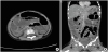
The patient was resuscitated using normal saline and received conservative management. At that time, cefotaxime was empirically administered intravenously. The following day, his condition rapidly deteriorated, and he developed a state of septic shock. Upon examination, the patient's vital signs now were: blood pressure (BP) 87/39 mmHg, heart rate 170 bpm, respiratory rate 56, temperature 38.1°C, and oxygen saturation 93% on room air. Our surgical team urgently came to the patient and performed physical examination; his abdomen was soft with mild tenderness and slight muscle guarding on palpation. They recommended continuing medical supportive care, but frequent physical examinations needed to be performed to detect subtle changes in the patient's signs and symptoms. The patient's laboratory tests were repeated and revealed: WBCs 5,300 cells/μL, Hb 9.7 g/dL, platelets 186,000/μL, CRP 28.96 mg/dL, sodium 129 mmol/L, potassium 3.7 mmol/L, chloride 95 mmol/L, and albumin 2.6 g/dL. Upon review, the coagulation tests revealed an abnormally prolonged prothrombin time at 21.5 seconds and an activated partial thromboplastin time at 56 seconds. The patient's venous blood gas analysis demonstrated metabolic acidosis: pH 7.28 and bicarbonate 15 mmol/L. Upon review, other blood tests remained within reference range. A plain abdominal X-ray was repeated and showed worsening of small bowel ileus. At that time, an urgent contrast-enhanced abdominal CT was performed in order to confirm the presence of a surgical abdomen. The imaging revealed diffuse dilatation of the large and small bowel loops (suggestive of paralytic ileus) with uniform enhancement of bowel wall and no lead point of bowel obstruction, mesenteric lymphadenitis, and mild ascites (Fig. 2). However, the radiologists were of the view that obstructive ileus could not be completely ruled out in this case. They did not find an abnormal appendix, but the condition of an acute appendicitis could not be excluded either. The patient was maintained on a nil per os status, and fluid resuscitation was continued. We tried rectal tube decompression, however, his condition did not show much improvement except the passage of small amount of rectal gas. He received albumin, packed red blood cells, and fresh frozen plasma intravenously. At that time, Metronidazole was additionally administered empirically, and his vital signs were closely monitored for any significant changes.
Fig 2
(A) Axial and (B) coronal computed tomography, showing diffuse dilatation of large and small bowel loops (suggestive of paralytic ileus), mesenteric lymphadenitis and scanty ascites.
On the third hospital day, the patient became more lethargic despite medical therapy. His BP was observed to have normalized; however, it was noted that tachycardia and tachypnea persisted. On repeated physical examination, his abdomen now showed distention with hypoactive bowel sounds, however, muscle guarding or tenderness could not be clearly ascertained on palpation. The patient developed generalized edema and oliguria. Based on the patient's clinical condition, we decided to perform an urgent exploratory laparotomy at 44 hours after admission.
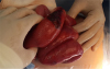
Upon opening the peritoneum, approximately 400 mL of ascitic fluid was drained. Next, a segment of markedly dilated and infarcted small bowel was identified 30 cm proximal to the ileocecal valve (Fig. 3). It is noted that the total length of the necrotic small bowel was approximately 60 cm. The omentum and the colon appeared to be intact. Upon review, the small bowel intussusception or volvulus was not identified. Even at laparotomy, our surgical team could not find the lead point of bowel obstruction. The appendix appeared unremarkable macroscopically. In this case, after decompression of the bowel following an incision on an infarcted small bowel segment, 110 cm of the distal small bowel containing the necrotic segment was resected and end-to-end small bowel anastomosis was performed in addition to a prophylactic appendectomy.
Fig. 3
Intraoperative findings, showing small intestinal necrosis at 30 cm superior from ileocecal valve.
Histopathological examination of the surgical specimen revealed multiple areas of mucosal necrosis with diffuse congestion. The patient blood cultures did not reveal any bacteria, and stool cultures did not reveal Salmonella or Shigella. His postoperative course was considered to be uneventful, and oral nutrition could be initiated on postoperative day 5. The patient was discharged in a good general condition on postoperative day 11. After discharge, the patient was noted as asymptomatic and did not report any serious residual complications.
DISCUSSION
In general, it is not easy to differentiate between a surgical abdomen and self-limited conditions in young children who are unable to describe their symptoms well and articulate their areas of pain or concern to a healthcare professional or provider. Therefore, imaging studies such as abdominal ultrasonography and CT have been helpful for clinicians working with children, to determine whether surgical intervention is essential in those cases. However, in this case, we could not establish a conclusive diagnosis and were required to verify the need for an immediate surgical intervention based on the radiological findings. Considering the clinical situation, we could not rule out strangulation of mechanical SBO in this case. Over the first 2 days of hospitalization, our surgical team closely followed the patient's condition, and repeated careful physical examinations to decide whether a surgical procedure was warranted at that time. Eventually, on the third hospital day, we decided to proceed with performing surgical exploration based on changes in clinical condition of the patient, but not solely based on the image findings, which proved to be the right decision because intraoperative findings confirmed the diagnosis of small bowel necrosis. A delay in performing surgical exploration (based on the negative findings observed on imaging studies) would have led to a significant worsening of the patient's condition, and a deteriorating hospital course for the patient in that case. From our experience in this case, in children with suspected strangulation of SBO, we empathize that timely decision for exploratory surgical approach should be the chosen intervention and procedure, based on changes in the clinical condition of the patient, through carefully repeated physical examinations and other evaluations including laboratory studies, especially when image finding is inconclusive but clinical suspicion is high for this type of occurrence and diagnosis.
It is important to note that SBO is a common clinical condition that occurs secondary to mechanical or functional obstruction of the small bowel, preventing the normal transit of its contents in the patient [4]. The prevailing discipline notes that several conditions including congenital disorders (malrotation with volvulus, Hirschsprung's disease, duplication cyst, and hernia), infectious/inflammatory conditions (appendicitis, intussusception, inflammatory bowel disease, and necrotizing enterocolitis), iatrogenic conditions (postoperative adhesions), vascular causes (ischemia), as well as neoplastic and other rare causes contribute to the occurrence of this condition in children [4]. Broadly speaking, patients with strangulated SBO tend to commonly present with atypical presentations including tachycardia or hypotension, which may be secondary to the condition of shock. Evidently, strangulation can cause increased luminal pressure leading to increased blood vessel permeability and bacterial translocation in the intestine, resulting in circulatory failure and hypotension in the patient [5]. In this patient, an abdominal ultrasonography was performed based on a high index of suspicion for mechanical SBO; however, it is noted that the ultrasonography only revealed the incidence of an acute enteritis without evidence of intussusception or other causes of mechanical obstruction. In fact, even the contrast-enhanced abdominal CT performed on the second day of hospitalization did not reveal any evidence of a mechanical obstruction. For example, it was noted that there was no lead point for SBO and no decreased enhancement of small bowel wall in the abdominal CT as reviewed at that time. Additionally, previous studies have reported sensitivity of contrast-enhanced CT ranging from 73% to 100% and specificity from 61% to 100% in detecting the early signs of strangulated SBO [36]. A recent retrospective study showed that adding unenhanced images to contrast-enhanced CT improved the sensitivity, diagnostic confidence, and interobserver agreement of the diagnosis of bowel ischemia in a patient with mechanical SBO [7]. In this case, we did not obtain unenhanced CT images in order to reduce the radiation dose and shorten the scan time; thus, the radiologists who followed this case might have been limited in evaluating whether or not bowel necrosis had actually occurred in this patient.
In this case, we could not demonstrate the cause of the patient's small bowel necrosis, even intraoperatively. We cannot exclude the possibility that an undiagnosed strangulation of SBO such as that occurring with intussusception, had been reduced spontaneously by the time we performed the laparotomy procedure on the patient. It is emphasized that several authors have reported that the risk of complications is higher in patients with SBO who are symptomatic over >48 hours [8]. Therefore, given that our patient underwent the surgical intervention only after 2 days of hospitalization, we have to consider the possibility of a missed diagnosis that could have led to strangulation of the small bowel that occurred during that time period. However, spontaneously reduced small bowel intussusception is presented as a benign disease in most patients, and usually is not associated with bowel ischemia or necrosis [9]. As has been noted, Meckel's diverticulum (MD), a vitelline duct remnant, shows no symptoms in most cases but rather causes complications in the patient such as hemorrhage, intestinal obstruction, inflammation, and perforation in less than 5% of cases [1011] Considering its frequency, MD could be thought as a possible cause of SBO in this patient. However, we could not find any evidence of the existence of MD intraoperatively and even pathologically in this case. There are a few case reports that have described the occurrence of nonocclusive mesenteric ischemia (NOMI) induced by diminished mesenteric blood flow (hypotension, hypovolemia, decreased cardiac output, or the use of vasopressors), or intestinal vasospasm without thromboembolic occlusion in children or adolescents with familial dysautonomia, Addison's disease, burns, and the administration of chemotherapy [2]. However, to the best of our knowledge, NOMI without an underlying disease has not been reported in children to date. In our case, a histopathological examination did not confirm the presence of a mesenteric vascular occlusion, although the presentation of the patient in this case could possibly be considered that of NOMI in a previous healthy child.
Despite being categorized as an emergency, only a few studies have described when to suspect strangulation and decide upon the optimal timing to perform surgical exploration in children with SBO. For adult patients, the guideline of diagnostic laparoscopy has been developed by the Society of American Gastrointestinal and Endoscopic Surgeons [12], and we could consider the general appropriateness of applying this guideline to pediatric patients. According to the guideline, the main indication to perform a diagnostic laparoscopy in the intensive care unit has been in the case of an unexplained sepsis, systemic inflammatory response syndrome, and in the case of a multisystem organ failure. Additionally, the procedure has been used for abdominal pain or tenderness associated with signs of patients diagnosed with sepsis, fever and/or leukocytosis in an obtunded or sedated patient not explained by another identifiable problem, metabolic acidosis not explained by another process (such as cardiogenic shock), and increased abdominal distention that is not a consequence of an observed bowel obstruction. Evidently, a multivariate regression analysis performed in 192 adults who underwent an operation for acute SBO showed that peritoneal signs (muscle guarding) and an elevated WBC count are the independent variables that serve as predictors of identifying bowel strangulation in a patient [6]. Recently, a retrospective study was conducted for 69 pediatric patients who underwent operation for acute SBO, to evaluate the clinical risk factors of strangulation; 4 clinical signs (severe continuous abdominal pain, tachycardia, WBC 13,600/mm3, and abdominal distention) offer the variables to determine an independent predictive value for the need for emergency operative treatment in these cases. Each of these signs is considered to be nonspecific and cannot be used in isolation [13]. Based on the observations of the afore mentioned studies, we found that the surgical exploration was truly necessary in this case considering that the patient developed tachycardia, abdominal distention and muscle guarding on physical examination and a laboratory study revealed the presence of metabolic acidosis. In this sense, more investigation is required to determine the risk factors for strangulation in children with suspected SBO.
In conclusion, in children with suspected strangulation of SBO, we ought to consider choosing the exploratory surgical approach based on changes in the noted clinical condition of the patient through carefully repeated physical examinations and other evaluations, especially in cases when imaging studies do not provide a conclusive diagnosis indicating the need for a necessary surgical intervention.
 XML Download