

 PDF
PDF ePub
ePub Citation
 Print
Print


INTRODUCTION
Pancreatic surgery has been markedly refined, which has appreciably decreased mortality. The mortality rate is now 0%–5% [1]. However, the rate of morbidity following pancreaticoduodenectomy (PD) remains high at 30%–40% [12345]. Among the various complications, postpancreatectomy hemorrhage (PPH) is obviously not 100% fatal, but is one of the most serious causes of mortality following PD. It is associated with 10%–38% of the overall mortalities [4678].
In particular, late PPH is mainly caused by pseudoaneurysm rupture. Reoperation or intervention have been considered as treatment of delayed arterial hemorrhage following PD. Recently, good outcomes of endovascular management for pseudoaneurysm rupture such as embolization or insertion of stentgraft have been reported [249101112131415161718]. Although there are some drawbacks of the endovascular technique, including irreversible ischemic change of the end organ, rebleeding or stent occlusion, diagnostic angiography followed by endovascular management is preferred over surgery and has become the standard therapeutic treatment for pseudoaneurysm rupture.
Despite this, the long-term clinical outcomes of endovascular management are not well known. We have hypothesized that the long-term clinical outcome of patients undergoing endovascular treatment after pseudoaneurysm rupture may be acceptable. Presently, we evaluated comprehensive results of endovascular management for ruptured pseudoaneurysm in patients undergoing PD during long-term follow-up.
METHODS
Patient selection
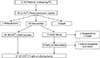
The Institutional Review Board approved the exemption of this retrospective study (approval number: 2016-10-087). The process of receiving informed consent from the patient was exempted from the IRB. Between October 1994 and December 2016, the medical records of 2,783 patients who underwent PD with curative intent at a single center were prospectively collected and retrospectively reviewed. We excluded patients with enculeation and distal pancratectomy. Among 2,783 patients, pseudoaneurysm rupture developed in 66 patients (2.4%). Of the 66 patients, 2 underwent immediate relaparotomy and 2 died of hemorrhagic shock before any treatment. Besides, 2 patients underwent relaparotomy after initial failure of intervention, 2 patients experienced death after initial failure of intervention, and 1 patient repeated failure of intervention prior to death.
Of the 62 patients who experienced endovascular treatment, success of hemostasis was accomplished in the first attempt in 50 patients (80.6%) and on the second attempt in 7 (11.3%). Of the seven patients who had success in the second attempt, the second trial was conducted for recurrent bleeding. The 3 patients experienced rebleeding from gastroduodenal artery (GDA) stump and 2 experienced rebleeding from blocked common hepatic artery (CHA). Two patients experienced endoleak from GDA and proper hepatic artery (PHA).
Finally, 57 patients (91.9%) who ultimately achieved successful endovascular management were included in this study (Fig. 1). The patients were divided into three groups based on type of intervention: (embolization only [EMB], n = 30), (stent-graft placement only [STENT], n = 19) and (both embolization and stent-graft placement simultaneously or different times [EMB + STENT], n = 8).
Exposure or causes of this study are as follows: age, sex, body mass index, operating time, pancreas texture, diameter of p-duct, Clinically relevant postoperative pancreatic fistula (CR-POPF), intra-abdominal infection, postoperative bile leakage, time to rupture after operation, time to intervention, location of ruptured pseudonaeurysm, diameter of target vessels, lengths of ruptured segment, diameter of stent-graft, lengths of stentgraft, in-hospital days and use of anticoagulant/antiplatelet.
Initial assessment and endovascular management
Vascular access for angiographic intervention was accomplished by puncturing the common femoral artery and inserting a 5F angiographic catheter (Yashiro, Terumo, Tokyo, Japan; RH catheter, Cook, Bloomington, IN, USA). To discover the bleeding focus, selective celiac trunk, common hepatic, and superior mesenteric arteriographies were routinely done.
Stent-graft placement was performed as the first option unless the stent-graft approach to the bleeding point was technically difficult. Furthermore, if embolization is expected to result in serious complications such as liver ischemia and failure, we first attempted stent-graft placement instead. These cases are as follows: (1) No intrahepatic collateral (IHc) is identified on angiogram. (2) No replaced hepatic artery is identified on angiogram. (3) Short GDA stump is likely to cause coil migration after coil embolization.
The embolization was performed only when the stent-graft placement was expected to be technically very difficult. These cases are as follows: (1) If a vasospasm caused by a hypovolemic shock makes the stent-graft unable to reach the site of bleeding point. (2) When target artery including bleeding point is tortuous and forms acute angle.
Stent-graft placement with embolization was performed only in special cases. If the bleeding site is adjacent to the bifurcation, back flow is often formed. Therefore, exclusion through stent-graft alone is not sufficient for hemostasis. For instance, 1 patient had bleeding at the proximal site of left hepatic artery (LHA). Stent-graft was inserted covering from right hepatic artery (RHA) to CHA initially. The direct stentgraft insertion of the LHA is technically impossible because the acute angle is formed at the point where the LHA is branched from the hepatic artery. However, since the back flow from LHA may flow to the rupture site at proximal LHA, additional embolization at LHA was performed.
Among the 27 patients who received stent-graft placement at least once more, 31 stent-grafts were used. Thirty were self-expandable stent-grafts, either the Niti-S Comvi (Taewoong Medical, Seoul, Korea) in 7 cases or the VIABAHN (W.L.Gore and Associates, Flagstaff, AZ, USA) in 23 cases. The remaining stent-graft was a balloon-expandable stent-graft (JOSTENT GRAFTMASTER, Abbott Vascular, Menlo Park, CA, USA). The anti-platelet agents after stent-graft were not used routinely. Eighteen patients (66.7%) received anticoagulant and/or antiplatelet medication.
In case of embolization, 3F microcatheter (Microferret, Cook, Bloomington, IN, USA) entered the hepatic artery to exclude the bleeding focus, and then embolic material was placed in the corresponding target artery including bleeding point. Among the 38 patients who underwent embolization at least once more, 40 procedures were conducted. Thirty-four (85.0%) were done by coil material mainly using suitably sized platinum coils (Tornado/Nester, Cook) 0.018 or 0.035 inches in diameter and/or Interlock Fibered IDC occlusion system (Boston Scientific, Natick, MA, USA), 4 procedures (10.0%) used glue (Histoacryl; B. Braun, Tuttlingen, Germany) and 2 procedures (5.0%) were carried out using gelfoam (Cali-Gel; Alicon, Hangzhou, China).
Anticoagulation/antiplatelet therapy was performed after stent-graft placement if the possibility of rebleeding due to the use of anticoagulants was not high. The risk of rebleeding was considered clinically and no anticoagulant was used in the following cases: (1) If rebleeding occurs before 24 hours after the procedure. (2) If the vital signs are expected to be unstable for a long time due to severe hemoperitoneum. Of 27 patients who received stent-graft placement, 18 patients (66.7%) received anticoagulant and/or antiplatelet medication. There was no established protocol in our institute to decide to stop using anticoagulants. This was interrupted by each surgeon according to his clinical judgment.
Schedule and dosage of anticoagulant use are as follows: (1) Dual antiplatelet therapy with aspirin and clopidogrel (DAPT) - Aspirin 100 mg PO with clopidogrel 75 mg per os (PO) during hospital stay followed by maintain treatment for a certain period in outpatient setting, (2) DAPT - Aspirin 100 mg PO with clopidogrel 75 mg PO during hospital stay and certain period in outpatient setting, followed by aspirin lifelong therapy alone, and (3) Aspirin 100 mg PO alone during hospital day, followed by maintain treatment for a certain period in outpatient setting. The duration of drug maintenance varied according to the judgment of individual surgeons.
Parameters and follow-up
Postoperative pancreatic fistula (POPF) was defined according to the criteria of the International Study Group of Pancreatic Fistulas as any measurable amount of fluid through an operatively placed drain, with an amylase content >3 times the upper normal serum value on or after postoperative day 3 [19].
The diameter of the target vessel and length of ruptured segment were measured in the tubogram view during the course of the angiogram. Considering the stent-graft placement, average value of longest diameter and shortest diameter in the ruptured segment was defined as the target vessel diameter. The length of the ruptured segment was defines as follows: (1) the lengths of the base of the pseudoaneurysmal sac or (2) the lengths of the lesion where the broad vessels irregularity is observed.
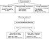
The endpoints, outcomes and follow-up methods were summarized in Fig. 2. The primary endpoints are primary patency, stent-graft related complications and clinical complication. The primary patency was defined as no evidence of stent occlusion on follow-up CT scan. Stent-graft related complications include partial stent thrombosis, stent occlusion and stent migration. Clinical complication is as follows: (1) liver ischemia or infarction, (2) liver abscess, (3) biliary necrosis, and (4) small bowel ischemia. The stent-graft related complication and clinical complication were subdivided into short term and longterm complication according to timing. Short- and long-term complications were defined as occurring within 30 days or more. The secondary endpoints are 30-day mortality.
After an endovascular intervention, follow-up CT scans and a laboratory test were performed at intervals of 1 to 3 months in the first year, and then performed at intervals of 3 to 6 months.
Statistical analyses
Chi-square and Fisher exact test were used to compare categorical variables. Student t-test was used for continuous variables. In the univariate analysis, P < 0.05 was considered significant. Parameters with P < 0.1 were included in a multivariate analysis using logistic regression or Cox proportional hazards regression to identify risk factor for development of complication and survival. Statistical significance was indicated at P < 0.05 in the multivariate analysis. Sensitivity analysis was performed to identify robustness of analysis. Data were analyzed using IBM SPSS Statistics ver. 23.0 (IBM Co., Armonk, NY, USA).
RESULTS
Cohort characteristics
The clinicopathological findings in the 57 patients (44 males, 13 females; median age 70 years) are summarized in Table 1. The median time interval between symptoms onset and intervention was 7.0 hours (0.5–168.0 hours). Color change of intrabdominal fluid through drainage was observed in 17 patients (29.8%) before diagnosis of the pseudoaneurysm rupture. Twenry-six patients (35.6%) experienced rupture at GDA stump, the most common site of bleeding. The mean diameter and lengths of target vessels were 5.1 (1.5 – 8.7) and 8.0 mm (1.0–40.0 mm). There were 8 cases (14.0%) of 30 daymortality caused by endovascular management. The median follow-up duration after final intervention was 26.4 months (1.0–140.0 months).
Analysis of collateral pathway to liver
By reviewing angiography for diagnosis of pseudoaneurysm rupture and follow-up abdominal CT, present investigation analyzed the pattern of collateral feeding vessels to liver which have already been at the time of angiography or were identified on follow-up CT. According to site of ruptured pseudoaneurysm and type of intervention, various collateral vessels developed (Table 2). Collateral formation from left gastric artery (LGA) is the most frequent collateral vessel (18 cases). We could see infraphrenic artery (IPha) in 17 cases and IHc in 6 cases.
Primary endpoint (short-term outcomes)
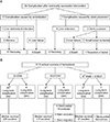
Fig. 3A demonstrates short-term primary endpoint after successful hemostasis. Among 57 patients, stent-graft related complications developed in 3 patients (5.3%) and clinical complication developed in 18 patients (31.5%). All 3 stent-graft related complications were stent-graft occlusion. Among 18 patients who had short-term clinical complications, 15 (83.3%) experienced liver ischemia or infarction, 2 (11.1%) experienced liver abscess, and 1 (5.6%) experienced small bowel ischemia.
All 2 patients who had liver abscess recovered by conservative treatment like antibiotics therapy. Three cases of stent occlusion led to 2 cases of liver failure eventually death and one case of bowel ischemia.
There were 4 patients who experienced liver infarct after stent insertion without stent occlusions. They experienced bleeding occurring between the middle and proximal portions of the PHA. To prevent backflow, the stent-graft was inserted covering from RHA and PHA. Thereafter, blood supply to the left lobe of liver through the LHA was blocked. As a result, it seems that IHc flow alone did not provide sufficient blood flow to the left lobe of the liver and liver ischemia at left love of liver developed.
Of 17 cases of complication after embolization, formation of collateral vessels to liver occurred in 11 cases (64.7%) and no collateral formation happened in all 6 mortality cases within 30 days after final hemostasis.
Primary endpoint (long-term outcomes)
Long-term primary end point is summarized in Fig. 3B. Among the 27 patients who received stent-graft placement at least once more, stent-graft related complications developed in 9 patients (33.3%) and clinical complication developed in 1 patient (3.7%). The 9 stent-graft related complications include 5 cases of partial stent thrombosis, 3 cases of stent occlusion, and 1 case of stent migration. Table 3 shows clinical data about long-term stent-graft related complication. All stent-graft related complications developed in patients with stent-graft placement at hepatic artery.
Of 5 cases with partial stent thrombosis, 4 received anticoagulant and/or antiplatelet treatment after stent-graft placement. The anticoagulant and/or antiplatelet treatment started one day after intervention. Despite partial thrombosis, no significant clinical events, including liver ischemic change, were observed and stent patency was intact in all 5 patients with partial thrombosis. After first detection, 2 of these 5 patients did not show any remarkable change in follow-up CT scan. However, in the other 3 patients, no further CT examinations were done after the first detection of partial thrombosis; of these patients, 2 died of recurrent cancer and the other remains alive. After partial stent thrombosis or occlusion, anticoagulant therapy was done in 2 patients. The remaining 3 were not treated for partial thrombosis.
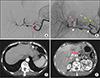
Among 3 patients who experienced stent occlusion, 2 did not present noticeable clinical complications. Fig. 4 shows a series of image of long-term stent occlusion at the hepatic artery. We found one mortality case after stent occlusion at the hepatic artery, in which a patient died of septic shock caused by biliary necrosis caused by stent occlusion 84 days after the intervention.
The median survival time of patients with no long-term primary outcomes between the EMB, STENT, and EMB + STENT groups was 25, 17, and 7 months, respectively (P = 0.080).
There were no significant differences in recurrence rate of tumor between the EMB, STENT, and EMB + STENT groups (14 of 30 [46.7%] vs. 9 of 19 [47.4%] vs. 3 of 8 [37.5%], P = 0.883). Of 26 patients with recurrence, no statistical differences in 5-year survival rate were observed (13.3% vs. 0% vs. 0%; P = 0.911).
Primary endpoints (analysis of stent patency)
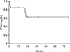
The stent graft primary patency rate, the primary endpoint, was 88.9% after 1 month, 84.2% after 1 year, and 63.2% after 2 years (Fig. 5). On image analysis and follow-up, stent graft occlusions were found in 6 of 27 cases (22.2%). Of 6 patients who experienced occlusion, 3 experienced occlusions in longterm follow-up period (64, 660, and 676 days of follow-up) in abdominal CT scans. In contrast, 21 of 27 patients (77.8%) maintained patency of the stent-graft on the last follow-up CT scan. The longest stent graft patency was 77 months; at the time of the last follow-up examination, this patient represented normal liver parenchymal perfusion without abnormal laboratory findings.
Analysis of risk factor for primary outcomes and sensitivity analysis
There was no independent significant risk factor for long-term partial stent thrombosis or occlusion. The diameter of stent-graft and discontinuation of anticoagulant and/or antiplatelet treatment was found to risk factor with marginal significance in univariable analysis (hazard ratio [HR], 0.194; 95% confidence interval [CI], 0.031–1.239; P = 0.083 and HR, 7.778; 95% CI, 0.795–76.088; P = 0.090).
Concerning stent occlusion in long-term period, there were no significant risk factors identified in the univariate analysis. However, patients with smaller diameter stent-graft (<7.5 mm) more often experienced occlusion (HR, 1.300; 95% CI, 0.965–1.751; P = 0.098). All 3 cases of occlusion developed with smaller diameter of stent-graft.
Given these, we conducted sensitivity analysis only for the risk factors of long-term partial stent thrombosis or occlusion. First, we evaluate ‘confounding effect.’ Among the unmeasured variable, history of hypertension (HTN) and number of endovascular managements were marginally significant (HR, 0.194; 95% CI, 0.031–1.239; P = 0.082 and HR, 0.600; 95% CI, 0.420–0.858; P = 0.057). These were added stepwise to the existing logistic regression model to see if there was any change in the hazard ratio and significance. Model 2 (existing model 1 + HTN): there was no change in significance of 2 risk factors (HR, 0.146; 95% CI, 0.016–1.326; P = 0.087 and HR, 8.196; 95% CI, 0.678–99.080; P = 0.098). Model 3 (model 2 + number of endovascular managements): there was changes of significance compared to existing model 1 and model 2 (HR, 0.243; 95% CI, 0.025–2.331; P = 0.243 and HR, 6.036; 95% CI, 0.419–86.934; P = 0.187).
Second, we performed subgroup analysis based on gender and age to confirm if there was no change in hazard ratio in subpopulation. Of 27 patients with stent-graft placement, 23 were male and 4 were female patients, 17 were over 66 years old and 10 were under 66 years old. Of 23 male patients, diameter of stent-graft were significant risk factor for long-term partial stent thrombosis or occlusion in unvariable analysis (HR, 0.083; 95% CI, 0.008–0.907; P = 0.035). However, discontinuation of anticoagulant and/or antiplatelet treatment were not significant factor (HR, 5.625; 95% CI, 0.537–58.909; P = 0.144). Of 4 female patietns, diameter of stent-graft and discontinuation of anticoagulant and/or antiplatelet treatment were not significant risk factor in univariable analysis (HR, 0.333; 95% CI, 0.067–1.653; P = 0.500 and HR, 3.000; 95% CI, 0.606–14.864; P = 0.500). These 2 factors were not significant risk factors in the age-based subgroup analysis (diameter of stent-graft: HR, 0.286; 95% CI, 0.023–3.528; P = 0.335 in the patients over 66 years old and HR, 0.067; 95% CI, 0.003–1.509; P = 0.190 in the patients under 66 years old; discontinuation of anticoagulant and/or antiplatelet treatment: HR, 1.667; 95% CI, 1.005–2.765; P = 0.103 in the patients over 66 years old and HR, 3.000; 95% CI, 0.188–47.963; P = 0.571 in the patients under 66 years old).
DISCUSSION
Ultimate goal of endovascular treatment for pseudoaneurysm rupture is to increase chance of successful intervention and reduce complication. The initial success rate reported previously by other centers ranges from 77.7% to 100% [131718]. These results are comparable with our center. It seems to reflect the development of endovascular intervention technique in our institute during 2 decades. As tertiary hospital, our division of interventional radiology experienced various cases other than pseudoaneurysm rupture as follows: transcatheter arterial choemoradiation for hepatocellular carcinoma, stent placement for aortoiliac arterial occlusive disease, embolization therapy for peripheral arteriovenous malformations and bronchial artery embolization for controlling of emergent hemoptysis. Accumulation of above techniques may have a good influence on each case in need of intervention. Together with advancement of sophisticated endovascular techniques, progress in postintervention management based on long period of experience might contribute to good outcome.
Although various recent studies showed high success rate of endovascular treatment after pseudoaneurysm rupture, reliable assessment of long-term complication of intervention has not yet been made. In the study of Pedersoli et al. [17], only 1 patient of total 10 patients who received eventual successful stent insertion experienced stent occlusion at the point of 3 days after intervention. However, the mean follow-up time was just 51 days. Furthermore, they divided complication into “early” and “delayed” complication, but did not mention exact criteria distinguishing between 2 periods. Wang et al. [18] reported acceptable success rate of stent-graft placement. Except for 3 mortality cases, 6 patients had no long-term complication related stent insertion. However, this research included small number of patients (9 cases) and the mean follow-up time was 10.5 months (range, 4–16 months), which were relatively shorter than 26.4 months (1.0–140.0 months) of our study. Besides, some limits on obtaining accurate results of test existed because they used Doppler ultrasound mainly for checking out stent function instead of using CT scan.
Stent-graft placement was considered as the first option unless the stent-graft approach to the bleeding point was technically difficult [13171820212223]. Nonetheless, in long-term follow-up, stent-graft is always accompanied by a potential risk of stent occlusion. In the current study, all long-term complications developed in patients with stent-graft placement. Complications associated with embolization were observed only in the short-term period, as this is an immediate response to interruption of blood supply to the liver. However, stent-graft placement did not lead to fatal complications in long-term period. Among 9 patients with long-term complications, only 1 patient experienced fatal complication such as biliary necrosis caused by stent occlusion within 2 months after stent-graft placement. By contrast, no clinical complications were observed 2 patients with stent occlusion 21 and 22 months after stentgraft placement. These results may be related to the presence of collateral. If enough time for formation of prominent collateral feeding vessels to liver is present, disastrous results, such as liver failure, will not happen frequently.
In multivariable analysis, no significant independent risk factors for primary outcomes were found. This may be related to small sample size of primary outcomes. In univariable analysis, the smaller diameter of stent-graft was associated with long-term partial stent thrombosis/occlusions and stent occlusions with marginal significance (P = 0.082 and P = 0.098). Indeed, the diameter of stent-graft inserted in superior mesenteric artery (SMA) was relatively larger than that of the hepatic artery in this study (Table 1). This may explain why no long-term complications happened in 5 cases of stent-graft placement targeting the SMA, which is relatively larger than the hepatic artery.
The duration of DAPT for the prevention of thromboocclusive events after stent-graft placement at coronary artery has been studied for more than a decade and guideline recommendations have been published [24]. According to this guideline, the fundamental principle of duration for DAPT is to consider the tradeoff between decreasing ischemic risk and increasing bleeding risk. In our univariable analysis, discontinuation of anticoagulant and/or antiplatelet treatment was associated with long term stent partial thrombosis or occlusion with marginal significance (HR, 7.778; 95% CI, 0.795–76.088; P = 0.090). Seven of 8 patients (87.5%) who experienced long-term thrombo-occlusive events were not taking anticoagulant and/or antiplatelet drugs at the time when partial thrombosis was found on follow-up CT, although they had received medication for a certain period (from 1 week to 3 months) after the procedure. However, considering the results that significant adverse clinical outcome rarely developed even after stent occlusion, the mandatory life-long anticoagulant treatment is still questionable. So, further studies to find out adequate duration of DAPT after stent-graft placement is requested.
There are several limitations in our study. First, because of the detection of collateral formation conducted by abdominal CT in long-term follow-up period, the accuracy of evaluation for collateral may be lower than using CT angiography or angiogram. Second, the diversity of duration and medication for prevention of occlusive event after stent insertion may have affected the long-term outcomes of this procedure. Third, there is a question about the robustness of this study through sensitivity analysis. Confounding effect was confirmed, and a change of significance was observed in subgroup analysis. Fourth, the small number of patients and lack of randomization make it difficult to deduce statistically significant results. Multicenter or nationwide investigations to analyze more cases would be useful.
In conclusion, after recovery from initial complication, most of patients did not experience fatal clinical complication during long-term follow-up. Once patients had passed the critical period for short-term complications, long-term clinical complications were rare and most of patients followed a natural course of disease in the long-term follow-up duration. Endovascular management is an effective and safe management of pseudoaneurysm rupture after PD in terms of long-term safety.
 XML Download