

 PDF
PDF Citation
 Print
Print


INTRODUCTION
Though it has been studied about intracranial hemorrhage model in dogs, A few study on spinal epidural hemmorrhage model has been reported in dogs [12]. The variable magnetic resonance (MR) imaging characteristic of epidural hemorrhage in dogs is possibly related with the stage of the hemorrhage and the quantity of accompanying inflammatory changes. However, the epidural hemorrhage may not be same with the degradation alterations as observed in brain hemorrhage.
Typically, a hematoma with several hours to several days old classically looks hypointense until conversion of deoxyhemoglobin to methemoglobin on T1- and T2-weighted spin echo images using a high strength magnet. Conversion to methemoglobin commonly happens after the first several days after hemorrhage and intracellular methemoglobin brings obvious shortening of T1 tissue relaxation times. Consequently, hemorrhages appears high signal on T1 weighted images [3].
Associated hemorrhage or inflammation may affect the MR imaging appearance of the disc extrusion. Some of these complications that have been described have been mainly considered as a consequence of hemorrhage, but the specific MR imaging features of such complications have not been fully described [4].
In addition, the time-dependent evaluation of MR-imaging patterns has not yet been defined in dogs, especially using low magnetic fields. The aims of this study were 1) to evaluate and define signal intensity changes in canine spinal epidural hemorrhage on MR images and 2) to find proper MR sequences to detect spinal epidural hemorrhage using 0.25T low magnetic fields.
MATERIALS AND METHODS
The study was approved by the Institutional Animal Care and Use Committees of Chonbuk National University. Experimental spinal epidural hemorrhage was induced in eight healthy female beagle dogs with a mean age of 6 years and a mean body weight of 9 kg. A general and neurological examination and complete blood work was performed prior to hemorrhage induction. A venous catheter was placed in the cephalic vein, general anesthesia was induced with DZ (combination of Tomidin; Provet Veterinary Products Ltd., Turkey) and Zoletil 50 (Virbac Laboratories, France; 0.02 mL/kg intravenously). After endotracheal intubation, anesthesia was maintained via 1.5%–2% isoflurane (Hana Pharm. Co., Korea) and oxygen. For creating a canine spinal epidural hemorrhage model, the balloon compression technique modified with balloon compression using hemilaminectomy was adapted [5]. Using an aseptic technique, hemilaminectomy was performed in the region between L5 and L6. After the hemilaminectomy was done, a balloon catheter was inserted into the epidural space and advanced to T12-L1 region (Fig. 1). A 2–3 mL blood sample was collected from the jugular vein of each dog and immediately injected into the balloon to pop off. The hemilaminectomy hole was then sealed with bone wax. The percentage O2 saturation and blood pH were measured using a Vet stat electrolyte and blood gas analyzer (IDEXX, USA). The PT and APTT were measured with a cassette-based coagulation analyzer (DX Coag; IDEXX). All animals initially showed ipsilateral hind limb paralysis, but there was no improvement in the paralysis. Pain management and medical support was sustained during the study. All animals were euthanized after 30-day imaging, the histopathology exam on this lesion was not unavailable in this study.
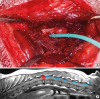
Fig. 1
Intrapinal hemorrhage induction. (A) Picture of hemilaminectomy at the level of L5–6 in a dog. (B) Diagram on magnetic resonance T2 Weighted sagittal image for balloon (red circle) catheter (a blue line) insertion to produce intrapinal hemorrhage at the level of T13–L1 through the hemilaminectomy site.
MR scanning was performed using a 0.25T (Vet Grande; Esaote, Italy) system and a solenoid knee coil. Five sequences were used to acquire images at the level of T12 to L2 spine on days 1–5 (Table 1), and thereafter at 5-day intervals for 30 days. The sequences included transverse T2-weighted (T2WI: turbo spin echo [4260/90; TR/TE]), T1-weighted (T1WI: spin echo [860/18; TR/TE]), T2*-gradient echo (1105/22; TR/TE), fluid-attenuated inversion recovery (FLAIR) (7140/90/17500; TR/TE/IR), and short tau inversion recovery (STIR) (5250/80/120; TR/TE/IR). Acquisition parameters are shown in Table 1. The slice thickness were 3.5 mm for transverse plane images with a 0.4-mm gap. Digital Imaging and Communications in Medicine (DICOM) images were obtained every 5 days from day 0 to day 30. The signal intensity was subjectively appraised as null, hypo-intense, isointense, or hyper-intense. Homogeneity of signal intensity and size change were also appraised. One radiologist compared the DICOM images from days 0 to 30 side by side using PACS software (INFINITT Healthcare, Korea) to estimate visual variances in signal intensity. The window width and level was selected subjectively to make an appropriate assessment at each sequence.
Table 1
Acquisition parameters for the transverse imaging

RESULTS
Spinal epidural and intramedullary hemorrhagic lesions in the T12 to L2 spine were successfully created in 8 beagles. The subjective assessments of chronological MR characteristics of the eight canine spinal epidural hemorrhages are summarized in Table 2.
Table 2
Time dependent magnetic resonance appearance of intramedullary & epidural hemorrhage in dogs

On T2 weighted MR images, the spinal cord lesions were hyper-intense compatible with hemorrhage (Figs. 2A and 3) during the whole study. Epidural hemorrhage with hyperintensity was observed initially but disappeared from day 10 (Fig. 3).
Fig. 2
Representative magnetic resonance images of epidural hemorrhage of T13–L1 region at day 1 in a same dog. (A) Remarkable hyper-intense (arrow) in T2WI, (B) Faint hyper-intense (arrow) in T1WI, (C) hyper-intense (arrow) in FLAIR, (D) Strong hyper-intense (arrow) in STIR, and (E) GRE image.
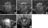
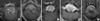
Fig. 3
Serial T2W magnetic resonance images at the level of T13–L1 in a same dog. Spinal lesion was observed with hyperintensity on T2W images compatible with hemorrhage throughout the study (arrow). Epidural hemorrhage was observed with hyperintensity on T2 weighted images (asterisk) on day 1 and 5 but not seen from day 10 and afterward. (A) Day 1, (B) Day 3, (C) Day 10, (D) Day 20, (E) Day 30.
On T1 weighted MR images, slightly hyper-intense was found initially (Fig. 2B) and since then mixed signal intensities were observed in spinal cord.
On FLAIR images, the spinal cord and epidural lesions were consistently hyperintense during the study period (Figs. 2C and 4) and more remarkable hyperinstensity on day 5 (Fig. 4C).
Fig. 4
Serial fluid-attenuated inversion recovery magnetic resonance images at the level of T13–L1 in a same dog. (A) Mild hyperintensity was observed initially (arrow) with hypo and hyper intense at epidural region ventrally. (C) Rmarkable hyperintensity was noticeable on day 5 (asterisk). (A) Day 1, (B) Day 3, (C) Day 5, (D) Day 20, (E) Day 30.

STIR images showed noticeable hyperintensity of spinal cord and paraspinal region initially (Fig. 2D) and hyper-intense signal in spinal cord remained during the entire study (Fig. 5).
Fig. 5
Serial short tau inversion recovery magnetic resonance images at the level of T13–L1 in a same dog. (A, B) Note the high intensity spinal cord and adjacent region initially. Hyper-intense signal in spinal cord and epidural space remained. (A) Day 1, (B) Day 3, (C) Day 10, (E) Day 30.

T2*-GRE images presented no significant hypointense signal compatible with hemorrhage was observed during MR study (Fig. 2E).
Low resolution imaging made it difficult to evaluate the lesion size change and signal intensity over time. There was no significant lesion size change during the study period.
DISCUSSION
Thoracolumbar disc herniation is commonly evaluated using magnetic resonance (MR) imaging in veterinary medicine. T1-hyperintensity is a classic appearance of epidural hematoma when an acute presentation occurs [6]. T1-hyperintensity was found in only seven of the 32 dogs and was not related to the clinical onset or time to imaging. It is suspected that he presence of blood in the epidural space and changes associated with it are not the sole cause of the signal intensity of the epidural lesions [7]. Besides hemorrhage, disc material in the vertebral canal produces inflammatory changes in the meninges, epidural fat and dorsal longitudinal ligament, and the extruded disc material itself [8].
The MR appearance of canine spinal epidural hemorrhage with a 0.25T low-field magnet was presented in this study. In this study, the results of time changes in MR appearance of canine spinal epidural hemorrhage in low magnetic fields did not agree with prior descriptions [39].
The induced hemorrhagic lesions had slightly different shapes, sizes, and distributions. In our experience, this is because the volume of backdraft in the blood injection at induction was slightly different in each experiment. The induced hemorrhagic lesion was distributed in the intramedullary and epidural space. The original intent was to induce extradural hemorrhage but creating solitary epidural hemorrhage was not easy. This wide distribution is a limitation of animal models. To mimic venous sinus rupture, venous blood was used for hemorrhage induction. This study observation from T2W images in intramedullary and epidural hemorrhage was not surprising considering the similar findings of hyperintense signal on T2W images from previous studies [4]. There were no significant signal intensity changes visible during study. Thus, hyperintense signals at the intramedullary and epidural region on T2W in the clinic may not rule out the possibility of hemorrhage.
Interestingly, significant signal intensity changes in the epidural hemorrhagic lesion were not observed on T1W in this study. Mild hyperintensity of intramedullary hemorrhage on T1W was not described prior reports [34]. Hemoglobin to methemoglobin degradation after 1–3 days may shorten the T1 time, so T1 hyperintensity can be observed in a hemorrhagic lesion [4]. In this study, unlike cerebral hemorrhage, spinal intramedullary hemorrhage consistently showed hyperintensity compared to normal spinal cord parenchyma. Motion artifacts caused by respiration are the key variants when scanning the thoracolumbar region. FLAIR and STIR sequences are considered to be better for detection or evaluation of hemorrhagic lesions due to high signal intensity, but long scan time resulted in more motion artifacts compared to other sequences. It was reported that STIR sequence could identify the inflammatory changes of spinal cord lesion with a high specificity suggesting that the sequences are a beneficial tool to evaluate the suspected inflammatory spinal cord disease in dogs [10]. In human medicine, STIR images were subjectively valuable in detection of the spinal and paraspinal lesions [11]. In this study, the high intensity was observed at the spinal cord and adjacent region initially compatible inflammatory changes but the paraspinal high signal lesion was reduced considerably. Still, hyper-intense signal in intramedullary and epidural space remained but hard to differentiate between the subarachnoid space containing cerebrospinal fluid and parenchyma. It was difficult to interpret and demarcate the lesion precisely because image quality was poor due to severe motion artifacts as a result of long scan time. Therefore clinicians should be aware of the scanning time especially for critical patients. In particular, low field magnet systems require additional scan times than high field systems, and inevitably have disadvantages for hemorrhage detection.
Differences in signal intensity change between spinal cord hemorrhage and epidural hemorrhage may originate from structural differences. The epidural space has epidural fat and dorsal longitudinal ligaments. It is apparent that the blood itself and changes related with it at the epidural space are the source of the signal intensity of the epidural lesions [412]. Aside from hemorrhage, disc material in the vertebral canal can yields inflammatory changes in the epidural space structures (epidural fat, dorsal longitudinal ligament), meninges, and even the extruded disc material itself resulting in signal intensity alteration on MR images [713]. The reason why it did not fit the classical MR appearance of epidural hemorrhage with extruded disk was because this study design did not include extruded disk material. Thus, further study is still needed.
In the present study, hemorrhagic lesions in T2*-GRE showed hyperintensity compared to the spinal cord. In human reports, this contradiction was expected because of limitations of the low field magnet, but we changed the sequence parameters during the study period in order to enhance the susceptibility artifact [1415], however, this attempt was unsuccessful. T2*-GRE sequences in intracranial hemorrhage were reported to show susceptibility artifacts of hemorrhagic lesions in both 1.5T magnetic fields and 0.6T magnetic fields, in contrast to the results of the present study [16]. This may be because susceptibility effects are weaker at low fields than at high fields [17]. Hemorrhage detection on T2*-GRE in a 0.25T low magnetic field may not be feasible compared to detection in a high field. In addition, the general image quality is poor due to the inherent low signal to noise ratio in low field magnet machine. Additionally, the relationship between histopathologic and imaging changes is still unclear. It is uncertain whether there is a direct correlation between severity of histopathologic changes and MR signal. Further studies are needed to determine a specific relationship.
In conclusion, this study suggests that generally hyper intensity was observed on T2W, FLAIR and STIR for 30 days though there was no significant discrimination of stages with time in this study. T2*-GRE sequence imaging of hemorrhagic lesions was less useful in this study, especially with a 0.25T low field, compared to previous reports [2]. FLAIR and STIR sequences with longer scan times compared to other sequences might not be helpful in detecting spinal epidural hemorrhage due to unavoidable thoracic movement by respiration. Conclusively this study could contribute to increases the understanding of low-field MR characteristics of canine spinal epidural hemorrhage caused by intervertebral disk extrusion or vascular injury and provides guidelines for MR interpretation of hemorrhage over time when low field magnetic device is applied.
 XML Download