

 PDF
PDF ePub
ePub Citation
 Print
Print


Dear Editor,
Cellular cannibalism, emperipolesis, and entosis are similar yet subtly different phenomena related to cell engulfment. Cellular cannibalism refers to one cell engulfing another living cell of its own or another type; emperipolesis refers to the presence of viable, undamaged hematopoietic cells in the cytoplasm of a host cell; and in entosis, a cell engulfs another cell of the same type, leading to the death of the engulfed cell [1]. Of these, cellular cannibalism is a characteristic morphological feature observed exclusively in aggressive malignancies that was first described in tumor cells in 1891 [2]. Cannibalistic cells ingesting other cells were noticed initially on cytological smears showing a vacuole containing the ingested cell [3]. Cellular cannibalism has been described in various types of tumors [456] and is closely related to the aggressiveness, degree of anaplasia, invasiveness, and metastatic potential of the tumor [1]. Cellular cannibalism in tissue sections [6] and malignant effusions of body fluids [7] has been reported to indicate poor prognosis, but there is no such a report regarding peripheral blood (PB). We report the first case, to our knowledge, of cellular cannibalism in small-cell carcinoma of the bladder detected in PB. This retrospective study was approved by the Institutional Review Board/Ethics Committee of Dongsan Medical Center, Daegu, Korea, which waived the need for informed consent.
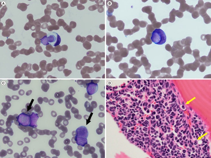
The patient was a 66-year-old man with small-cell carcinoma of the urinary bladder that had been diagnosed nine months previously. He had received five cycles of chemotherapy. He also had been recently diagnosed as having anemia and thrombocytopenia without any clinical symptoms. He was admitted to the hematology department of Dongsan Medical Center in January 2018 for transfusion and further evaluation to determine the cause of cytopenia. His complete blood count showed the following: white blood cells, 5.45×10⁹/L; Hb, 98 g/L; and platelets, 24×109/L. A PB smear showed a few suspicious tumor cells (5% of total nucleated cells) exhibiting cellular cannibalism, which is characterized by a typically large cell with a crescentic nucleus engulfing another smaller cell with a round-to-oval nucleus rimmed by a halo of phagocytic vacuoles (Fig. 1A–C). Some flattening or “molding” of adjacent cells was observed (Fig. 1C), which is a feature of small-cell carcinoma [8]. Subsequent bone marrow (BM) aspiration and biopsy revealed a markedly increased number of suspicious tumor cells exhibiting cellular cannibalism (Fig. 1D). The cells were large with a high nucleus-to-cytoplasm ratio, fine chromatin pattern, and scant cytoplasm. Myeloperoxidase and periodic acid-Schiff staining were negative. Flow cytometric analysis of the suspicious tumor cells in BM aspirate sample showed that the population was negative for CD45, CD34, HLA-DR, TdT, and all lymphoid/myeloid markers. Chromogranin A staining on both PB smear and tissue section confirmed the suspicious tumor cells as small-cell carcinoma.
Twenty-six hours after BM examination, the patient had a sudden-onset mental change. Brain computed tomography revealed intracerebral hemorrhage. The patient could not undergo brain surgery because of the low platelet count, and he died six hours after the mental change.
Small-cell carcinoma of the bladder is generally diagnosed at an advanced stage and is believed to be highly metastatic [9]. By the time our patient was diagnosed as having small-cell carcinoma of the bladder, the cancer had already metastasized to multiple lymph nodes. As cannibalistic cells are related to the aggressiveness, invasiveness, and metastatic disposition of the malignancy [1610], if cellular cannibalism is observed on a PB smear, the patient should be diagnosed precisely and treated intensively. More cases should be evaluated to confirm its precise nature and clinical significance.
 XML Download