

 PDF
PDF ePub
ePub Citation
 Print
Print


Penetrating wounds in the maxillofacial region are rare and poorly reported.12 However, they can result in serious complications that are difficult to resolve and may compromise the patient's quality of life, especially when large blood vessels or other vital structures are involved.234
Injuries to blood vessels may occur at the time of the trauma or during the surgical procedure to remove the penetrating foreign body, which commonly remains entrapped inside the tissue, meaning that the removal process should be planned carefully. Thus, it is essential to determine the extent of the affected blood vessels and the proximity of the retained object to the anatomical structures of the involved region.5
For these purposes, various imaging exams are useful, including computed tomography, X-ray angiography, digital subtraction angiography (DSA), computed tomography angiography (angiotomography), magnetic resonance imaging angiography (angioresonance), and color Doppler ultrasonography.567
This report describes the usefulness of DSA analysis in a case of facial penetrating trauma that involved the superficial and deep anatomical planes.
Case Report
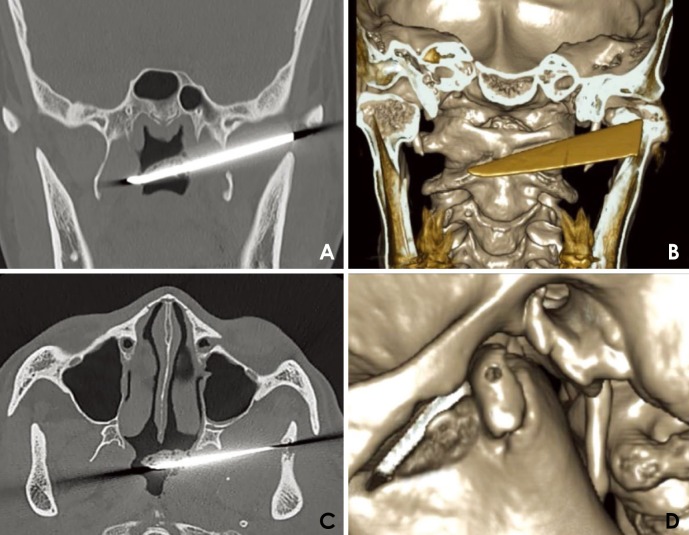
A 47-year-old male patient presented to the Division of Maxillofacial and Traumatology Surgery of the Federal University of Minas Gerais with a history of a knife attack 3 months previously. He complained of pain in the left temporomandibular joint and limited mouth opening. His current medical condition and medical history included rheumatoid arthritis and hepatitis during childhood. Upon physical examination, a scar was observed in the left preauricular region, and significant limitation of the buccal opening was noted, with an interincisal distance of 13 mm. An intraoral evaluation revealed no changes in the hard or soft tissues. Computed tomography revealed the presence of a large knife fragment in the infratemporal fossa. A perforation path was observed, extending from the pre-auricular region to the infratemporal fossa, passing through the zygomatic arch and the sigmoid notch of the mandible until the nasopharynx (Fig. 1). An upper left condylar fracture was also observed (Fig. 1D).
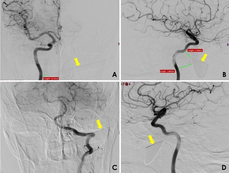
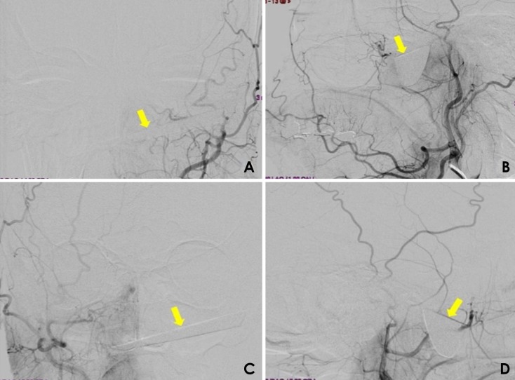
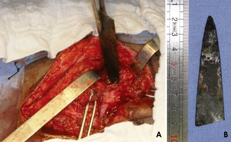
The images suggested moderate proximity of the foreign body to the internal and external carotid arteries; therefore, DSA was requested, since it is selective for the internal and external carotid arteries bilaterally (Figs. 2 and 3). DSA showed no extravasation of contrast, pseudoaneurysm, or arteriovenous fistula, suggesting that the small branches of the left external carotid artery had been transected. DSA also showed that the tip of the knife fragment had an intimate relationship with the right internal carotid artery (1.1 cm×1.9 cm×2.1 cm), but without any signs of extravasation. As a therapeutic plan, it was decided to surgically remove the knife, with conservative treatment of the condylar fracture. The procedure was performed jointly with the Head and Neck Surgery Service of the Federal University of Minas Gerais due the imminent risk of involvement of large vessels during the removal of the impacted blade. Under general anesthesia, extensive left pre-auricular surgical access was obtained, and after careful dissection of the facial tissues, the knife fragment was located and gently removed with the use of a needle holder (Fig. 4).
During the procedure, no intense bleeding was observed, and there was no need for vessel ligation or replacement of blood products. The patient received tetanus antitoxin and prophylactic antibiotics in the perioperative period. In the immediate postoperative period, the patient experienced complete remission of pain symptoms and recovered normal mouth opening. At 3 years after surgery, he is receiving ambulatory follow-up, and has shown a good clinical course.
Discussion
Penetrating injuries in the maxillofacial region are uncommon;8 therefore few cases in the literature have been documented.9 In a review of 37 penetrating injuries involving this region, only 4 were caused by a knife.10
Considering the high vascularization of the face, the successful treatment of penetrating injuries in the maxillofacial region requires accurate localization of the foreign body. Furthermore, determining the extent of the penetrating injury and its relationship with the relevant anatomical structures decreases the likelihood of severe complications, especially hemorrhage and cranial nerve damage. It is possible for these structures to be injured during the facial trauma itself or the removal process, turning it into a critical situation.51011
Therefore, imaging exams play a crucial role in mapping the involved region and preventing certain severe complications. The imaging modalities reported in the literature include computed tomography, conventional angiography, DSA, angiotomography, angioresonance, and color Doppler ultrasound.56712
Carotid angiography is a valuable exam that allows the identification of possible anatomical alterations in the pathway of blood vessel flow in the skull, face, head and neck; however, the injection of radiopaque contrast is necessary. 567
The use of angiography has been questioned in the literature. Some authors have argued that, even though angiography has traditionally been considered the gold standard method for identifying vascular injuries, a small rate of false positive results of this test have been reported in the literature.4567 Therefore, it has been argued that angiography should not automatically be performed in cases in which no evidence of vascular injury is observed on the physical examination and a radiographic examination does not suggest the presence of ongoing bleeding.13
However, other authors have argued that the importance of angiography, as a diagnostic tool in maxillofacial trauma, is underestimated.5 They recommended the selective use of emergency angiography for patients who are victims of penetrating injuries in the maxillofacial region.3 They considered angiography to be mandatory specifically in cases of excessive bleeding, bruising and expansion, or penetrating lacerations closer to large vessels.4
A study showed that angiography was highly reliable for the diagnosis of vascular alterations after penetrating injuries; in 31 patients who underwent angiography, the findings of preoperative angiography coincided with intraoperative findings in 26 cases. In 2 of the remaining patients, angiography underestimated the magnitude of the hemorrhage, and in 3 patients, it did not identify an existing venous lesion. Nonetheless, even in those 5 cases, angiography helped in planning and executing the surgical procedure, meaning that no higher rate of morbidity was associated with the failure of the exam.14
In cases of facial injury where the penetrating object remains in the tissues, angiography lends itself both to the investigation of possible vascular lesions related to the trauma and to establishing the proximity of the object to the larger blood vessels. This information helps prevent hemorrhage during surgical removal of the foreign body and reduces the likelihood of the formation of pseudoaneurysms, which may develop when an impacted object strikes the artery.3511
Angiotomography is a reliable, sensitive, rapid, and non-invasive examination that has wide applications in the assessment of trauma involving vascular and non-vascular structures of the neck, such as the upper digestive tract and cervical spine. According to some authors, the use of this modality led to a significant reduction in negative surgical explorations.1215 However, as well as requiring another imaging examination to be conducted, angiotomography also has certain limitations, such as the possibility of image artifacts when metallic objects are present in the area under investigation.4
The imaging exams requested in this case were computed tomography and digital subtraction angiography of the carotid arteries. The left condylar fracture was demonstrated by computed tomography, while angiography revealed a close relationship between the tip of the knife and the right internal carotid artery, along with no damage of the important great blood vessels of the face and neck. A rupture of some smaller branches of the left external carotid artery was observed, and this rupture most likely occurred at the moment of trauma.
DSA was chosen because the knife could yield metal artifacts in other imaging methods (Figs. 1A and B). Such artifacts would have compromised the quality of the images and the interpretation thereof.
We suggest that selective angiography is routinely indicated in cases of deep lesions caused by a penetrating object that is retained in the maxillofacial region, with or without vascular impairment on the physical examination. Pseudoaneurysms without a clinical manifestation can be detected using this modality, and they must be treated surgically before they rupture. In addition, when removal of the foreign body is indicated, angiography assists in planning the operation by establishing the proximity of the object to blood vessels in the region.
Fortunately, in this case, the knife did not cause important injuries upon tissue penetration, which allowed the surgical treatment to be delayed. However, the blade was fractured, leaving a large fragment lodged in the left infratemporal fossa, which caused pain and restriction of mandibular movement. This concern led us to remove the fragment under general anesthesia, since preservation of a foreign body close to vascular structures could predispose the patient to the subsequent development of pseudoaneurysms.3
Penetrating lesions in the maxillofacial region can vary significantly in terms of the affected structures and severity. Thus, a multidisciplinary therapeutic approach has been recommended by several authors.416 Our case was conducted in partnership with the Service of Vascular Surgery and the Head and Neck Surgery Service, both of the Federal University of Minas Gerais, due to the relatively high likelihood of involvement of large vessels during surgical removal of the impacted blade.
Postoperative clinical follow-up of the patient is essential to manage possible future complications and ensure treatment success.10 At 3 years after surgery, our patient did not present any type of alteration related to the injury that he had suffered.
When the removal of retained penetrating objects in the maxillofacial region is indicated, surgery should be meticulously planned. The use of specific imaging exams, such as carotid angiography by digital subtraction, enables a proper map of the surgical field to be made, reducing the chances of vascular damage during the surgical procedure.
 XML Download