

 PDF
PDF ePub
ePub Citation
 Print
Print


Byung-Yoon Roh1 , Jeong-Uk Seo1, Chang-Gyum Kim1, Chang-Un Choi2, Won-Joon Lee2, Sang-Seob Lee1
, Jeong-Uk Seo1, Chang-Gyum Kim1, Chang-Un Choi2, Won-Joon Lee2, Sang-Seob Lee1
Abstract
There have been many radiographic studies on age estimation that evaluate reduction in size of dental pulp cavity with secondary dentin formation. The Paewinsky method reported high accuracy in estimating ages by measuring the width of the pulp cavity in panoramic radiographs. The aim of this study was to evaluate the application of the Paewinsky method to digital periapical radiographs. This study was conducted on 103 cases that reported to the Section of Human Identification of the National Forensic Service. The age was calculated by applying the Paewinsky method that measures the root and pulp canal width at three points in a tooth. The estimation results were compared with those calculated by the Johanson method. When the Paewinsky models were applied to digital periapical radiographs, the errors were significantly greater as compared to the original study. The errors of the maxillary second premolar and mandibular lateral incisor were greater than those of the maxillary central incisor, lateral incisor, mandibular canine, and first premolar. Furthermore, errors of the age estimation models in level C were greater than those in levels A and B. This study could be a reference for the application of the Paewinsky method to digital periapical radiographs.
Figures and Tables
Fig. 1
Paewinsky method application for measurements in periapical radiographs. A, root and pulp width at the cementoenamel junction (CEJ); B, root and pulp width midway between measurement levels A and C; C, root and pulp width midway between the apex and CEJ.

Fig. 2
Box-plot representing the difference between estimated ages based on the Paewinsky method and Johanson method (maxillary central incisor, lateral incisor, and second premolar). The box-plot shows median and interquartile range, and whiskers indicate the range.
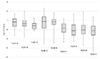
Fig. 3
Box-plot representing the difference between estimated ages based on the Paewinsky method and Johanson method (mandibular lateral incisor, canine, and first premolar).
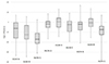
Table 4
Standard errors of estimation and descriptive statistics of errors using the models of Paewinsky et al.'s [12] (maxillary central incisor, lateral incisor, and second premolar)
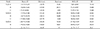
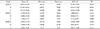
Table 5
Standard errors of estimation and descriptive statistics of errors using the models of Paewinsky et al.'s [12] (mandibular lateral incisor, canine, and first premolar)
References
1. Kwon C, Byun JS, Jung JK, et al. An analysis of age estimation cases in Korea from the view of social aspects. Korean J Oral Med. 2013; 38:235–246.

2. Roh BY, Lee WJ, Seo JU, et al. Analysis of forensic odontological examinations at the National Forensic Service of Korea from 2011 to 2015. Leg Med (Tokyo). 2018; 32:37–42.
3. Mansour H, Fuhrmann A, Paradowski I, et al. The role of forensic medicine and forensic dentistry in estimating the chronological age of living individuals in Hamburg, Germany. Int J Legal Med. 2017; 131:593–601.
4. Schmeling A, Olze A, Reisinger W, et al. Statistical analysis and verification of forensic age estimation of living persons in the Institute of Legal Medicine of the Berlin University Hospital Charite. Leg Med (Tokyo). 2003; 5:Suppl 1. S367–S371.
5. Willems G. A review of the most commonly used dental age estimation techniques. J Forensic Odontostomatol. 2001; 19:9–17.
6. Lee SS, Byun YS, Park MJ, et al. The chronology of second and third molar development in Koreans and its application to forensic age estimation. Int J Legal Med. 2010; 124:659–665.
7. Lee SS, Kim D, Lee S, et al. Validity of Demirjian's and modified Demirjian's methods in age estimation for Korean juveniles and adolescents. Forensic Sci Int. 2011; 211:41–46.
8. Gustafson G, Malmo DO. Age determinations on teeth. J Am Dent Assoc. 1950; 41:45–54.
9. Lamendin H, Baccino E, Humbert JF, et al. A simple technique for age estimation in adult corpses: the two criteria dental method. J Forensic Sci. 1992; 37:1373–1379.
10. Marroquin TY, Karkhanis S, Kvaal SI, et al. Age estimation in adults by dental imaging assessment systematic review. Forensic Sci Int. 2017; 275:203–211.
11. Kvaal SI, Kolltveit KM, Thomsen IO, et al. Age estimation of adults from dental radiographs. Forensic Sci Int. 1995; 74:175–185.
12. Paewinsky E, Pfeiffer H, Brinkmann B. Quantification of secondary dentine formation from orthopantomograms: a contribution to forensic age estimation methods in adults. Int J Legal Med. 2005; 119:27–30.
13. Cameriere R, Ferrante L, Belcastro MG, et al. Age estimation by pulp/tooth ratio in canines by peri-apical X-rays. J Forensic Sci. 2007; 52:166–170.
14. Johanson G. Age determinations from human teeth: a critical evaluation with special consideration of changes after fourteen years of age. Lund: Berlingska Boktryckeriet;1971.
15. Jeon HM, Jang SM, Kim KH, et al. Age estimation based on pulp chamber size of mandibular first molars from intraoral periapical radiographs in Korean. Korean J Leg Med. 2018; 42:56–61.
16. Jeon HM, Jeon JW, Kim SY, et al. An assessment of radiological age estimation method using mandibular first molars in Korean adults. Korean J Leg Med. 2017; 41:7–11.
17. Jeong EG, Heo JY, Ok SM, et al. Drusini's and Takei's methods for age estimation in Korean adults. Korean J Leg Med. 2015; 39:1–5.
18. Roh BY, Lee WJ, Ryu JW, et al. The application of the Kvaal method to estimate the age of live Korean subjects using digital panoramic radiographs. Int J Legal Med. 2018; 132:1161–1166.
19. Erbudak HO, Ozbek M, Uysal S, et al. Application of Kvaal et al.'s age estimation method to panoramic radiographs from Turkish individuals. Forensic Sci Int. 2012; 219:141–146.
20. Meinl A, Tangl S, Pernicka E, et al. On the applicability of secondary dentin formation to radiological age estimation in young adults. J Forensic Sci. 2007; 52:438–441.
21. Sert S, Bayirli GS. Evaluation of the root canal configurations of the mandibular and maxillary permanent teeth by gender in the Turkish population. J Endod. 2004; 30:391–398.
22. Vertucci FJ. Root canal morphology and its relationship to endodontic procedures. Endod Topics. 2005; 10:3–29.
 XML Download