

 PDF
PDF ePub
ePub Citation
 Print
Print


Abstract
Evaluation of diaphragm function is challenging because no single test has a high diagnostic yield. We describe ultrasound findings in three cases with acquired unilateral diaphragmatic elevation. These cases confirm that sonographic evaluation is a valid tool for identifying dia-phragm dysfunction. In addition, ultrasound measurements of diaphragm thickness and the contractility can be used to determine if a diaphragm is paralyzed and suggest the duration of paralysis (i.e., acute or chronic).
References
1. Sarwal A, Walker FO, Cartwright MS. Neuromuscular ultrasound for evaluation of the diaphragm. Muscle Nerve. 2013; 47:319–329.

2. Markand ON, Kincaid JC, Pourmand RA, Moorthy SS, King RD, Mahomed Y, et al. Electrophysiologic evaluation of diaphragm by transcutaneous phrenic nerve stimulation. Neurology. 1984; 34:604–614.
3. Seok JI, Kim SY, Walker FO, Kwak SG, Kwon DH. Ultrasonographic findings of the normal diaphragm: thickness and contractility. Ann Clin Neurophysiol. 2017; 19:131–135.
4. Baldwin CE, Paratz JD, Bersten AD. Diaphragm and peripheral muscle thickness on ultrasound: intra-rater reliability and variability of a methodology using non-standard recumbent positions. Respirology. 2011; 16:1136–1143.
5. Boon AJ, Harper CJ, Ghahfarokhi LS, Strommen JA, Watson JC, Sorenson EJ. Two-dimensional ultrasound imaging of the diaphragm: quantitative values in normal subjects. Muscle Nerve. 2013; 47:884–889.
6. Gottesman E, McCool FD. Ultrasound evaluation of the paralyzed diaphragm. Am J Respir Crit Care Med. 1997; 155:15701574.
7. Summerhill EM, El-Sameed YA, Glidden TJ, McCool FD. Monitoring recovery from diaphragm paralysis with ultrasound. Chest. 2008; 133:737743.
Fig. 1.
Ultrasound of the right (A and C) and left (B and D) diaphragm of patient 2. The distance between the two markings (+) is the diaphragm thickness. (A, B) At the end of quiet expiration, the diaphragm is seen as a hypoechoic structure between two layers of hyperechoic fascia. (C) At maximal inspiration, the right diaphragm is seen ‘peeling away' from the chest wall and thickens markedly. (D) At maximal inspiration, the left diaphragm slightly thickens. D, diaphragm; IC, intercostal muscle.
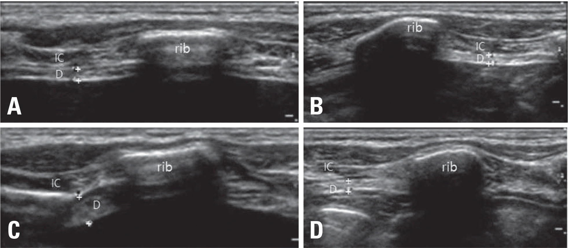
Table 1.
Clinical characteristics, NCS findings, and ultrasound findings of the three patients
| Patient 1 | Patient 2 | Patient 3 | ||||
|---|---|---|---|---|---|---|
| Age | 58 | 71 | 58 | |||
| Sex | Male | Male | Female | |||
| Symptom | None | None | None | |||
| Duration | About 10 years | 1–4 months | 2–10 months | |||
| Chest X-ray | ||||||
| Elevation side | Left | Left | Right | |||
| Diaphragm ultrasound | Right | Left | Right | Left | Right | Left |
| Resting thickness (cm) | 0.328 | 0.183 | 0.203 | 0.157 | 0.171 | 0.184 |
| Side-to-side ratioa | 0.56d | 0.77 | 0.93 | |||
| Maximal thickness (cm) | 0.427 | 0.19 | 0.58 | 0.177 | 0.178 | 0.57 |
| DTFb (%) | 30 | 4d | 186 | 13d | 4d | 210 |
| NCSc | Right | Left | Right | Left | Right | Left |
| Latency (ms) | 12.6d | 11.7d | 8.6 | 14.2d | 8.65d | 6.98 |
| CMAP amplitude (mV) | 0.1d | 0.1d | 0.1d | 0.1d | 0.4 | 1.2 |
 XML Download