

 PDF
PDF ePub
ePub Citation
 Print
Print


INTRODUCTION
Diarrheal disease continues to be an important cause of morbidity and mortality among young children in developing countries [1]. It is characterized by diarrhea, vomiting and abdominal pain that may lead to hypovolemic shock and dehydration, and subsequently death in severe cases [1]. Worldwide, gastroenteritis affects 3 to 5 billion children each year, and accounts for 1.5 to 2.5 million deaths per year or 12% of all deaths among children less than 5 years of age [2]. In developed countries acute gastroenteritis (AGE) accounts for 300 deaths per year [2].
Globally, rotavirus (RV) is the most common etiological agent for AGE [3]. Data from the coordinated global network for RV surveillance of the World Health Organization at 185 sentinel sites in 64 countries showed RV detection rates of 37-53% in children hospitalized with diarrhea in 2011 in regions of the world where vaccination has not been widely implemented [4] and RV is one of the most common causes of moderate to severe diarrhea during the first 2 years of life [5]. RV vaccination has been widely available for children since 2006 [6] and is now recommended worldwide, a growing body of evidence supports the benefits of its vaccination in the developed and developing world [7].
The protozoan intestinal parasite Entamoeba histolytica is the agent of human amoebiasis. Infection by this parasite is a major cause of morbidity and mortality; it is also responsible for 50 million cases of invasive disease [8] and about 70,000 deaths annually in the world [9]. Both E. histolytica and Giardia lamblia have been listed by the National Institute of Health as category B priority biodefense pathogens due to their low infectious doses and potential for dissemination through compromised food and water supplies in the United States [10]. In developing countries, parasitic diseases represent a social and economic problem with more severe disease associated with young age, malnutrition, and immunosuppression [11].
Despite the heavy burden of AGE especially in children below five years of age, improved prevention is achievable. Personal and food hygiene, including the use of clean water sources, are key measures to prevent transmission of these diseases [12]. Breastfeeding, especially under 6 months of age, also effectively protects infants [12].
A previous recent study in Lebanon showed an increasing prevalence of amoebiasis as the leading cause of hospitalized children for AGE in comparison to others Enteropathogens with data representing mainly the population of Beirut [13]. Our SAGE study prospectively enrolled and tested children admitted for AGE to our hospital, Ragheb Harb Hospital located in Nabatieh, south Lebanon, serving a population of 350,000 habitants and about 35,000 Syrian refugees, during the summer period of 2014, to determine the prevalence and etiology of infectious gastroenteritis by using the routine available tests performed in our hospital laboratory. To our knowledge, few studies have been performed before and reported information regarding the percentage, the age distribution, the risk factors and the protective factors among hospitalized children with AGE in Southern's Lebanon.
MATERIALS AND METHODS
Patients
During the summer of 2014, from July to mid of September, children were admitted to the pediatric department of a tertiary care private hospital located in Toul, Nabatieh district, Lebanon. Children aged from 1 month to 10 years old were prospectively included in the study if the admission diagnosis was either: AGE or diarrhea that was defined as the occurrence of three or more of watery stool of any nature during the last twenty-four hours and in the absence of fever, vomiting, nausea, abdominal cramps or macroscopic blood and mucus in the stool. We excluded patients with chronic diarrhea, malnutrition, immunodeficiency or patients with multiple malformation.
A trained healthcare person followed each case from the admission until discharge and collected data prospectively including: 1) Demographics: age, sex, residency area, family size, nutrition, breast feeding and the vaccination history to determine if any dose of the two RV vaccines (Rotarix from GlaxoSmithKline Biologicals, Rixensart, Belgium; or Rotateq from Merck Sharp & Dohme Corp, Whitestation, NJ, USA) that are available and approved in Lebanon, was given. 2) Clinical data: the presence of fever, vomiting, diarrhea, associated co-infections, urine output, and the presence of blood or mucus in stool. 3) Laboratory tests values: the results of stool analysis, microscopy for ova and parasites, the quick identification tests for adenovirus (CerTest; Biotec, Zaragoza, Spain) and RV (CerTest) and if available the results of stool culture, leukocytes counts, C-reactive protein (CRP) level, electrolytes, blood sugar and transaminases level. 4) Also therapeutic interventions as intravenous bolus requirement, antibiotics use, antiemetics and different antidiarrheic agents used during the whole hospitalization length.
The study was approved by the ethical and scientific committee of the Ragheb Harb Hospital (ethical approval reference number 14/2014, date: 05/06/2014).
Laboratory methods and studies
Fresh stool samples were used to detect the probable infectious agents. Stool samples were analyzed within less than one hour after being sent to our laboratory, analysis included specific preparation and direct observation under light microscopy for white blood cells, red blood cells, ova and parasites using the trichrome stain method.
Stool cultures were not performed unless requested by medical order, by the direct methods using the solid media plates ethyl methyl blue agar and the indirect culture where it is incubated in broth selenite medium agar for 24 hours at 37℃ then it was sub-cultured on Salmonella-Shigella agar for 48 hours, if positive results, colonies are identified based on morphologic characteristics followed by the API20E biochemical tests (BioMérieux, Paris, France) identification if there is bacterial growth.
For viral detection, we used the rotavirus and adenovirus kit tests (CerTest) which provides a single, rapid and highly sensitive (Sensitivity >99% and Specificity >98%) screening assay to make the diagnosis of RV infection or adenovirus infection by a qualitative immuno-chromatographic assay method. Manufacturer instructions were followed to identify positive results which will be available in about 10 minutes. In these tests, the membrane in the test band was first coated with mouse monoclonal antibodies against viral antigens. During the test, a pre-colored conjugate that had previously been dried was reacted with the sample. Afterwards, the mixture moved forward on the membrane by means of capillary action. The colored particles were replaced as the sample moved along the test membrane. If the result was positive, the specific antibodies on the membrane captured the colored particles.
Statistical analysis
Statistical analyses were conducted using IBM SPSS Statistics ver. 20.0 (IBM Co., Armonk, NY, USA). The level of significance was set at p<0.05 for all statistical analyses. Descriptive analyses were based on frequencies and percentages. The demographic and the clinical characteristics among the five studied groups (E. histolytica group I, RV group II, adenovirus group III, mixed group IV, unidentified group V) were tabulated. Baseline comparisons between groups were performed using Kruskall-Wallis test for continuous variables. The chi-square test was used to assess any significant difference between the categorical variables. Logistic regression was used to determine the associations between the infectious agents (yes/no) as independent variables and breast-feed and rota-vaccine as dependent variables. Four separate models were performed for each of the following infectious agents as dependent measures (unidentified pathogens, RV, E. histolytica, and adenovirus).
RESULTS
From July 2014 to mid-September 2014, we had 222 cases of AGE admitted to the pediatric wards at the Ragheb Harb Hospital, which represented 40.4% (222/549) of total admissions in this period. We enrolled 89.2% (198 of 222 cases) of all gastroenteritis cases admitted at that time, a number that reflects the high burden of AGE on the hospital occupancy rate and on the general health system overall.
Demographic characteristics among the five studied groups
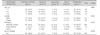
Of the one hundred ninety eight pediatric patients hospitalized for AGE, we identified the causative pathogen in 57.6% of cases (114/198), and these pathogens were subsequently divided into five main groups: Group I (E. histolytica group, n=52), group II (RV group, n=37), group III (adenovirus group, n=12), group IV (mixed group, n=13), group V (unidentified group, n=84) (Table 1).
The distribution of age reveals that 92% of our patients are under 5 years with a range of age from 1 month up to 10 years old and an age average of 2 years and 5 months overall. The percentage of cases under 1 year of age was significantly higher in unidentified group than in other 4 group (p=0.001). However, when considering the frequency of E. histolytica among different age groups, it shows that E. histolytica was more frequent more than 1 year of age. In contrast, RV was more frequent below 5 years of age.
Concerning the sex distribution of our patients, 57.1% were male gender and 42.9% were female. Also to be noted that the percentage of exclusive breast feeding for more than 1 month duration was 40% in comparison to 33% for the partial breast feeding of more than 1 month duration (Table 1).
Clinical characteristics among the five studied groups
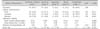
The most common presenting symptoms of patients admitted were diarrhea which was present in 93.4% followed by fever in 76.8% then vomiting in 71.2% of cases admitted with AGE; we only included these three objectives symptoms for clinical assessment (Table 2). High grade fever was more frequent in E. histolytica and unidentified groups.
Laboratory findings of all hospitalized AGE cases were also studied among the five main groups. Laboratory findings identified significantly higher mean CRP in E. histolytica group than the other four groups (p=0.0001). Also, there was a significant difference in mean leukocytosis among the five groups (p=0.024). However, there was no significant difference in the average of age and the hospital stay for the total number of patients among the five studied groups.
It is important to note that we found a significant difference between the age of patients admitted for RV gastroenteritis in vaccinated and non-vaccinated group (p=0.049), meaning that vaccinated patients has a protective effects of five months more than non-vaccinated patients (Data not shown here) (Table 2).
Effect of breast-feeding and RV vaccination on different pathogens
We analyzed the protective factors from RV AGE; precisely we determined the percentage of RV cases of AGE whom had received any dose of any RV vaccines and we found the percentage to be only 18.9%.
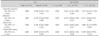
Results showed that in general, breast-fed children were less prone to unknown-pathogens, RV (p=0.041), E. histolytica and adenovirus. On the other hand, children who had received the rota-vaccine were more prone to unidentified pathogens (p=0.015) and E. histolytica but were less prone to RV (p=0.032) and adenovirus (Table 3).
Pathogens predisposition according to age and sex
Analysis of patient demographics identified that, males are more prone to unidentified pathogens (p=0.030), RV (p=0.042), E. histolytica and adenovirus than female (Table 4). Concerning age, E. histolytica infections are more prevalent in patients older than one year.
DISCUSSION
Diarrheal diseases are a major public health problem for children in Lebanon, as in other developing countries. Knowledge of the enteropathogens responsible for diarrheal illnesses is essential for implementation of appropriate public and hospital health measures to control these diseases [14]. This single center study was conducted in the summer period of the year 2014 during the peak season of diarrheal diseases. It is one of the few studies conducted in Lebanon to determine the enteropathogens implicated in the pathogenesis of acute diarrheal diseases in hospitalized children using the common laboratory methods [13]. In this study we enrolled 198 pediatric cases with AGE representing 89% of all children admitted to our hospital with AGE for the entire period of the study that constituted about 40% of the total pediatric admissions.
In our study we identified the enteropathogens in about 58% of AGE cases by using the routine laboratory methods which is slightly below the 66%, the number identified by Youssef et al. [14] in Jordan in 2000, but they were using a combination of traditional and molecular diagnostic techniques. Of the 58% of detected pathogens, 51% (101 of 198) were single pathogens cases of AGE and 7% (13 of 198) were due to mixed pathogens, which is a little lower than the number of 9.8% of co-infection reported in a Bulgarian study in 2015 [15].
Our study showed that the percentage of E. histolytica AGE was 26.3% (52 of 198) which was in correlation with the previous reported prevalence of 22% reported at the Makassed General Hospital in Beirut in 2013 [13] and very similar to the reported prevalence of 25.9% reported also in Tajikistan by in 2011 [16]. Moreover, the age distribution of amoebiasis was the following: 34% (18/52) were below one year old and constituted 20% as shown in Table 1, which is an unusual presentation in this age group because E. histolytica is usually transmitted via fecal oral route with contaminated food and water, so young infants are less likely to develop intestinal amebiasis very often [17]. That means that the drinking water used for milk preparation and the tap water used for daily home and body hygiene was contaminated and could be orally aspirated or ingested during bathing or face washing especially when parents were holding their baby in face down position, a procedure that could be easily adjusted by preventing the water coming into the mouth of the baby or by using clean water for daily facial wash; to be noted that the prevalence of E. histolytica in stool was increasing with age especially after the age of five years.
The second major enteropathogen found was RV, causing 19 % of AGE admissions to our hospital as a single pathogen, which was slightly lower than the previous reported results in 2013 in Lebanon [18]. Many countries in the world have reported that RV is a leading cause of pediatric AGE, accounting for 27-51% of all diarrhea cases in children less than 5 years of age [19].
As found in different studies from the Eastern Mediterranean region, RV AGE disease burden in our study was highest in children less than 5 years of age 92% of our reported cases [20]. The prevalence of RV infection during the summer months was found in our study was an interesting observation; In general, RVs causes diarrheal illnesses around the year, but predominantly during the winter months in countries with temperate climates. Temperature and relatively humid weather conditions have been proposed to contribute to increased RV infection. However, a few of the virus characteristics such as environmental stability, heat resistance and easy transmission by the fecal-oral route foster the spread of infections also during summer season. Diarrheal viruses such as RV, spread mainly through contact with infected persons, contaminated environmental surfaces or via ingestion of contaminated food and water. The emergence of a new virus variant in the susceptible child population may potentially be factors that favor the spread of virus gastroenteritis agents during the summer months [15].
Adenoviruses are ubiquitous agents that infect people of all ages, we identified the presence of adenovirus as a single agent in 6% of cases, this percentage will be doubled if we take into account both single and mixed infections, Adenoviruses have long been suspected to cause diarrhea and there is a long term asymptomatic shedding of certain serotypes from the stools after adenovirus respiratory infection, the group F adenoviruses (serotypes 40 and 41) are strongly associated with diarrheal disease in children; diarrhea induced by adenovirus type 41 may be more protracted than disease caused by adenovirus type 40 [21].
In our study, we have 42.4% of cases with unidentified causes or pathogens, a remarkable percentage that is when added to the negative stool cultures performed for only 17 cases, we could initiate further needed steps to improve our ability for more pathogens identification by increasing the number of cultures performed for hospitalized children with suspected invasive AGE in order to cultivate more pathogenic bacteria.
Concerning the clinical manifestations, we studied three clinical parameters among them: fever in 76.8%, vomiting in 71.2% and diarrhea in 93.4% of patients as the most common presenting symptoms in our study population which are not informative as it will be if we considered the determination of Vesikari score, used for severity estimation of AGE. We noted that in both the amebiasis and the unidentified pathogens group, there was an elevated CRP in comparison to viral groups which means that in the unidentified pathogens group the possibility an invasive infections is highly suggestive.
The average length of hospital stay (LOS) was 3.5 days and it was quite similar for the four groups and when compared with the two RV group based on either vaccinated and not vaccinated patients, the LOS did not differ between them in accordance to previous results reported in a Lebanese study in 2016 [22].
Limitation
The SAGE study was conducted in our hospital (Ragheb Harb Hospital, Nabatieh [South Lebanon]) was among the few prospective studies in Lebanon that studied the etio-pathogenesis of AGE in children, it has some limitations that include:
It was of short duration representing the summer months only, but a fair number of patients 198 for such period.
It was run in a single center so may not reflect the whole regional of country data and included only Lebanese population.
The diagnostic tests were limited to the routine ones which are dependent on personnel experience of our lab workers.
Most of the time, the stool culture was not requested routinely on admission.
Another point to emphasis is the importance to consider the Vesikari score for AGE severity in any future study.
We found that amebiasis was the major cause of invasive entero-colitis in hospitalized children. RV AGE ranks second in the list of enteropathogens, with a protective effect of vaccination on the rate of hospitalization. We have an important group of unidentified pathogens that should solicit us to expand our diagnostic arsenal.
The vaccination against RV infection as it is well known to be the most protective act against RV GE, it should be promoted in the public and private health sectors as early as possible. Providing a clean, uncontaminated water supply and food is a national paramount for a healthier life of our children.

 XML Download