

 PDF
PDF ePub
ePub Citation
 Print
Print


Dear Editor:
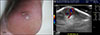
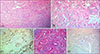
A 33-year-old male visited our clinic for painful mass on the heel of his right foot. The patient did not know when occurred. A slight blue to skin-colored nodule about 1×1 cm in size was found on his right heel (Fig. 1A). Ultrasonography was performed and a well-demarcated oval hyperechoic mass in the subcutaneous fat layer, with prominent vascularity was indicated (Fig. 1B). A vascular leiomyoma, hemangioma, glomus tumor or complicated epidermoid cyst was suspected. The tumor was completely resected and the microscopic examination exposed numerous tortuous vascular channels with proliferation of spindle-shaped cells displaying an interlacing band-like pattern. Elongated spindle cells with abundant brightly eosinophilic cytoplasm were observed without necrosis, pleomorphism, mitosis or nuclear atypia (Fig. 2A; H&E, ×40). With these results, fibrous components with edematous stroma were seen (Fig. 2B; H&E, ×200). Immunohistochemistry revealed the spindle-shaped tumor cells were diffusely positive with desmine (Fig. 2C, ×100). Tissue were stained with Masson's trichrome (Fig. 2D, ×100). S-100 stain was negative (Fig. 2E, ×100). The final diagnosis appeared to be vascular leiomyoma.
Leiomyoma is a benign smooth muscle tumor which usually occurs in the extra-skeletal area, such as ovaries, uterus, bladder, lung and gastrointestinal tract1. Occasionally skin and subcutaneous soft tissues are involved2. Enzinger and Weiss explained that leiomyomas can be classified into three types; vascular, cutaneous and deep soft tissue3. Vascular leiomyomas, featured by a painful solitary tumor occurring most frequently in the extremities, originate from the tunica media layer of a vein23. Pain is accompanied in approximately 60% of patients whether there is tenderness4. It typically affects middle aged females in the third and fourth decades but may occur at any age124. It usually presents as a solitary mass. Its differential diagnosis includes tender tumors such as neuroma, neurilemmoma, and eccrine spiradenoma because pain is the most characteristic subjective symptom. Etiology is still unknown. Morimoto suggested that pain may be caused by the contraction of vessels which lead to local ischemia5. X-ray findings are mostly normal, however, rarely, dystrophic calcifications might be seen3. The essential histologic features include bundles and masses of smooth muscle fibers, irregularly separated by strands of collagen fibers1. A variety of immunohistochemical stainings such as desmin, vimentin, S-100 protein, neuron specific enolase, actin and factor VIII-related protein could be performed for differentiation or an indication of muscle origin and endothelical cell13. The most satisfactory treatment is complete excision1. In the Korean dermatology literature, there have been several reported cases on lower extremities, cheek, lip and multiple locations involving shoulders, upper chest, upper back, arm, and neck1. Few studies analyzing vascular leiomyoma have been conducted in Korea1. Little has been reported on the heel in young adult male patient, just like our case. Vascular leiomyomas are rarely diagnosed clinically before biopsy, but can occur anywhere in various ages. Although vascular leiomyoma is an infrequent soft tissue tumor, it is worth consideration as the differential diagnosis of nodule with pain on the heel.
 XML Download