

 PDF
PDF ePub
ePub Citation
 Print
Print


Abstract
Objective
Neuralgic amyotrophy (NA) is a distinct clinical syndrome of unknown etiology involving the brachial plexus, which is characterized by the acute onset of shoulder and arm pain followed by weakness, and sensory loss. Diagnosis with neurophysiologic studies and a conventional MRI of brachial plexus is very difficult in acute stage. The magnetic resonance neurography (MRN) is considered to be more sensitive than MRI for the peripheral neuropathies. The objective of this study is to describe the MRN findings and its usability for patients with neuralgic amyotrophy in acute stage.
Methods
The authors have treated 10 patients with NA between 2006 and 2009. All the patients had clinical and neurophysiologic evidence of acute brachial plexopathy without a definable cause. Recently, three patients were evaluated in acute stage using a MRN with a 1.5-T scanner and had positive findings for NA. Imaging sequences for brachial plexus included axial, sagittal, and coronal conventional spin echo sequences, and gadolinium was administered for axial and coronal images, employing short tau inversion recovery sequences.
Results
All patients were checked the brachial plexus MRN within 3 weeks after onset. In conventional MRI, the authors did not find any evidences of plexopathy, mass lesion, muscle atrophy or rotator cuff tear. However, brachial plexus MRN of all the patients showed thickened and hyperintense trunks of the brachial plexus in lesion site consistent with plexitis.
Figures and Tables
FIGURE 1
These pictures of No. 1 patient show weakness of deltoid and biceps in the right side and normal motor function of palmar interossei.
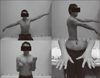
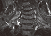
FIGURE 2
Short tau inversion recovery image of the magnetic resonance neurography of patient No. 3 shows a diffuse thickening and increased signal intensity in the right lower trunk of the brachial plexus (white arrow).
References
1. Aagaard BD, Maravilla KR, Kliot M. MR neurography. MR imaging of peripheral nerves. Magn Reson Imaging Clin N Am. 1998; 6:179–194.
2. Dill-Macky MJ, Song S, Silbert PL. Magnetic resonance imaging features of subacute idiopathic brachial neuritis. Australas Radiol. 2000; 44:98–100.

3. Du R, Auguste KI, Chin CT, Engstrom JW, Weinstein PR. Magnetic resonance neurography for the evaluation of peripheral nerve, brachial plexus, and nerve root disorders. J Neurosurg. 2010; 112:362–371.
4. Duman I, Guvenc I, Kalyon TA. Neuralgic amyotrophy, diagnosed with magnetic resonance neurography in acute stage: a case report and review of the literature. Neurologist. 2007; 13:219–221.
5. Favero KJ, Hawkins RH, Jones MW. Neuralgic amyotrophy. J Bone Joint Surg Br. 1987; 69:195–198.
6. Flaggman PD, Kelly JJ Jr. Brachial plexus neuropathy. An electrophysiologic evaluation. Arch Neurol. 1980; 37:160–164.
7. Helms CA, Martinez S, Speer KP. Acute brachial neuritis (Parsonage-Turner syndrome): MR imaging appearance--report of three cases. Radiology. 1998; 207:255–259.
8. Mullins GM, O'Sullivan SS, Neligan A, Daly S, Galvin RJ, Sweeney BJ, et al. Non-traumatic brachial plexopathies, clinical, radiological and neurophysiological findings from a tertiary centre. Clin Neurol Neurosurg. 2007; 109:661–666.
9. Parsonage MJ, Turner JW. Neuralgic amyotrophy; the shoulder-girdle syndrome. Lancet. 1948; 1:973–978.
10. Patel M, Mahajan A, Desai S. Neuralgic amyotrophy: a long term follow-up of four cases. J Postgrad Med. 1990; 36:112–114.
11. Qayyum A, MacVicar AD, Padhani AR, Revell P, Husband JE. Symptomatic brachial plexopathy following treatment for breast cancer: utility of MR imaging with surface-coil techniques. Radiology. 2000; 214:837–842.
12. Rix GD, Rothman EH, Robinson AW. Idiopathic neuralgic amyotrophy: an illustrative case report. J Manipulative Physiol Ther. 2006; 29:52–59.
13. Rubin DI. Neuralgic amyotrophy: clinical features and diagnostic evaluation. Neurologist. 2001; 7:350–356.
14. Sarikaya S, Sumer M, Ozdolap S, Erdem CZ. Magnetic resonance neurography diagnosed brachial plexitis: a case report. Arch Phys Med Rehabil. 2005; 86:1058–1059.
15. Scalf RE, Wenger DE, Frick MA, Mandrekar JN, Adkins MC. MRI findings of 26 patients with Parsonage-Turner syndrome. AJR Am J Roentgenol. 2007; 189:W39–W44.
16. Tsairis P, Dyck PJ, Mulder DW. Natural history of brachial plexus neuropathy. Report on 99 patients. Arch Neurol. 1972; 27:109–117.
17. van Alfen N. The neuralgic amyotrophy consultation. J Neurol. 2007; 254:695–704.
18. van Alfen N, van Engelen BG. The clinical spectrum of neuralgic amyotrophy in 246 cases. Brain. 2006; 129:438–450.
19. Vargas MI, Viallon M, Nguyen D, Beaulieu JY, Delavelle J, Becker M. New approaches in imaging of the brachial plexus. Eur J Radiol. 2010; 74:403–410.
 XML Download