

 PDF
PDF ePub
ePub Citation
 Print
Print


Dear Editor:
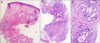
A 51-year-old woman presented with nodule on the nose detected 2 years prior. Physical examination revealed a solitary, 1-cm-sized, soft, skin-colored nodule on the nose with a central erythematous pore (Fig. 1). There was no specific medical or trauma history. Under the presumed diagnosis of epidermal cyst, a 3-mm punch biopsy was done. The histological findings revealed unremarkable findings in the epidermis and upper and mid dermis (Fig. 2A). Separate tissue from deep dermis revealed multiple cholesterol clefts surrounded by lymphohistiocytes, giant cells and fibrosis (Fig. 2B, C). Based on these clinical and histological findings, the lesion was diagnosed as a cholesterol granuloma. After diagnosis, laboratory examination was done. Fasting serum glucose, cholesterol and triglycerides were within normal limits. She denied any family history of hypercholesterolemia. There were no specific symptoms or complications, and the size of the lesion decreased after biopsy. We recommend regular follow-up without excision.
Since it has been reported rarely, cholesterol granuloma presenting as a cutaneous nodule has been described in various ways, including as a subcutaneous cholesterol granuloma, subcutaneous cholesterol nodule, subcutaneous cholesterol crystal (deposition), and cholesterol tophus. It does not always occur in the joint area. Therefore, the term tophus or crystal is considered to inappropriate. Eleven previous cases of subcutaneous cholesterol granuloma were reported1. Some cases presented with multiple lesions and had concomitant dyslipidemia, preceding skin disease, or rheumatological disease. Our case is unique in that cholesterol granuloma located in not subcutaneous area, but dermis without preceding skin disease. The pathogenesis of cholesterol cleft deposition is not well known, and local microtrauma is considered the primary cause2. In our case, the central pore of the lesion suggested the possibility of unrecognized trauma. Cholesterol cleft and surrounding granulomatous reaction are the most characteristic histological findings of cholesterol granuloma. The ghost-like, biconvex, and needle-like shape of the cholesterol cleft on pathology was due to tissue processing for pathologic diagnosis, which includes the dissolution of lipids3. Cholesterol clefts could be seen in skin biopsy specimens of xanthoma, cutaneous cholesterol embolization, necrobiosis lipoidica diabeticorum, and necrobiotic xanthogranuloma. Rarely, cholesterol clefts have also been observed in some cutaneous tumors such as epidermal cysts, trichilemmal cysts, pilomatricomas and basal cell carcinomas34. In cutaneous cholesterol embolization, livedo reticularis is the most common clinical finding, and histopathological findings of cholesterol crystals within the lumen of small arteries in the deep dermis are distinct. Foam cells and Touton giant cells are characteristic of xanthoma and necrobiotic xanthogranuloma. Distinctive histopathological findings of necrobiosis lipoidica diabeticorum and necrobiotic xanthogranuloma include hyaline necrobiosis, foreign body giant cells and lymphoid nodules5. Cholesterol granulomas do not routinely require excision, however excision may be considered based on the size, location, symptoms and the need for diagnostic confirmation. Dermatologists should consider a diagnosis of cholesterol granuloma in patients with cutaneous nodules with histopathological findings of cholesterol cleft and granulomatous reaction.

 XML Download