

 PDF
PDF ePub
ePub Citation
 Print
Print


INTRODUCTION
Phytophotodermatitis is a phototoxic reaction which is caused by contact with photosensitizing substances (e.g., psoralens, furocoumarins) present in plants and subsequent exposure to sunlight. Among many plants that can cause phytophotodermatitis, celery is known as the most common causative plant followed by citrus fruits, such as lime, lemon and grapefruit.
Here, we present two cases of phytophotodermatitis presenting with asymptomatic hyperpigmentation without preceding inflammation on the hand after contact with citrus fruits and subsequent sun exposure.
CASE REPORT
Case 1
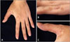
A 30-year-old female patient presented with sudden appearance of brown colored patches on her left hand (Fig. 1). They were partly reticulated, but most of the pigmentation showed irregular shaped, homogeneous, brown colored patches with sharp borders. They were located mostly on the lateral sides of the fingers and the interdigital areas. She reported that the lesion appeared abruptly without any preceding changes such as erythema or erosion. She was right-handed, and didn't recall any history of trauma or irritation. There were no symptoms.
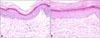
In history taking, she returned from Thailand a week before the skin lesions appeared. During her trip, she attended a local cooking class in which she squeezed limes with her bare hands. She also spent a lot of time on a beach. There was no history of a similar skin rash in the past and she was otherwise systemically well. She had no familial history of similar skin lesion. Three millimeters punch biopsy was performed, and histopathologic examination revealed orthohyperkeratosis and slight increase in the number of basal melanocytes (Fig. 3A). On follow-up, the lesions resolved spontaneously without any treatment. There was no recurrence at 5-months' follow-up.
Case 2
A 36-year-old female patient presented with sudden development of multiple homogeneous brown colored patches with sharp and straight borders on the dorsum of left hand (Fig. 2). They were mostly located on the interdigital areas. The patient was right-handed, and like the patient in case 1, the pigmentation appeared suddenly and spontaneously without preceding trauma or irritation to the best of her knowledge. She didn't complain of any symptom.
A week before development of the lesions, she returned from her trip to California where she spent a lot of time outdoors and was exposed to sunlight. And during her trip, she used to peel fruits such as oranges and grapefruits with her bare hands. There was no history of similar skin lesions in the past and she was otherwise systemically well. There was no familial history of similar skin lesions. Three millimeters punch biopsy was performed, and histopathologic examination revealed orthohyperkeratosis, a slight increase in the basal pigmentation (Fig. 3B). Her skin lesions resolved spontaneously without any treatment.
DISCUSSION
Phytophotodermatitis occurs by contact with plants containing furocoumarins or psoralens that induce phototoxicity when activated by sunlight, particularly ultraviolet A (UVA) radiation (320 nm to 400 nm)1. Typical presentation of phytophotodermatitis starts with erythematous swelling at the site of contact and exposure, and vesicles and large blisters can also be present with itching and burning sensation. It usually leaves brown pigmentation on inflamed areas but the spontaneous disappearance of the pigmentation could be a diagnostic clue of phytophotodermatitis2.
However, pigmentation without preceding erythema or blistering could be the only clinical finding of phytophotodermatitis, depending on the amount of contacting substance, skin color of the affected patient and intensity of photo-exposure2. Lime is the most well-known culprit for asymptomatic hyperpigmentation as clinical feature of phytophotodermatitis. There have been several reports in which waiters who prepare cocktails with lime, such as mojito, developed spontaneous, usually asymptomatic pigmentation on their dorsum of hands. Galvañ-Pérez Del Pulgar et al.3 proposed a new term “dorsal acropigmentation secondary to mojito preparation” to define a variant of occupational phytophotodermatitis. In most of these asymptomatic pigmentation induced by contact with lime, an interval of 7 to 14 days between exposure and onset of lesion was a common finding.
Two patients in this report shared several clinical features. Both developed spontaneous pigmentation on the hands, usually the most contact-prone body part. The pigmentations were of homogeneous brown color with distinct borders. Most importantly, they travelled to countries with strong sunlight about a week prior to development of hyperpigmentation and they both had histories of contacts with citrus fruits with their hands. Lastly, hyperpigmentation faded away without any treatment.
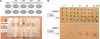
To prove whether lime can induce photodermatitis and hyperpigmentation after UV irradiation, we designed a simple provocation test. We first applied lime extract in finn chambers on the back of a healthy adult male volunteer for 2 hours and then various doses of UVA (5, 10, 20, and 30 J/cm2) were irradiated on lime-applied areas as well as untreated areas as seen Fig. 4A. Three days after UVA irradiation, mild erythema developed only on the lime extract-applied skin following UVA irradiation with energy level of 10 J/cm2 or greater. Six days after UVA irradiation, erythema subsided leaving homogeneous brown pigmentation (Fig. 4B). One month after the UVA irradiation, the pigmentation was gone without any treatment.
Unlike typical phytophotodermatitis which can develop as fast as a few hours after contact with photosensitizing substance( s), phytophotodermatitis presenting only as pigmentation have a window of 1 to 2 weeks between exposure and development of pigmentation. Therefore, it could be difficult to connect those two seemingly unrelated events. Since they are not caused by immune reaction, previous sensitization is not required and therefore anyone can be affected. In this report, hyperpigmentation has been developed in both patients, who had history of traveling to the countries where sunlight is generally stronger than Korea. According to previous reports, California, where the patient of case 2 traveled, was an endemic area of phytophotodermatitis4. In the report, most of 10 children with phytophotodermatitis had histories of contact with citrus fruits and exposure of sunlight, and 4 out of 10 children presented only pigmentation without preceding signs of inflammation.
As to the reason why the lesion developed only on the one side, we suggest there could have been difference in the amount of exposure to the causative agent(s) between both hands. Since the development of phytophotodermatitis is related with the amount and concentration of photosensitizer and/or the energy level of the light, we speculate that uneven contact with photosensitizer(s), or the effect of protection from sunlight such as clothing could have been attributed to the unilateral development of phytophotodermatitis. The same cause can be applied to why the skin lesions developed on their non-dominant hand. Since phytophotodermatitis develops on the area where the required conditions are met, there might be a slightly higher change of developing phytophotodermatitis on the patient's dominant hand, but it is not always case since opportunistic contact with photosensitizer and subsequent sun exposure can happen at any body site.
Therefore, dermatologists should consider a possibility of phytophotodermatitis in patients who have abrupt asymptomatic hyperpigmentation on exposed areas if they have history of traveling to countries with strong sunlight, or spending a lot of time outdoors together with contact with citrus fruits within 1 or 2 weeks prior to development of pigmentation. In addition, as hyperpigmentation as a clinical feature of phytophotodermatitis usually resolves without treatment, dermatologists should avoid unnecessary test and treatment.

 XML Download