

 PDF
PDF ePub
ePub Citation
 Print
Print


Abstract
Intrahepatic duct (IHD) stone is the presence of calculi within the intrahepatic bile duct specifically located proximal to the confluence of the left and right hepatic ducts. This stone is characterized by its intractable nature and frequent recurrence, requiring multiple therapeutic interventions. Without proper treatment, biliary strictures and retained stones can lead to repeated episodes of cholangitis, liver abscesses, secondary biliary cirrhosis, portal hypertension, and death from sepsis or hepatic failure. The ultimate treatment goals for IHD stones are complete removal of the stone, the correction of the associated strictures, and the prevention of recurrent cholangitis. A surgical resection can satisfy the goal of treatment for hepatolithiasis, i.e., complete removal of the IHD stones, stricture, and the risk of cholangiocarcinogenesis. On the other hand, in some cases, such as bilateral IHD stones, surgery alone cannot achieve these goals. Therefore, the optimal treatments require a multidisciplinary approach, including endoscopic and radiologic interventional procedures before and/or after surgery. Percutaneous transhepatic cholangioscopic lithotomy (PTCS-L) is particularly suited for patients at poor surgical risk or who refuse surgery and those with previous biliary surgery or stones distributed in multiple segments. PTCS-L is relatively safe and effective for the treatment of IHD stones, and complete stone clearance is mandatory to reduce the sequelae of IHD stones. An IHD stricture is the main factor contributing to incomplete clearance and stone recurrence. Long-term follow-up is required because of the overall high recurrence rate of IHD stones and the association with cholangiocarcinoma.
References
1. Choi TK. Intrahepatic stones. Br J Surg. 1989; 76:213–214.

2. Tazuma S. Gallstone disease: epidemiology, pathogenesis, and classification of biliary stones (common bile duct and intrahepatic). Best Pract Res Clin Gastroenterol. 2006; 20:1075–1083.
3. Kim MH, Lim BC, Myung SJ, et al. Epidemiological study on Korean gallstone disease: a nationwide cooperative study. Dig Dis Sci. 1999; 44:1674–1683.
4. Park YH, Park SJ, Jang JY, et al. Changing patterns of gallstone disease in Korea. World J Surg. 2004; 28:206–210.
5. Tsunoda T, Tsuchiya R, Harada N, et al. Longterm results of surgical treatment for intrahepatic stones. Jpn J Surg. 1985; 15:455–462.
6. Lee SK, Seo DW, Myung SJ, et al. Percutaneous transhepatic cholangioscopic treatment for hepatolithiasis: an evaluation of long-term results and risk factors for recurrence. Gastrointest Endosc. 2001; 53:318–323.
7. Feng X, Zheng S, Xia F, et al. Classification and management of hepatolithiasis: a high-volume, single-center's experience. Intractable Rare Dis Res. 2012; 1:151–156.
8. Suzuki Y, Mori T, Yokoyama M, et al. Hepatolithiasis: analysis of Japanese nationwide surveys over a period of 40 years. J Hepatobiliary Pancreat Sci. 2014; 21:617–622.
9. Shim CS, Neuhaus H, Tamada K. Direct cholangioscopy. Endoscopy. 2003; 35:752–758.
10. Mori T, Sugiyama M, Atomi Y. Gallstone disease: management of intrahepatic stones. Best Pract Res Clin Gastroenterol. 2006; 20:1117–1137.
11. Okugawa T, Tsuyuguchi T, K C S, et al. Peroral cholangioscopic treatment of hepatolithiasis: long-term results. Gastrointest Endosc. 2002; 56:366–371.
12. Liu R, Zhang B, Liu D. Peroral cholangioscopy-guided laser lithotripsy to treat regional hepatolithiasis without stricture. Dig Endosc. 2018 Mar 25. [Epub ahead of print].
13. Matsumoto K, Tsutsumi K, Kato H, et al. Effectiveness of peroral direct cholangioscopy using an ultraslim endoscope for the treatment of hepatolithiasis in patients with hepaticojejunostomy (with video). Surg Endosc. 2016; 30:1249–1254.
14. Huang MH, Chen CH, Yang JC, et al. Longterm outcome of percutaneous transhepatic cholangioscopic lithotomy for hepatolithiasis. Am J Gastroenterol. 2003; 98:2655–2662.
15. Kim JH, Lee SK, Kim MH, et al. Percutaneous transhepatic cholangioscopic treatment of patients with benign bilioenteric anastomotic strictures. Gastrointest Endosc. 2003; 58:733–738.
16. Moon JH, Cho YD, Ryu CB, et al. The role of percutaneous transhepatic papillary balloon dilation in percutaneous choledocho-scopic lithotomy. Gastrointest Endosc. 2001; 54:232–236.
17. Oh HC, Lee SK, Lee TY, et al. Analysis of percutaneous transhepatic cholangioscopy-related complications and the risk factors for those complications. Endoscopy. 2007; 39:731–736.
18. Lee TY, Chen YL, Chang HC, et al. Outcomes of hepatectomy for hepatolithiasis. World J Surg. 2007; 31:479–482.
19. Uenishi T, Hamba H, Takemura S, et al. Outcomes of hepatic resection for hepatolithiasis. Am J Surg. 2009; 198:199–202.
20. Park JS, Han HS, Hwang DW, et al. Current status of laparoscopic liver resection in Korea. J Korean Med Sci. 2012; 27:767–771.
21. Uchiyama K, Kawai M, Ueno M, Ozawa S, Tani M, Yamaue H. Reducing residual and recurrent stones by hepatectomy for hepatolithiasis. J Gastrointest Surg. 2007; 11:626–630.
22. Cheon YK, Cho YD, Moon JH, Lee JS, Shim CS. Evaluation of long-term results and recurrent factors after operative and non-operative treatment for hepatolithiasis. Surgery. 2009; 146:843–853.
23. Kim HJ, Kim JS, Joo MK, et al. Hepatolithiasis and intrahepatic cholangiocarcinoma: a review. World J Gastroenterol. 2015; 21:13418–13431.
24. Kusano T, Isa T, Ohtsubo M, Yasaka T, Furukawa M. Natural progression of untreated hepatolithiasis that shows no clinical signs at its initial presentation. J Clin Gastroenterol. 2001; 33:114–117.
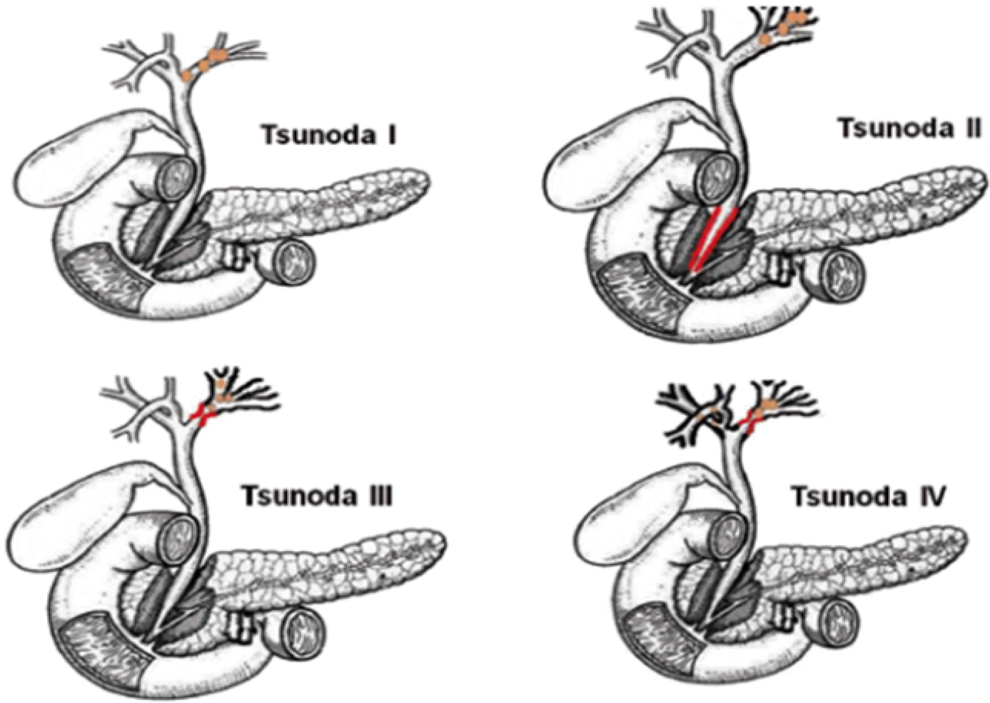
Fig. 1.
Tsunoda classification.5 Type I, no marked dilatation or strictures of intrahepatic bile ducts. Type II, diffuse dilatation of the intrahepatic biliary tree without intrahepatic duct strictures and frequently a stricture of the distal common bile duct. Type III, unilateral solitary or multiple cystic intrahepatic dilatation, frequently accompanied by stenosis of the left or right intrahepatic bile ducts. Type IV, the same attributes as type III but with bilateral involvement of hepatic lobes.
 XML Download