

 PDF
PDF ePub
ePub Citation
 Print
Print


INTRODUCTION
Behçet's disease (BD) was first described by the Turkish dermatologist Hulusi Behçet1 in 1937. BD is a systemic disorder causing extensive vascular inflammation of unknown etiology, and is mainly characterized by mucocutaneous lesions and ocular, articular, neurological, and vascular involvement2. As reported previously, cardiovascular disease is significantly higher in patients with chronic inflammatory diseases, such as rheumatoid arthritis (RA) or systemic lupus erythematosus (SLE)345. BD is also associated with acute and chronic inflammatory processes, which may cause increases in arterial stiffness and vascular damage. However, until now, little research has been reported on cardiovascular disease in association with BD.
The assessments of coronary artery disease and generalized atherosclerosis, as well as the detection of carotid artery intima-media thickness (CIMT) by using high-resolution B-mode ultrasound, are good indicators of the increased risk of subclinical atherosclerosis678910.
The ankle-brachial pressure index (ABPI), which is a noninvasive technique used to assess peripheral arteries in the extremities, is easily measured in the clinical setting1112. Currently, coronary artery calcium score (CACaS) determination by using multidetector computed tomography angiography has been used as a noninvasive method for the assessment of coronary atherosclerosis13. Calcification of the coronary artery is an important part of the process of atherosclerotic degeneration in the arterial walls1415.
The serum glycoprotein fetuin-A is synthesized mainly by hepatocytes. A consensus has not been reached concerning the role of fetuin-A in atherosclerosis16.
Thus, we aimed to investigate the association among the serum fetuin-A level, CIMT, ABPI, and CACaS in subclinical atherosclerosis in BD. To our knowledge, this is the first clinical study to investigate CACaS and fetuin-A levels in BD for the evaluation of vascular involvement.
MATERIALS AND METHODS
Patients and controls
The present research is a prospective cross-sectional, case-control study of 26 patients who attended our dermatology clinic and who had a diagnosis of BD between July 2012 and April 2014, according to the criteria of the International Study Group17. A control group of 25 healthy volunteers, matched for age, sex, and body mass index, was recruited.
In all patients and volunteers, age, sex, body mass index, blood pressure, and the CIMT, ABPI, and CACaS were recorded. Complete blood count, erythrocyte sedimentation rate (ESR), C-reactive protein (CRP), serum urea, creatinine, uric acid, aspartate aminotransferase (AST), alanine aminotransferase (ALT), serum glucose, total cholesterol, high-density lipoprotein (HDL) cholesterol, low-density lipoprotein (LDL) cholesterol, and triglycerides (fasting overnight determinations) were assessed in all patients and healthy volunteers at the time of the study. All patients were evaluated for ocular manifestations of BD.
The study used the following exclusion criteria for preventing negative effects on cardiovascular status: history of smoking, hypertension (systolic blood pressure >140 mmHg and/or diastolic blood pressure >90 mmHg), body mass index >30 kg/m2, coronary artery disease, dyslipidemia (total cholesterol and/or triglyceride levels in fasting plasma >240 mg/dl and >160 mg/dl, respectively), diabetes mellitus, chronic renal failure, thyroid disease, rheumatic disease, and pregnancy.
The study was approved by the ethics committee of the Sifa University (IRB No. 13-26), and was conducted according to the ethical principles of the Declaration of Helsinki. All patients provided informed consent before their participation in the study.
Carotid intima media thicknesses
The intima media thicknesses (IMT) of the common carotid artery (CCA) was obtained by using a real-time ultrasound scanner (Siemens, Acuson Antares, Germany) with a 7.5-MHz, 50-mm linear transducer. CIMT measurements were obtained with the patient lying in the supine position, with the neck rotated to the opposite side of the examiner. CCA images were obtained to measure IMT by using three differently angled views for each vessel. Initially, a transversal scanning view of the CCA was obtained in the longest extension possible from the base of the neck to the carotid bulb. At least three IMT points were measured in the near and far walls in the most thickened area of each vessel. Lateral wall measurements were also taken when thickening was evident and accurate images were possible. Subsequently, the vessel was scanned in two longitudinal views: posterolateral, with the transducer positioned parallel to the posterior border of the sternocleidomastoid muscle, and anterolateral, with the transducer positioned parallel to the anterior border of the sternocleidomastoid muscle. At least three IMT measurements were obtained for each near and far wall. Optimal B-mode settings of gain, depth, focal zone placement, and compression were individually adjusted for each vessel to enhance the arterial wall structures and image quality. IMT was measured manually by using electronic calipers, similar to the method of Sidhu and Desai18. The maximum IMT value was selected for each angle. For further data analyses, the maximum value of either the right or the left carotid artery was used. An IMT >1.0 mm was considered an abnormal finding19.
Coronary artery calcium scoring
CACaS was performed with a dual-source (256-slice) computed tomography scanner (SOMATOM Definition Flash; Siemens Healthcare, Forchheim, Germany). CACaS was performed in a longitudinal scan field from the tracheal carina down to the diaphragm. The corresponding images for calcium scoring were reconstructed with a slice width of 2.5~3 mm, slice interval of 1.25~1.5 mm, and tube voltage of 120 kVp. The total calcium score was calculated by using a dedicated Leonardo-X 20 (Siemens Medical Solutions, Forchheim, Germany). On the basis of the Agatston method, the calcium score was defined as the presence of a lesion with an area >1 mm2 and a peak intensity >130 Hounsfield units, which was automatically identified and marked in color by the software20. All lesions were added to calculate the total calcium score by using the Agatston method. CACaS values ≤10 were evaluated as normal.
Ankle-brachial pressure index
The pressure was taken after the patient had rested for at least 10 min. A sphygmomanometer cuff was placed around each arm and each leg. For the arms, a continuous wave pencil probe Doppler was placed over the brachial artery in the cubital fossa. When the signal was heard, the cuff was inflated to occlude the brachial artery. It was then slowly released, and the pressure at which the signal returned was recorded. This procedure was repeated with the other arm. For the legs, the cuff was placed around each limb just above the level of the malleolus. Both the posterior tibial artery and the dorsalis pedis were insonated with the wave pencil probe. The cuff and systolic measurements were recorded in the same manner as for the brachial arteries.
The ABPI was calculated as follows: ABPI=ankle pressure (posterior tibial artery or dorsalis pedis)/highest branchial pressure (right or left).
Biochemical parameters
For the sample preparation, 8 mL venous blood was collected in serum tubes (Vacuette-Z Serum Sep Clot Activator; Greiner bio-one GmbH, Kremsmünster, Austria) and centrifuged after clotting at 2,000g for 10 min at room temperature (22~24℃). Serum samples were stored at -80℃ until assayed in June 2014. Fetuin-A was measured with a human enzyme-linked immunosorbent assay kit (analytical sensitivity, 0.37 ng/ml) (alpha-2-Heremans Schmid glycoprotein; Uscn Life Science Inc., Wuhan, China).
Statistical analyses
The normality of the data was analyzed by using the Kolmogorov-Smirnov test. All numerical variables with a normal distribution were expressed as mean±standard deviation, whereas data that were not normally distributed were expressed as medians with interquartile ranges. Continuous variables were compared by using t-test or the Mann-Whitney U-test. Correlations among the CIMT, ABPI, CACaS, and fetuin-A were tested through the estimation of Pearson's partial correlation. p-values <0.05 were considered to indicate statistical significance. The statistical analyses were carried out by using the IBM SPSS Statistics ver. 20.0 (IBM Co., Armonk, NY, USA).
RESULTS
The demographic characteristics of patients and controls, including age, sex, and body mass index, were comparable. The total cholesterol, LDL, HDL, triglyceride, serum glucose, AST, ALT, urea, and creatinine levels were not statistically significant in the BD group and the control group. The ESR and CRP levels were found to be significantly higher in the BD group than in controls (both p-values were <0.001).
The mean CIMT was significantly higher in patients with BD than in the controls (patients with BD, 1.02±0.30 mm; controls, 0.60±0.16 mm; p<0.001). The mean CACaS was significantly higher in patients with BD than in the controls (patients with BD, 83.61±108.05; controls, 10.00±0.00; p<0.001). The mean ABPI was significantly lower in patients with BD than in the controls (patients with BD, 0.87±0.12; controls, 1.16±0.24; p<0.001). The mean serum concentration of fetuin-A did not show statistically significant differences between the two groups (patients with BD, 7.30±5.43 ng/mL; controls, 3.67±6.61 ng/ml; p=0.064). The demographics, laboratory findings, fetuin-A levels, CIMT levels, ABPI, and CACaS of the BD and control groups are presented Table 1.
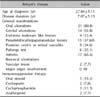
The results showed positive significant correlations between the CACaS and CIMT (r=0.33, p=0.003) and negative correlations between the CACaS and ABPI (r=-0.62, p=0.001). The correlations between fetuin-A levels and the CIMT (r=0.42; p=0.045), and between fetuin-A levels and the CACaS (r=0.09; p=0.040) were significant. The correlations of the CACaS with the CIMT, ABPI, and fetuin-A for all patients and controls are presented Table 2. The general characteristics and clinical manifestations of BD in patients at the time of enrollment in the study are presented in Table 3.
DISCUSSION
BD is a chronic, systemic inflammatory disorder with a spectrum of clinical manifestations, including mucocutaneous, ocular, vascular, musculoskeletal, gastrointestinal, and central nervous system involvement2. Cardiovascular disease is significantly higher in patients with systemic inflammatory rheumatic diseases such as RA or SLE345. Systemic vasculitis also plays an important role in cardiovascular disease, such as Takayasu arteritis, Wegener's granulomatosis, Churg-Strauss syndrome, polyarteritis nodosa, Henoch-Schonlein purpura, and BD24.
Vascular involvement in BD is usually recognized as an unclassified vasculitis, and it involves veins and arteries of all sizes. Acute systemic inflammation and chronic systemic vasculitis are known to be related to endothelial cell dysfunction25. Endothelial cell dysfunction and subsequent systemic vascular inflammation lead to arterial stiffness, which, in this case, directly indicates a risk for coronary artery disease. The assessment of coronary artery disease and generalized atherosclerosis, detection of CIMT and CACaS, and assessment of peripheral arterial vascular disease according to the ABPI are noninvasive and reliable methods11122627.
In this study, we demonstrated that the CIMT and CACaS were significantly higher, and the ABPI was significantly lower in BD patients than in the healthy controls.
Previous studies also demonstrated that the CIMT was significantly higher in BD patients than in the healthy controls25282930. In line with the results of our study, Alan et al. reported that the CIMT values were significantly higher and the carotid artery distensibility was significantly lower in patients with BD than in the controls29. They also reported that although the CIMT values were even higher in BD patients with vascular involvement than in those without vascular involvement, arterial distensibility was not significant in patients with vascular involvement, compared with those without vascular involvement29. Because vascular involvement was detected in only two patients in our study group, we did not evaluate this condition.
Hong et al.25 found that CIMT was significantly higher in BD patients with retinal vasculitis or posterior uveitis than in BD patients without these disorders. In our study, the CIMT, CACaS, and fetuin-A levels were found to be higher in BD patients with posterior uveitis or retinal vasculitis than in BD patients without these disorders. The ABPI was found to be lower in BD patients with posterior uveitis or retinal vasculitis than in BD patients without these disorders; however, the difference was not statistically significant. Hong et al.'s study25 found no significant differences in CIMT in the other subgroups of BD in relation to vascular involvement and other severe manifestations, such as major organ involvement and a history of immunosuppressive therapy.
In contrast, Korean and Brazilian studies demonstrated that BD patients had significantly increased arterial stiffness compared with control subjects. However, the CIMT of patients with BD did not differ from that of the control group313233. Yılmaz et al.34 reported that in patients with active and inactive BD, the 24-h day and night pulse wave velocity values were higher in patients with active BD than in those with inactive BD. Yılmaz et al.34 did not include controls. Seyahi et al.35 reported no differences between BD patients and controls in CIMT and femoral IMT in a study involving 239 patients with BD.
As a multisystemic inflammatory disorder characterized by remission and exacerbation, BD has a wide spectrum of clinical manifestations. Therefore, we think that the reasons for obtaining different values of arterial stiffness and CIMT in previous studies of BD may be related to the different stages of vascular involvement of the disease, the severity of BD, and the potential variance depending on racial characteristics. Therefore, further studies with a large series of patients classified according to their clinical manifestations are needed to explain these different results.
Because of the high sensitivity and specificity of the ABPI in detecting peripheral arterial vascular disease in the extremities, we chose this parameter for the evaluation of peripheral arterial stiffness. The ABPI is a good indicator of the future risk of coronary arterial disease1112. Normally, foot arterial pressure is slightly higher than arm pressure, and the ABPI is measured between 0.9 and 1.2. In the case of abnormal calcification and hardening of the arterial walls, these values increase to >1.2. In the case of decreased arterial stiffness due to inflammation and loss of vascular tone, these values decrease to <0.9. In this study, we demonstrated that the ABPI was significantly lower in BD patients than in healthy controls (0.87±0.12, 1.16±0.24, p<0.001). On the basis of these results, low ABPI values in BD patients could indicate the risk for peripheral arterial disease.
In the literature, the average CIMT has been found to vary by age from 0.5 mm (500 microns) in younger patients to 0.8 mm (800 microns) in elderly patients in large cross-sectional screenings36. In our study, whereas only two (8.0%) patients in the control group had CIMT >0.8 mm, in the BD group, 20 (76.9%) patients had CIMT >0.8 mm. Calcification of the coronary artery is an important part of the process of atherosclerotic degeneration in the arterial walls3637. An Agatston score >400 is indicative of the presence of significant and extensive obstructive coronary artery disease in at least one coronary artery20. It has been repeatedly observed that a calcification score of <100 also can be associated with significant coronary stenosis. Moreover, it was shown that the absence of coronary artery calcification could not definitely rule out significant stenosis of the coronary arteries3839. In our study, whereas no subject in the control group had CACaS >10, in the BD group, 1 patient had CACaS >400, 8 patients had CACaS >100, 13 patients had CACaS between 10 and 100, and 4 patients had CACaS <10. On the basis of these results, the CACaS and CIMT are noninvasive diagnostic methods that are useful in determining cardiovascular disease risk in BD patients.
In our study, although the mean serum concentration of fetuin-A was higher in BD patients than in healthy controls, it was not statistically significant. Fetuin-A seems to play a role in vascular disease through two distinct effects: by increasing insulin resistance and dyslipidemia and by reducing ectopic calcification. Mori et al. reported that different fetuin-A levels were obtained depending on the severity of the vascular disease16.
Ix et al.40 reported that among 1,375 community-living persons without prevalent clinical cardiovascular disease, lower fetuin-A levels were independently associated with greater severity of coronary artery calcification but not with peripheral arterial disease or CIMT. They found that fetuin-A might mark calcium deposition within the vasculature but not atherosclerosis. Furthermore, low fetuin-A might predispose patients to greater calcium deposition but not necessarily to the burden or progression of atherosclerosis. Therefore, further studies with a large series of patients classified according to their clinical manifestations are needed to explain the impact of fetuin-A levels in BD.
CRP is an important predictor of cardiovascular disease. An elevated CRP level is a very important sign of endothelial cell dysfunction41. In line with the results of Hong et al.25, our study showed that the CIMT in BD patients did not correlate with CRP and ESR. However, the CACaS in BD patients was positively correlated with CRP and ESR (Table 2). Had we used a high-sensitivity CRP (hsCRP) assay, we might have found a positive correlation between
CIMT and hsCRP.
This study has some limitations. The number of patients was relatively low. Moreover, BD in our cohort of patients was not very severe. Finally, most patients in this study were in remission.
On the basis of these results, the CACaS and CIMT are noninvasive diagnostic methods that are useful in determining the cardiovascular disease risk in BD patients. However, further studies with a larger series of patients are needed to assess the risk of cardiovascular disease and to compare the diagnostic methods in BD patients.


 XML Download