

 PDF
PDF ePub
ePub Citation
 Print
Print


Introduction
Steroid cell tumors are rare sex cord-stromal tumors of the ovary. These tumors were previously classified as "lipid" or "lipoid" cell tumors. The term "steroid cell tumors" was initiated by Scully in 1979. Steroid cell tumors account for less than 0.1% of all ovarian tumors. There are three steroid cell tumor subtypes: steroid cell tumor not otherwise specified (NOS), stromal luteoma and Leydig cell tumor. Steroid cell tumors, NOS, account for approximately 60% of steroid cell tumors. The proportion of tumors that are clinically malignant has ranged from 25% to 43%. Therefore, steroid cell tumors, NOS, are managed surgically like other ovarian stromal tumors.1 Additionally, a gonadotropin releasing hormone agonist (GnRHa) could be used as postoperative adjuvant therapy.2 This report describes a case of a steroid cell tumor, NOS, in an adult woman who presented with metrorrhagia and hirsutism.
Case Report
A 35-year-old Korean woman (gravida 5, para 2) visited hospital with the complaint of metrorrhagia. The patient reported that she had been suffering from metrorrhagia since 7 months ago. Her past medical history revealed left salpingectomy due to ectopic pregnancy. She was 154 cm in height and 70 kg in weight. Her body mass index was 29.5 and she was overweight. Physical examination revealed increased pubic hair and normal external genitalia.
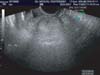
Transvaginal ultrasound indentified a 4.9 × 3.4 cm, well-circumscribed and solid left ovarian tumor (Fig. 1). Laboratory analyses showed normal blood counts and normal serum values of cancer antigen 125 (CA-125). Serum value of dehydroepiandrosterone-sulfate (DHEA-S) was 548.9 µg/dl (reference value, 35-430 µg/dL).
Considering transvaginal ultrasound and laboratory analyses, laparoscopic left oophorectomy was performed. During the operation, the left ovary ruptured due to adhesion caused by previous operation.
Grossly, cut surface was vaguely lobulated, bright yellow and soft. No hemorrhage or necrosis was noted. Microscopically, the tumor consisted of irregular cords and nests of large rounded to polygonal cells having abundant clear vacuolated cytoplasm and centrally located monotonous nuclei with prominent nucleoli (Fig. 2). No cellular atypia was noted and mitotic figures were rare (less than 1 per 10 high-power fields). It was diagnosed as steroid cell tumor, NOS.
Two weeks postoperatively, a computerized tomography (CT) scan and endometrial biopsy were performed and no remarkable findings were found.
As she had completed childbearing, complete surgical staging including peritoneal washing, total abdominal hysterectomy, right salpingo-oophorectomy, appendectomy, bilateral pelvic lymph node dissection and infracolic omentectomy was performed after 4 weeks from the first laparoscopic surgery. Microscopically, no evidence of metastasis was found. As the left ovary rupture at initial laparoscopic surgery, the patient was treated with GnRHa monthly for 6 months. After 43 months, she has had no evidence of recurrence.
Discussion
Steroid cell tumors were previously classified as lipid or lipoid cell tumors. This prior nomenclature was misleading in that some tumors have little or no lipid present. The term steroid cell tumor more accurately describes the morphological appearance of these tumors and provides insight into their clinical manifestations. Among three steroid cell tumor subtypes, steroid cell tumor, NOS, is the most common type and associated with androgenic changes in approximately half of cases. These tumors usually occur in younger individuals (mean age, 43 years) and, in contrast to other steroid cell tumors, occasionally occur before puberty.3 Hirsutism and virilization are the most common symptoms occurring in 56-77% of patients. In our patient, increased pubic hair was found and serum value of DHEAS was elevated.
Hormones other than testosterone can be elevated in steroid cell tumors, NOS. Estradiol secretion by these tumors is not uncommon, occurring in 6-23% of patients. This excess estrogen production can result in menorrhagia and postmenopausal bleeding; in addition, endometrial adenocarcinoma has been described.4 Our patient presented metrorrhagia, but serum value of estradiol and endometrial biopsy were normal. Steroid cell tumors, NOS, have also been associated with Cushing's syndrome in 6-10% of cases, but approximately 25% of steroid cell tumors, NOS, do not produce hormones.5
The proportion of tumors that are clinically malignant has ranged from 25% to 43%. Extraovarian spread of tumor is noted at the time of operation in 20% of the patients. The following pathologic features indicate malignancy : two or more mitotic figures per 10 high-power fields associated with 92% malignancy, necrosis with 86% of malignancy; a diameter of >7 cm with 78% of malignancy; hemorrhage with 77% of malignancy; and grade 2 or 3 nuclear atypia with 64% of malignancy.5 Pathologically, biopsy revealed a benign tumor in our patient.
There are few collected series in the literature. In addition, since most of these tumors are diagnosed in an early stage and do not recur or metastasize, even less is known about their response to therapy. Although the clinical course of steroid cell tumors is not exactly known, it is recommended that ovarian steroid cell tumors, NOS, are managed surgically like other ovarian stromal tumors. Conservative surgery with unilateral oophorectomy and proper staging should be performed in women with stage I disease who desire future fertility. Complete surgical staging including total abdominal hysterectomy and bilateral salpingo-oophorectomy is an accepted treatment for older women who do not want to preserve their fertility,6 as was the case here.
We could find 5 recently reported cases of ovarian steroid cell tumor in Korea.7,8,9,10,11 Conservative surgery was performed in 3 patients aged under 25 years old. Complete surgical staging was performed in 2 patients who completed childbearing (Table 1). 4 patients were premenopausal women7,8,9,10 and 1 patient was a postmenopausal woman with elevated CA-125.11 So, steroid cell tumor should be considered in postmenopausal women having ovarian tumors. Increasing use of ultrasound has resulted in increasing detection of ovarian cysts in postmenopausal women. When these cysts remain stable on follow-up, ovarian rete cyst should be considered.12
The clinical course of steroid cell tumors of the ovary is unknown, because there are few collected series in the literature. Too few cases exist to have good information as to the role of adjuvant therapy. Gonadotropin has been known to suppress androgen secretion by ovarian virilizing tumor. Pascal et al.13 suggested that various ovarian androgen-secreting tumors, as well as hyperthecosis, were not autonomous but apparently depended upon continuous gonadotropin stimulation. Imai et al.14 also noted a direct suppressive effect of GnRHa on ovarian steroidogenesis. Wang et al.2 suggested that GnRHa might be an alternative choice as adjuvant therapy for managing recurrence of ovarian steroid cell tumor. As the tumor ruptured during the first laparoscopic surgery and we could not expect the clinical course, we administered GnRHa in our patient to prevent recurrence on the basis of these evidences.
As far as we know, there is no previously reported case of ovarian steroid cell tumor treated with complete surgical staging followed by GnRHa in Korea (Table 1). Our patient has had no evidence of recurrence for 43 months. GnRHa should be considered as one of adjuvant therapy in ovarian steroid cell tumors.

 XML Download