

 PDF
PDF ePub
ePub Citation
 Print
Print


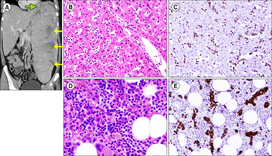
A 34-year-old male patient exhibited episodes of high-grade fever with chills and left upper quadrant pain for the past 1 year. The coronal computed tomography (CT) image shows large hepatosplenomegaly with heterogeneous, hypodense areas within the splenic parenchyma (A, yellow arrows). Similarly, heterogeneous enhancement (A, green arrow) with features of hepatosplenic fusion is also seen on the left liver lobe. Liver biopsy showed diffuse infiltration of atypical lymphoid cells within the dilated sinusoids (B). Immunohistochemical analysis revealed these atypical lymphoid cells as CD3 epsilon- and CD56-positive, and CD20-negative (C). Considering his pancytopenia and to stage the lymphoma, a bone marrow (BM) biopsy was conducted, which demonstrated sinusoidal infiltration of the lymphomatous cells in the background of trilineage hematopoiesis (D). These cells also showed similar immunohistochemistry with those exhibiting CD3 epsilon positivity (E), suggesting the lymphomatous involvement of the BM. Furthermore, cytogenetic analysis revealed the abnormality of isochromosome 7q, and a diagnosis of hepatosplenic lymphoma was confirmed. It is an uncommon variant of T-cell lymphoma that shows sinusoidal localization of tumor cells and is difficult to diagnose based on normal liver test results. Two-thirds of patients with hepatosplenic lymphoma present with BM involvement, and preferential sinusoidal infiltration challenges histological diagnosis.
 XML Download