

 PDF
PDF ePub
ePub Citation
 Print
Print


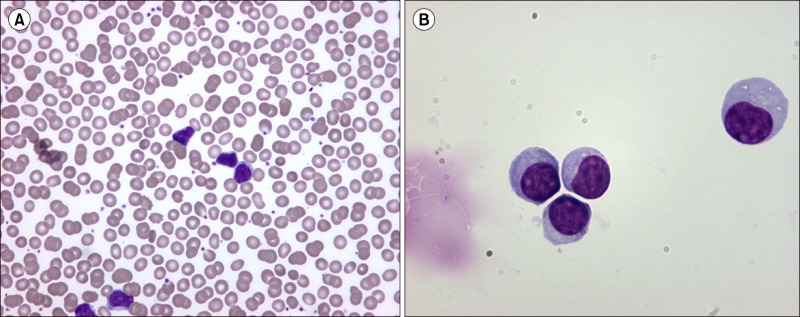
An 87-year-old woman, first diagnosed with CLL in 2003, presented with left peripheral facial palsy and binocular diplopia. Physical examination and computed tomography showed no lymphadenopathy. Neurological examination indicated right Babinski sign and left sixth cranial nerve palsy. The hemoglobin level was 10.2 g/dL; total leukocyte count, 16.8×10⁹/L (65% lymphocytes); and platelet count, 279×109/L. A peripheral blood smear showed mature lymphocytes with dense nuclei (A), partially aggregated chromatin, and Gumprecht shadows. The immunophenotype was consistent with CLL, with negative CD38 expression (6%). Cerebral magnetic resonance imaging findings were normal. Cerebrospinal fluid (CSF) examination revealed glucose levels of 3.2 mmol/L, protein levels of 1.87 g/L, a white cell count of 94/mm3, and 100% abnormal lymphocytes (B), but tests for bacteria and viruses were negative. Immunophenotyping of the CSF cells revealed clonal small B lymphocytes, corresponding to the systemic findings, except for a positive CD38 expression (34%). Nine courses of intrathecal chemotherapy of methotrexate, cytarabine, and hydrocortisone were administered; neurological symptoms resolved and CSF findings normalized. CNS involvement is uncommon in CLL cases and therefore may be underestimated. There was an unusual discrepancy in CD38 expression between peripheral blood and CNS clonal B cells.
 XML Download