

 PDF
PDF ePub
ePub Citation
 Print
Print


Dear Editor
Aberrations of the human chromosome 1 are common in hematologic malignancies such as multiple myeloma, myeloproliferative disorders, and myelodysplastic syndrome, highlighting their significance in carcinogenesis [1]. The isochromosome 1q [i(1)(q10) or i(1q)] is a distinctive structural chromosomal abnormality in hematologic malignancies [2], especially in childhood Burkitt lymphoma/leukemia (BL) [3]. We report the first Korean pediatric BL patient with i(1)(q10) as well as t(8;14)(q24;q32).
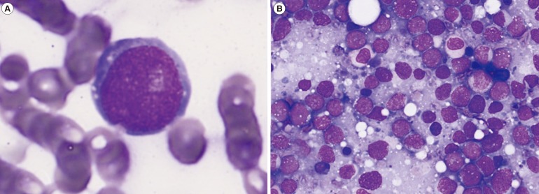
A 9-yr-old girl with fever for three days was referred to our hospital for abnormal complete blood count, which revealed the following: hemoglobin, 8.4 g/dL; platelets, 12×109/L; and leukocyte count, 4.46×10⁹/L with 9% lymphoma cells (Fig. 1A). Subsequent physical examinations revealed splenomegaly and several palpable lymph nodes. The patient also showed swelling at both submandibular areas, which developed the day before admission.
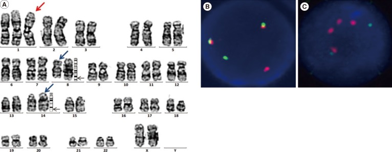
Even though the initial bone marrow specimen was dry-tapped and inadequate for accurate differential count, lymphoma cells with bluish cytoplasm and prominent nucleoli were heavily loaded on touch imprint preparation (Fig. 1B). Flow cytometric analysis performed with peripheral blood specimen presented that the lymphoma cells were positive for CD19 and CD10; but negative for CD20, terminal deoxynucleotidyl transferase (TdT), kappa, and lambda surface immunoglobulins. Chromosomal analysis of the bone marrow specimen using standard trypsin-Giemsa banding technique revealed an abnormal karyotype of 47,XX,+i(1)(q10),t(8;14)(q24;q32)[17]/49,idem,+6,+14[6]/46,XX[4] (Fig. 2A). FISH was performed to confirm the abnormality, which indicated the presence of i(1)(q10) (Fig. 2). She was diagnosed as having BL and treated with vincristine and daunorubicin. Although she presented jaundice and liver enzyme elevation that were considered as toxic side effects of the chemotherapy, the patient tolerated the induction and consolidation chemotherapy quite well. Follow-up bone marrow examination and cytogenetic analyses showed no residual lymphoma cells with the 46,XX[20] karyotype. Informed consent was obtained from the patient's parents for the case report.
Among various types of recurrent chromosomal aberrations reported in BL patients, chromosomes 1, 6, 7, 13, 17, and 22 are most frequently affected in up to 70% of the cases [4]. In comparison with other structural rearrangements within the long arm of chromosome 1 [5], i(1)(q10) is unique for genetic dosage gain, which results in a complete triplicate of 1q and distinctive morphologic feature. There were three cases of childhood BL with i(1)(q10) previously classified as ALL FAB L3 type (before the 2008 WHO classification) [67]. Additionally, there were eight case reports of i(1)(q10) in patients with BL [4]. Among these patients, six cases presented abnormal karyotypes, which included i(1)(q10) and t(8;14)(q24;q23) together.
An important question regarding isochromosomes is whether they represent primary or secondary chromosomal change. Isochromosome 1q was observed to be one of the secondary chromosomal changes in BL cell lines [5]. Furthermore, i(1)(q10) in BL was associated with unfavorable therapeutic responses in pediatric patients [48]. Interestingly, our patient showed atypical immature immunophenotype of BL, which was previously suggested to be associated with i(1)(q10) [4]. Apparently, atypical immunophenotype BL has poor prognosis compared with its typical counterpart [9]. High incidence of patients presenting an immunophenotype of immature B-cell arrest among BL with i(1) (q10) patients was recognized, which is consistent with our case. Although impaired immunoglobulin production in tumor cells due to MYC translocation during early B-cell maturation has been proposed as the underlying mechanism, further investigations are required for its complete elucidation.
Centromeric misdivision along the short, rather than the long, axis of a chromosome is one of the possible mechanisms to explain the origin of isochromosomes [10]. Therefore, it is plausible that the centromere of chromosome 1 is unusually susceptible to abnormal division in our patient's lymphoma cells. The genetic consequence of an isochromosome, in addition to the normal chromosome, is acquired isodisomy of 1p and quadrisomy of 1q. As a result, relevant oncogenes on 1q might manifest carcinogenesis, leading to various malignancies including BL [1].
In conclusion, we report the first Korean pediatric BL patient with i(1)(q10). This case provides additional insight into the wide spectrum of chromosomal structural abnormalities. Clinical attention to the appropriate detection of i(1)(q10) by chromosomal analysis and FISH is recommended. Since it is possibly associated with poor clinical outcomes, further studies are required to investigate the implications of i(1)(q10).
 XML Download