

 PDF
PDF ePub
ePub Citation
 Print
Print


Introduction
Tooth discoloration might compromise the esthetic appearance of patients, resulting in an increase in demand for esthetic dental procedures. Of all the therapies available, tooth bleaching has gained great popularity as an easy, cost-effective and conservative technique to treat discolored teeth.1 The in-office external tooth bleaching technique is used to whiten discolored teeth. In recent years, introduction of bleaching gels containing high concentrations of hydrogen peroxide (HP, 35 - 45%) have made in-office bleaching techniques easier and more favorable. The advantage of this bleaching modality is its favorable and immediate results with no need for further patient cooperation,2 in association with patient comfort and minimum chair time.
Various studies have been undertaken to assess the effects of peroxide-containing products on the physical and chemical properties of enamel, with some reporting no observable changes in enamel microhardness and morphology subsequent to bleaching procedures.3456 However, some others have reported loss of calcium,789 changes in surface morphology,10 and a decrease in enamel hardness9101112 and fracture resistance.13 Some previous studies have reported a decrease in enamel surface microhardness subsequent to bleaching procedures.1415 Based on a study, the decrease in microhardness of bleached enamel was inversely proportional to the concentration of HP.9 However, another study reported no alterations in enamel microhardness after bleaching with 30% HP.16 The discrepancies between the results of different studies might be attributed to differences in methodologies (in vivo or in vitro, time of evaluation, bleaching agents applied, duration of application, immersion of the specimens in artificial saliva between treatments, differences in storage protocols, pH of bleaching agent, use of fluoride and other factors).17 Peroxide penetrates into the tooth hard tissues and generates free radicals to oxidize organic chromophores, resulting in a decrease in discoloration, particularly in dentin.17 Despite its proven efficacy, concerns still exist in relation to the negative effect of HP on enamel. Various studies have reported that bleaching agents can induce chemical, structural and mechanical changes in enamel.1819 In addition, a decrease in the mineral content of bleached enamel is not confined to the enamel surface; it extends to the enamel subsurface, too.14 Therefore, it is absolutely necessary to minimize potential damages to enamel as a result of exposure to HP.20 In addition, inhibiting the detrimental effects of bleaching agents on enamel mineral content and use of remineralizing agents in bleaching peroxides might decrease enamel solubility and sensitivity due to deposition of minerals in enamel crystallites.2122
To minimize the detrimental effects of bleaching agents on enamel, fluoride, hydroxyapatite (HA), 45S5 bioglass, and casein phosphopeptide-amorphous calcium phosphate (CPP-ACP) have been incorporated into the bleaching agents in some studies to decrease demineralization and accelerate remineralization of enamel.1721222324 However, some studies have reported no improvements in fluoride uptake in bleached enamel25 and fluoridated bleaching gels might not be able to promote remineralization of predemineralized enamel.26 Current knowledge on the effects of in-office bleaching agents modified by remineralizing agents on the microhardness of enamel surface is inadequate and controversial.23 Therefore, this in vitro study was undertaken to investigate the effect of in-office bleaching technique in association with the use of three different nanobiomaterials as remineralizing agents, including ACP, HA, bioactive glass (BAG), on enamel hardness. The null hypothesis tested was that application of ACP, HA, and BAG with HP gel would not affect enamel microhardness.
Materials and Methods
This study was approved by the Ethics Committee for Research of Isfahan university of Medical Sciences (IRB No., 293154). A total of 24 extracted human molars were collected and stored in 0.2% thymol solution at 4℃. The teeth were subsequently stored in distilled water for 24 hours for thorough removal of thymol residues. The crowns were separated from the roots with the use of a cutting machine (Krupp Dental 759DRZ, Dentarapid Co., Hilzingen, Germany). The crown of each tooth was sectioned along the central line with a slow-speed water-cooled diamond saw (TC3000, Vafaei Industrial Co., Tehran, Iran). The tooth fragments were placed in acrylic resin (Dentsply Ltd., Surrey, England). The enamel surfaces were ground and polished sequentially with 600, 800, 1,500, and 3,000 grit silicon carbide abrasive papers (Silicium Carbide, Matador, Wasserfest 991A, Softflex, 3M ESPE Co., St. Paul, MN, USA). These procedures were carried out to achieve parallel planar surfaces, considered fundamental for microhardness testing. Sixty enamel specimens (2 × 3 × 4 mm) from all the tooth samples were selected, i.e. one or two specimens from each sample tooth embedded in acrylic resin, depending on the surface area.
The specimens were randomly divided into five groups, according to the bleaching procedure (n = 12). In Group 1, samples did not undergo any bleaching procedure (control) and were only stored in distilled water. In Group 2, samples were bleached with a 40% HP gel (Opalescence Xtra Boost, Ultradent Products Inc., South Jordan, UT, USA). In Groups 3, 4, and 5, samples were bleached with a 40% HP gel modified by incorporation of BAG (NovaBone Products LLC, Alachua, FL, USA), ACP (Sigma Aldrich Co., Madrid, Spain), and HA (Sigma Aldrich), respectively. Remineralizing agents ground and filtered to particles up to 50 nm were used. Then, remineralizing agents were mixed with distilled water in a ratio of 2 g powder to 1 mL liquid. After that, they were mixed with 1 mL of HP. The bleaching agents were applied for 60 minutes. After bleaching, the specimens were thoroughly rinsed with air-water spray for 15 seconds. The procedure was repeated twice a week for 2 weeks. All the samples were stored in distilled water at 37℃ between procedures.
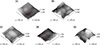
Knoop microhardness value was determined using a microhardness tester (Metallux 3, Leitz Co., Wetzlar, Germany) under a load of 100 g and an indentation time of 20 seconds. Three indentations were placed on the surface of each specimen, 100 µm apart from each other. The obtained values were averaged as the baseline microhardness value before treatment. After the treatment procedures, the specimens were tested for final microhardness measurements. The difference between baseline and final values was calculated. The differences in enamel surface microhardness values before and after treatment in each group were analyzed using one-way ANOVA, followed by post hoc Tukey tests at a 5% significance level. Atomic force microscopy (AFM, Brukernano scale 1.1, Bruker Co., Santa Barbara, CA, USA) images were presented in Figure 1.
Results
Average surface microhardness values before and after bleaching procedures in each group are presented in Table 1. In Group 2, AFM evaluation exhibited irregular surface topography with more structure removal than in Group 1. In Group 4, the surface of specimens looked more prominent and dense than Group 1 (Figure 1). There was a significant difference in the microhardness change (MHC) between the five study groups (p < 0.001, Table 1). The MHC of Groups 1, 3, 4, and 5 were significantly lower than that of Group 2. The MHC of the Group 1 and Group 4 were significantly different. In the Group 4, enamel microhardness increased significantly (p < 0.001, Table 1). However, there were no significant diffrences between Groups 1 and 5 (p = 0.834) and Groups 1 and 3 (p = 0.219).
Discussion
Tooth enamel is the hardest and stiffest mineralized biological tissue that contains approximately 96% mineral, 3% water, and 1% organic matter by weight.27 The superficial aprismatic enamel is normally hypermineralized and rich in fluorapatite; therefore, it is more resistant to demineralization.27 However, tooth bleaching with HP might give rise to calcium loss, alterations in the surface morphology, and a decrease in hardness and fracture resistance of enamel.78910111213 With accurate diagnosis and appropriate implementation of the technique, bleaching might prove a conservative and safe technique to whiten discolored teeth.28 Some in situ1129 and in vivo30 studies have shown a decrease in microhardness of bleached enamel. A decrease in microhardness might be attributed to the loss of mineral content due to demineralization.3132
Given the effects of bleaching products on mineral loss in tooth hard structures, studies on external bleaching often evaluate microhardness because it reflects the mineral content of the tooth.33 In the present study, the microhardness of all the samples were evaluated at baseline and after bleaching procedures. In order to minimize variations among teeth, the superficial layer of enamel samples was removed and polished to achieve a uniform mirror-like surface17 that is considered an appropriate surface for microhardness test. Based on the results, the use of each one of the three remineralizing agents, including BAG, ACP, and HA, during bleaching procedures with 40% HP in the in-office protocol resulted in higher mean enamel microhardness values, compared to the Group 2 without any remineralizing agents. This result was significant for Group 4, in which ACP was incorporated into the bleaching gel, resulting in an increase in microhardness compared to the control group. This might be attributed to the fact that the microporosities formed on the subsurface enamel due to bleaching gives rise to the areas conducive to re-deposition of these materials similar to that taking place in arrested caries.3
In the oral environment, ideal conditions exist for remineralization and demineralization of enamel, with more likelihood for remineralization. Subsequent to enamel demineralization induced by bleaching agents, ionic exchange is facilitated, resulting in greater absorption of minerals to replace those lost during bleaching.3 Two previous studies have suggested that the combined use of BAG, CPP-ACP, and bleaching agents did not interfere in tooth whitening efficacy.1734 In addition, incorporation of HA into toothpastes resulted in a marked increase in tooth whitening.35 In the present study, three remineralizing agents were used in nano sizes. An increase in remineralization effect has been reported when the particle size of HA is reduced down to the nano-metric levels because the interaction between the nanoparticles and dentin and enamel becomes more effective due to an increase in surface-to-volume ratio.36
Based on the results of this study, incorporation of ACP to HP resulted in better protective effect than HA, as they reduced the mineral loss of enamel more effectively. We used three different remineralizing agents during in-office bleaching procedure in this study. ACP might serve as a promising biomimetic adjunct in bleaching procedures to prevent/restore enamel damage induced by bleaching agents.17 No similar study is available in the literature. However, a previous study suggested that BAG can occlude dentinal tubules, inhibit dentin demineralization and promote dentin remineralization through interfacial apatite precipitation.37 In addition, BAG has been shown to be able to inhibit and reverse the initial caries progression in enamel.38 ACP, BAG, and HA, as alkaline salts, might buffer the acidity of HP and reduce demineralization after being mixed with HP. A study reported that BAG increased the pH of HP from 3.2 to 4.73 and decreased its acidity.
Furthermore, the BAG released more mineral ions in HP compared to that in distilled deionized water. This might be responsible for the enhanced protection of using BAG with HP.17 In the present study, BAG+HP increased enamel microhardness, with no significant differences from the control group. It is important to consider that BAG, as an inorganic biocompatible material, releases Ca2+, Na+, and PO43- in aqueous environments.1739 Furthermore, the rapid ionic exchange of Na+ with H+ or H3O+ at glass-liquid interface, as evidenced by the initial rapid increase of pH and Na+, allowed Ca2+ and PO43– to be released to result in a supersaturated ionic reservoir for the enamel apatite. Progression of these reactions leads to crystallization of enamel into carbonate-enriched hydroxyapatite.1740
In another study, HA increased the pH of HP solution from 3.2 to 5.4, making it less acidic.41 In addition, even adherence of HA particles to the enamel surface resulted in the formation of a protective layer for the underlying enamel, decreasing the direct contact of HP with enamel surface. Furthermore, the solution around the enamel surface might soon become supersaturated with respect to enamel apatite.42 All these effects of HA might give rise to more significant decrease in enamel demineralization induced by HP. HA facilitates the bleaching procedure by promoting the release of free radicals from HP.21 Therefore, HA nanoparticles incorporated into HP might improve the biocompatibility of the final product and increase the safety of the bleaching process. More recently the use of 6% HP with 2% nano-HA resulted in significantly less sensitivity than the bleaching product without nano-HA.43 In the present study the MHC between the Groups 1 and 5 was not significantly different.
Cunha et al. suggested the increased microhardness after bleaching with CPP-ACP and HP might indicate mineral deposition on enamel.32 Upon acid challenge, the attached CPP-ACP releases calcium and phosphate ions to maintain a supersaturated mineral environment, decrease demineralization and promote remineralization of enamel, as observed in clinical in situ studies.44 In the present study, there was a significant increase in microhardness after bleaching in the Group 4. ACP might improve safety45 and decrease tooth sensitivity during the bleaching process. However, further studies are necessary to determine the most effective concentration of the material and the incorporation procedure should be evaluated more precisely.
It should be emphasized that this study was carried out in vitro, and further studies are required to show that remineralizing agents can prevent a decrease in microhardness when they are used in association with high concentrations of bleaching agents.

 XML Download