

 PDF
PDF ePub
ePub Citation
 Print
Print


Introduction
Four types of materials are widely used for direct aesthetic restorations, including resin composites, polyacid-modified composite resin material (compomers), glass ionomers, and resin-modified glass ionomers. Resin-based composites introduced in 1970s are commonly used in clinical practice due to their improvements in esthetics, mechanical properties, bonding procedures, and material formulation.123 Their monomer structure or chemistry, filler amount, size and shape have undergone multiple refinements over the years to yield better physical properties.4 The changes in monomer chemistry were at first directed to improve the methacrylate-based systems, by modifying the Bowen's monomer (Bis-GMA, Bisphenol-A-glycidyl dimethacrylate). Aliphatic urethane-based dimethacrylate resins (UEDMA, urethane dimethacrylate; UDMA, urethane dimethacrylate) and partially aromatic urethane dimethacrylate were developed to create monomers with lower viscosity. Moreover, ring-opening systems and epoxy-based resins such as siloxane and oxirane, which are commonly called siloranes (a new type of monomer) were introduced in the market.567 Except monomer structures, the filler contents of materials have been continuously changed over the years. Recently, nanocomposite materials have been developed. These materials have submicrometer particles (nanofillers, approximately 40 - 50 nm) and provide excellent esthetics and polished surfaces by minimizing the filler size.89
Compomers (polyacid-modified resin composites) were introduced in 1995 by adding dimethacrylate monomers to conventional glass ionomer cements.1 These materials combine the benefits of resin composites and glass ionomers, but they behave more like resin composites.10 They contain acid-decomposable glass and acidic polymerizable monomers, such as acidic carboxylate groups and polymerizable methacrylate groups, which enable both free radical polymerization by light curing and an acid-base reaction in the presence of water.11
Extensive efforts were made over the years to develop the esthetic properties of dental restorative materials. To obtain the ideal esthetics, any restorative material must simulate the natural tooth in color, translucency, and surface texture, and also show color stability for long periods of time.12 Discoloration of restorations can be due to extrinsic (exogenous) or intrinsic (endogenous) causes. Extrinsic factors are related to the surface absorption of staining solutions from exogenous sources or through the accumulation of plaque and surface stains.13 In the oral environment, superficial degradation of the restorative materials and their adsorption of staining agents can cause discoloration.14 The characteristics of the inorganic fillers have a direct impact on composite resin surface property and their susceptibility to extrinsic staining.1516 On the other hand, intrinsic factors involve the discoloration of the resin material itself by alteration of the resin matrix and of the interface of the matrix and fillers via oxidation.15 Therefore, the photoinitiator systems, resin matrix, filler loading, etc., influence the color stability.17 Endogenous discolorations are irreversible, while the exogenous discolorations caused by adsorption of dyes or plaque can be easily removed by polishing.
Several studies have shown that composite resins are susceptible to color alteration when exposed to staining solutions like coffee, cola, tea, and wine.1314 Consumption of certain beverages such as coffee and tea may affect the aesthetic and physical properties of composite resins, thereby undermining the quality of the restoration.13 The consumption of aerated drinks like cola is high in young adults and children. The acidity of these drinks may be detrimental to the properties of restorative resins.13
Due to the common usage of tooth-colored restorative materials, it is important to determine which materials are susceptible to color changes. Thus, the objective of this study was to evaluate the color stability of a universal restorative composite, a silorane-based composite, a flowable composite, and a polyacid-modified composite resin material (compomer) after exposure to commonly consumed beverages like black tea, Coca cola, and water for 24 hours and 1 month, respectively. The null hypothesis tested was that different beverages would not affect the stainability of different types of restorative materials.
Materials and Methods
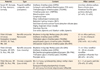
Eighty-four disc-shaped specimens were prepared from three different resin composites including Filtek Ultimate Universal Restorative, Filtek Ultimate Flowable, and Filtek Silorane (3M ESPE, St. Paul, MN, USA), and a compomer material (Dyract XP, Dentsply De Trey, Konstanz, Germany) of shade A2 (Table 1). The specimens were 8 mm in diameter and 2 mm in thickness and were fabricated using a cylindrical stainless steel mold. Materials were loaded into the molds and pressed between two glass slides lined with polyester film (Mylar Strip, SS White Co., Philadelphia, PA, USA). The top surfaces of all specimens were polymerized for 20 seconds using a standard LED light curing unit (Elipar S10, 3M ESPE) with a minimum output of 1,200 mW/cm2. A dental radiometer (Hilux Ledmax Light Curing Meter, Benlioğlu Dental Inc., Ankara, Turkey) was used to monitor the light intensity output to be at least 1,200 mW/cm2. Then the 21 discs of each group were divided into 3 subgroups that were submerged in black tea, Coca cola, and water (n = 7). Final contouring and finishing of the specimens were performed using 600 grit silicon carbide (SİC) paper for 20 seconds, after which the surfaces were polished under dry condition using Sof-Lex Polishing Disks (3M ESPE) from medium to superfine. Each disc was used for 30 seconds with a hand piece rotating at 10,000 rpm. Specimens were thoroughly rinsed with water for 10 seconds to remove the debris after each polishing step. Subsequently, the specimens were stored in distilled water for 24 hours at 37℃ before baseline color evaluation.
Baseline colors of the resin composites were measured with a VİTA Easyshade Compact (Model DEASYCHP, VITA Zahnfabrik, Bad Sackingen, Germany). Before measuring the color of the specimens, the Vita Easyshade was calibrated using its calibration block according to the manufacturer's instructions. The probe tip was then placed perpendicular at the center of each specimen and flushed into the surface of the specimens to obtain accurate measurements. The measurement procedures were repeated three times. All measurements were made on a white Plexiglass background in order to eliminate background light.
CIE Lab* is expressed by the L* coordinate representing color luminosity, varying from white to black, and the a* and b* coordinates representing the chromaticity of the color, with axes varying from green to red and blue to yellow, respectively. The means of the values obtained were calculated, and the L*, a*, and b* parameters were determined. The color changes (ΔE*) after 30 days were calculated from the changes in CIE L*, a*, and b* values (ΔL*, Δa*, Δb*) as follows:
Following the baseline measurements, the specimens of each restorative material were randomly divided into 3 subgroups and submerged in different drinks (water, black tea, and Coca cola) for 1 month (n = 7 each). The first subgroup was stored in the dark at 37 ± 1℃ in distilled water and served as the control. The water was changed daily for 30 days. The other subgroups of each composite resin were immersed in black tea and Coca cola. The black tea (Lipton yellow label tea, Unilever, Istanbul, Turkey) was prepared by immersing 5 g of tea granules in 250 mL hot water (95℃) for 3 minutes; 20 mL tea was freshly prepared daily prior to each testing and immersion for 30 days (pH, ~ 6.5, Smart pH meter, HANNA instruments, Ann Arbor, MI, USA). For Coca cola (The Coca-Cola Company, Istanbul, Turkey), a new bottle was used every day (pH, ~ 2.5). The temperatures were measured with a digital thermometer (TPI 310C, Test Products International, Beaverton, OR, USA). Immersions were carried out for 1 hour, three times a day at room temperature. After each immersion process, the specimens were washed with distilled water and stored in distilled water at room temperature during the 30 days cycle.1819
All statistical analyses were performed using a commercially available software package IBM SPSS 20.0 (SPSS Inc., Chicago, IL, USA). The distribution of data was first checked for normalcy with the Kolmogorov-Smirnov test, and then the Kruskal-Wallis test. One-way analysis of variance was done to determine statistically significant differences among the restorative materials within the groups immersed in each beverage. If a significant difference was observed in any material, Dunn's post hoc multiple comparison test was performed. All the statistical tests were performed at a significance level of α = 0.05.
Results
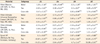
Changes in ΔL*, Δa*, Δb* after exposure to the three beverages for 30 days were summarized in Table 2. Also, color change (ΔE*) of the specimens after 30 days were shown in Table 2. Color changes of the resin materials were in the range of 1.31 - 15.28 ΔE* unit. After 30 days, the highest mean ΔE* values were observed in the Dyract XP immersed in Coca cola (15.28 ± 2.61) and black tea (12.22 ± 2.73), and these values were significantly different from the corresponding values for Filtek Ultimate Universal Restorative, Filtek Ultimate Flowable, and Filtek Silorane (p < 0.05).
The Filtek Silorane specimens stored in black tea (mean ΔE*, 3.69 ± 0.76) exhibited significantly more color changes than those stored in Coca cola (mean ΔE*, 1.74 ± 0.65) (p < 0.05), but showed no significant difference with Filtek Silorane specimens stored in water (mean ΔE*, 3.05 ± 1.01). Filtek Ultimate Universal Restorative and Filtek Ultimate Flowable showed significantly more color changes in tea, compared to in Coca cola (p < 0.05). Dyract XP showed significantly higher color changes in tea (12.22 ± 2.73) and Coca cola (15.28 ± 2.61), compared to water (4.01 ± 0.88) (p < 0.05).
In tea, Dyract XP (mean ΔE*, 12.22 ± 2.73) displayed significantly more color changes than did Filtek Ultimate Flowable (mean ΔE*, 4.71 ± 1.4) and Filtek Silorane (mean ΔE*, 3.69 ± 0.76) (p < 0.05). In Coca cola, mean ΔE* values of Dyract XP (15.28 ± 2.61) were significantly higher than those of Filtek Ultimate Universal Restorative (1.35 ± 0.34) and Filtek Ultimate Flowable (1.31 ± 0.43) (p < 0.05). In water, the mean ΔE* values of the materials were similar at the end of 30 days.
ΔL* (brightness) values
There was a significant change in the brightness (ΔL*) after a period of 30 days for Filtek Silorane and Filtek Ultimate Universal Restorative in water, Dyract XP and Filtek Ultimate Flowable in black tea, and Dyract XP and Filtek Silorane in Coca cola (p < 0.05). A positive ΔL* indicates that the specimens became lighter, whereas a negative ΔL* indicates that the specimens became darker. Filtek Ultimate Universal Restorative and Filtek Flowable displayed negative ΔL* in water. All the materials tested showed negative ΔL* in black tea. Moreover, all the materials except Filtek Silorane showed negative ΔL* in Coca cola. In all four materials studied, the maximum change in ΔL* was seen in Dyract XP (-10.87 ± 2.21 in black tea and -14.21 ± 2.81 in Coca cola).
Δa* (change along Red-Green axis) values
There was a significant change in Δa* after a period of 30 days for Filtek Ultimate Flowable and Filtek Ultimate Universal Restorative in water, Dyract XP and Filtek Silorane in black tea, and Filtek Ultimate Flowable, Dyract XP, and Filtek Silorane in Coca cola (p < 0.05). A negative Δa* indicates a shift towards green color, whereas a positive Δa* indicates a shift towards red color.13 All the materials showed positive Δa* in each beverage. The maximum change in Δa* was seen in Dyract XP (mean Δa*, 2.64 ± 0.37) after exposure to Coca cola.
Δb* (change along Yellow-Blue axis) values
A significant change was noted for all the materials after 30 days (p < 0.05) in every subgroup. A positive Δb* indicates a shift towards yellow color, while a negative Δb* denotes a shift towards blue color.13 All the materials tested showed negative Δb* in water and positive Δb* in black tea. Filtek Ultimate Universal Restorative and Filtek Ultimate Flowable showed negative Δb* in Coca cola. The maximum change in positive Δb* was seen in Dyract XP (mean Δb*, 5.12 ± 1.81) in black tea.
Discussion
In this study, color changes in resin composites were measured with a VİTA Easyshade Compact device. Anusavice et al. reported that instrumental colorimetry can potentially eliminate subjective errors in color assessment.20 Instrumental techniques for color measurement including colorimetry and spectrophotometry have been reported to be reliable techniques in dental material studies.21 The CIE L*a*b* color system used in this study is a recommended method for dental purposes.12 It characterizes the color based on human perception, and designates it according to 3 spatial coordinates, L*, a*, and b*. L* represents the brightness (value) of a shade, Δa* represents the amount of red-green color, and Δb* represents the amount of yellow-blue color. Absolute measurements are made in L*a*b* color parameters and the color change is calculated as ΔE.12 Hypothetically, if a material is completely color stable, no color difference will be detected after its exposure to the testing environment (ΔE* = 0).13
In the study, the ΔL* values ranged from -14.21 ± 2.81 to 1.26 ± 1.16, Δa* values ranged from 0 ± 0.76 to 2.64 ± 0.37, and Δb* values ranged from -3.87 ± 0.88 to 5.12 ± 1.81. Lightness (ΔL*) decreased in most materials, and Δa* shifted to the red direction for all tested materials. In water, all the materials shifted to the blue direction (Δb*), while the shift was towards the yellow direction in black tea. The findings of the study suggest that the type of restorative material and beverage significantly influence the color stability of the materials. Thus, null hypothesis was rejected.
Dyract XP showed significantly high color change (ΔE* > 12.22 ± 2.73) at the end of 30 days, while Filtek Silorane showed the least color change (ΔE* < 3.69 ± 0.76). It was reported that color difference values (ΔE*) ranging from 1 to 3 were perceptible with the naked eye, whereas values greater than 3.3 were clinically unacceptable for all composites studied.22 Thus, Filtek Silorane in water (ΔE* = 3.05 ± 1.01) and Coca cola (ΔE* = 1.74 ± 0.65), Filtek Flowable in Coca cola (ΔE* = 1.31 ± 0.43), and Filtek Ultimate Universal Restorative (enamel) in Coca cola (ΔE* = 1.35 ± 0.34) showed an acceptable color change after 1 month.
Consistent with the findings of this study, Bagheri et al. reported that compomer material F2000 displayed significantly more color change in tea compared to other materials (composite 'Charisma and Durafil'; glass ionomer cements 'Fuji II LC, Photac Fil, and Fuji IX').14 Janda et al. reported that Dyract AP compomer proved to be the most color-unstable material, compared to Durafill, Charisma, and Definite composite materials.23 According to Janda et al., acid-base reaction rather than the incomplete radical polymerization in the superficial layer of Dyract XP could be responsible for the excessive color change of the material.23 Also, the monomer structure and the relatively lower filler content of Dyract XP could be responsible for the excessive color change of the material. Reis et al. reported that high filler content in resin composites can decrease the monomer content and thus enhance color stability, whereas higher resin volume is reported to cause greater discoloration.24 The filler content of Dyract XP (47 wt%) was much lower than that of the other restorative materials (65 - 78 wt%) used in the study. Moreover, Dyract XP is the only material studied here that contains hydrophilic tetracarboxylic acid hydroxyethyl methacrylate ester (TCB) monomer, which causes higher water sorption responsible for the relatively severe color change than other resin composites.23
In this study, Filtek Ultimate Universal Restorative and Filtek Ultimate Flowable displayed similar color changes in each beverage. This may be related to the same filler particles of both materials (silica nanofiller and zirconia nanofiller). Also, both the nanocomposites displayed unacceptable color change in water and black tea at the end of 30 days. Color stability of nanocomposites is a controversial topic. In accordance with our findings, Yazici et al. reported that the nanocomposites (Filtek Supreme) showed higher color changes than the microhybrid composites (Clearfil AP-X) after 30 days in tea.25 Villalta et al. showed that the nanocomposite (Filtek Supreme) underwent more significant color change than the microhybrid composite (Esthet-X) in coffee or red wine solutions.26 In contrast, Reddy et al. reported that nanofilled composite resin showed less color change than microhybrid and hybrid composite resins.27 Nasim et al. reported that this finding can be expected, because nanocomposites with smaller particle sizes will have a smoother surface and will retain less surface stains.13
In our study, Filtek Silorane seemed relatively more resistant to discoloration than did Dyract XP and nanocomposites. Consistent with the findings of our study, Palin et al. and Eick et al. reported that silorane-based materials showed good chemical and hydrolytic stability when exposed to fluids, which could be an explanation to the higher color stability of the silorane.2829 It has been shown that hydrophilic materials have higher degree of water sorption and a relatively higher discoloration value with staining solutions than hydrophobic materials.24 Silorane exhibited decreased water sorption, solubility and diffusion coefficient, which may potentially improve hydrolytic stability compared with conventional methacrylate based materials.28 Also, the resin systems of Filtek Silorane do not contain bis-GMA. Bis-GMA is more vulnerable to staining than the other monomers. The low discoloration of Filtek Silorane may be related to its novel chemistry or lower water sorption rate.
All the materials stored in the water exhibited similar color changes after 30 days ranging between 3.05 ± 1.01 and 4.01 ± 0.88. According to our inference, the absorbed water in this time interval could cause filler-matrix debonding or hydrolytic degradation of the filler and consequently discoloration. In this study, specimens in black tea and water exhibited similar color changes for the resin composites. Garcia et al. reported that the color of composite resin changed over time, regardless of immersion media (saliva, coke, tea).19 Prodan et al. reported that specimens immersed in saliva showed color changes compared to baseline, and these color changes were assumed to be due to water absorption characteristics of the materials.30 The increase of the ΔE* values of the specimens in water can be explained by the water absorption. Water absorption is important because if the composite resin can absorb water, it is also capable of absorbing other fluids, such as coffee or tea, resulting in the discoloration of the material.31
In our study, the highest discoloration was generally observed in the specimens that were immersed in black tea except Dyract XP. Our results were in agreement with those of Nasim et al., Prodan et al., and Malekipour et al. who found that tea produced the greatest discoloration.133032 Garcia et al. reported that discoloration by tea at a high temperature might be due to absorption of polar colorants into the material surface.19 According to Nasim et al., the staining ability of tea could be due to the presence of tannic acid.13
This study demonstrated that specimens in Coca cola exhibited relatively low color change compared to tea and water. In accordance with our study, Bagheri et al. revealed that cola did not produce as much discoloration as coffee and tea, which may be explained by the lack of a yellow colorant in cola.14 Ortengren et al. reported that the pH of the solution seems to have an influence on the sorption and solubility of composite resin materials.33 Although the pH of cola is between 1.5 - 2, acceptable ΔE*Lab values were obtained from resin composites tested in the study. Only Dyract XP displayed an unacceptable color change in Coca cola. Differences in the chemical structure of the materials and the higher hydrolytic stability of the resin composites in comparison with the compomers could be the potential reasons for this conclusion.
Conclusions
All the materials immersed in black tea displayed unacceptable color changes at the end of 30 days. Dyract XP showed the most unacceptable color changes, and the Filtek Silorane was leastly affected. The two nanocomposites Filtek Universal Restorative Enamel and Filtek Flowable showed comparable color changes in all beverages. All the materials immersed in water displayed similar color changes at the end of one month. Filtek Silorane, Filtek Ultimate Universal Restorative Enamel, and Filtek Ultimate Flowable immersed in Coca cola displayed similar color changes, whereas Dyract XP displayed significantly higher color changes than other materials at the end of 1 month.
 XML Download