

 PDF
PDF ePub
ePub Citation
 Print
Print


Dermal filling injection is a technique extensively used in modern therapeutic approaches for cosmetic tissue augmentation and the correction of skin depressions1 in the maxillofacial area. The increased demand has led to the development of a variety of commercial cosmetic fillers,2 with polymethyl methacrylate microspheres suspended in a solution of bovine collagen23 being widely used. Although cosmetic fillers are non-toxic, non-immunogenic,4 and minimally invasive,1 complications are associated with their use, such as foreign body granuloma formation,35 which is rare and has a delayed onset.
The clinical presentation of foreign body granuloma varies, ranging from a painful firm swelling to a painful nodule,6 and patients usually seek treatment from dental care practitioners and oral surgeons. Therefore, clinicians in both of those categories should be prepared to evaluate these patients accurately. This report presents the unusual case of a 52-year-old woman with a facial granuloma mimicking a benign neoplasm, underscoring the importance of an appropriate diagnosis in order to avoid confounding it with a true pathological entity.
Case Report
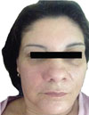
A 52-year-old woman visited a private dentist complaining of a painless swelling in the face characterised by unilateral volume of the nasolabial area. The swelling had been present for nearly 3 months and the patient reported that it had not increased in size since developing (Fig. 1). The patient's medical history and habits were non-informative. Upon an oral examination, the right and left central incisors, left maxillary lateral incisor, and canine responded to thermal and electric pulp testing within normal limits. Periodontal probing showed normal and healthy gingiva. On palpation, swelling was non-tender, firm in consistency, and not fixed. There was no detectable lesion on periapical radiographs. Clinically, a benign neoplasm of the soft tissue was hypothesised.
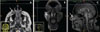
Magnetic resonance imaging (MRI) of the head was performed as an additional assessment, revealing a low-intensity rounded lesion on T2-weighted images (axial view) (Fig. 2A) and anterior wall involvement of the ipsilateral maxillary sinus with slight bulging. On the T1-weighted post-gadolinium images with spectral pre-saturation with inversion recovery (SPIR), the coronal scan showed a region with a low and homogeneous signal and well-circumscribed limits (Fig. 2B). The T1-weighted sagittal image with an iso-hypointense signal revealed an augmented mass in the medial canthal area (Fig. 2C). The signal characteristics and morphology conflicted with those of a tumour.
After MRI, the patient was further questioned about her medical history regarding the nasolabial folds, and she reported undergoing an aesthetic procedure with filling material performed by an aesthetic plastic surgeon 15 years ago, but she was unable to say which material was used.
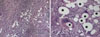
An incisional biopsy was performed and histopathological analysis showed a well-circumscribed granulomatous reaction without central necrosis, characterized by an epithelioid histiocytic organization, numerous multinucleated giant cells with peripheral disposition of the nuclei, and optically clear vacuoles in the cytoplasm, suggesting that polymethyl methacrylate was the foreign body. The peripheral areas of the granulomas were surrounded by a collagenous capsule with mononuclear inflammatory cell infiltration (Fig. 3).
The patient was scheduled for an excisional biopsy. A nodule, measuring 2.7 cm×1.6 cm×1.1 cm, of fibrous consistency, whitish colouration, lobular surface, and irregular growth was easily removed under local anaesthesia, including a small amount of healthy surrounding tissue. The post-operative period was uneventful, and her recovery was uncomplicated.
Discussion
Injectable cosmetic fillers are widely used in cosmetic surgery for their lasting effects and few complications.578 However, some complications such as inflammatory granuloma may occur at the injection site or other sites, even several years after the operation.7 Granulomas are caused by granulomatous inflammation after the aggregation of macrophages in response to large foreign bodies that cannot be phagocytosed by macrophages.9
Foreign body granulomas can arise following the injection of dermal fillers, manifesting with various clinical and histological features depending on the type of injected filler,7 frequently several years after the original cosmetic treatment.1 Therefore, because of the period elapsed between the surgical procedure and the complications, it is common that these patients do not remember the filler they received, or the origin of the lesion.1
A strong female tendency is evident among all previously published reports, possibly reflecting the tendency of women to seek cosmetic care more often than men,510 as in the present case. In addition, the patient's age and the location of the lesion reflect the fact that physiological lengthening and loss of volume are expected to occur with aging.10
The differential diagnosis may encompass a wide range of conditions. Labial cases presenting well-defined nodules suggest salivary gland cysts and tumours, in addition to soft-tissue neoplasms and cysts.1011 The MRI appearance of facial fillers varies according to the type of filler used.3 In this case, the T1- and T2-weighted MRI scans showed that the lesion was well-circumscribed and typically iso-intense or hypo-intense to the superficial and deep layers of the facial fat.
The characteristics of soft-tissue facial tumours on MRI depend on the histological grade of the tumour,12 but in general, these lesions present an intermediate signal on T1-weighted images and hyper-intensity on T2-weighted images with enhancement after contrast administration.1213
The advent of long-standing foreign body granulomas due to cosmetic fillers can cause confusion, as patients may not remember the previous facial filling treatment or when it occurred. The clinical features include erythematous and indurated painless nodules or painful swelling,26 but such features are non-specific. This means that they are often difficult to distinguish from other pathological conditions.
Imaging is important, not only to confirm the diagnosis of foreign body granuloma lesions, but also in the differential diagnosis of other lesions.514 Ultrasound has been reported to be useful for identifying granuloma lesions,14 but this procedure has weaknesses, such as the absence of certain anatomical landmarks, the lack of consolidated criteria to diagnose inflammatory reactions, and dependence on the operator's skill.1
MRI seems to be the best diagnostic tool, allowing a correct assessment of filler dislocation due to multiplanar acquisitions and determination of anatomical landmarks.14 Various studies have investigated MRI151415 as a diagnostic modality for accurately identifying the presence of foreign material. Grippaudo et al.14 showed that contrast-enhanced MRI enabled the identification of sub-cutaneous abscesses or granulomas characterized, respectively, by circular or diffuse enhancement.
In conclusion, clinicians should keep in mind that there are several clinical similarities between granulomatous reactions due to dermal fillers and salivary gland cysts or tumours. The integration of clinical examinations and imaging techniques, particularly MRI, enables a correct diagnosis to be made.
 XML Download