

 PDF
PDF ePub
ePub Citation
 Print
Print


The normal process of pneumatization of the skull base and paranasal sinuses starts in utero and develops through young adulthood.1,2 It is known that the red bone marrow is replaced by the fatty marrow prior to the normal pneumatization process of the paranasal sinuses, including the sphenoid bones.3,4 The process of marrow conversion occurs before epithelialization and the formation of the respiratory mucosa in the aerated sinus. In the sphenoid, the fatty conversion usually begins at around four months of age, and by 10-14 years of age, the fatty marrow is replaced by a fully pneumatized sinus lined by respiratory epithelium.5,6 Any deviation from the normal developmental process presents as a developmental variation, including the absence, hypoplasia, or arrested pneumatization of the sinuses, with the latter resulting in the persistence of atypical fatty marrow adjacent to the sinus into adulthood.7,8 Individuals with arrested pneumatization of the skull base are usually asymptomatic and the variant is most often discovered incidentally when imaging is performed for a separate cause. It is important to differentiate arrested pneumatization from more threatening conditions that may involve the central skull base in order to help the clinician choose the appropriate management approach. Some of the imaging features of this entity can closely mimic other benign fat-containing or ominous skull base lesions such as intraosseous lipoma, intraosseous hemangioma, hamartoma, fibrous dysplasia, and chordomas.8,9,10
Arrested pneumatization of the skull base is not well recognized among dentists and can often be confused with other pathologies associated with the skull base, which may lead to unnecessary invasive diagnostic procedures or interventions. As a result of greater appreciation of this entity by radiologists and higher resolution cone-beam computed tomography (CBCT), computed tomography (CT), and magnetic resonance image (MRI) scans, arrested pneumatization of the paranasal sinuses is now thought be significantly more common than previous estimates indicated and is thought to be underreported.
The aim of this study was to highlight the imaging features of arrested pneumatization of the skull base to aid the clinician in recognizing this developmental variant. The presenting characteristics of arrested pneumatization of the skull base and a list of differential diagnoses for similar bony changes within the skull base are also described.
Case Report
A 47-year-old female patient was referred to the advanced oral and maxillofacial imaging center of the University of Connecticut School of Dental Medicine for a presurgical evaluation of implant sites in the maxilla using CBCT.
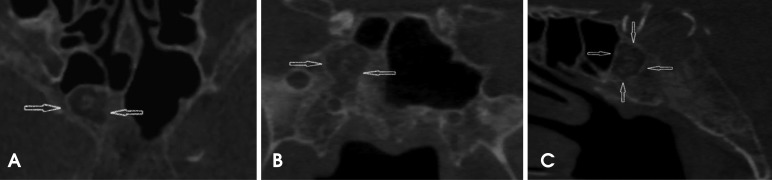
The CBCT scan was acquired with a Hitachi CBMercu-Ray CBCT unit (Hitachi Medical Corporation, Tokyo, Japan) with a nine-inch field of view at 120 kVp and 15 mA. During the radiographic interpretation of the acquired scan volume, an ellipsoid, well-demarcated, mixed-density lesion with multiple areas of mineralization was incidentally discovered in the greater sphenoid/basisphenoid area. The borders of this lesion were mildly hyperostotic. The lesion extended laterally from the midline to the right border of the greater sphenoid and sagittally to the basisphenoid, and did not communicate with the aerated portion of the remaining sphenoid sinus. A change in the normal pattern of bone trabeculae was noted inside the entity, along with multiple internal curvilinear calcifications. No mass effect was observed on the adjacent bony structures (Fig. 1). Given its location and non-aggressive radiographic appearance, the primary differential diagnoses were intraosseous lipoma, a benign fibro-osseous lesion, and arrested pneumatization.
Upon clinical examination, the patient did not show any neurological deficits or any other overt clinical symptoms. The patient's past medical history was unremarkable and no symptoms were reported of headache, deteriorating vision, or vertigo. The gray-scale CBCT values of the hypodense portion of the mass were measured. Using the grayscale values of the dental CBCT, Hounsfield units were derived using the comprehensive method described in the studies performed by Mah et al.11 and Reeves et al.12 The corrected Hounsfield units in the CBCT acquisition were characteristic of fatty tissue, suggesting the presence of fat surrounding the central, scattered curvilinear calcifications. The margins of the lesion were sharp and sclerotic, suggesting non-aggressive growth. At this time, the following working radiographic differential diagnoses were considered: intraosseous lipoma, a fibro-osseous lesion, and arrested pneumatization. Since the patient was asymptomatic and the lesion lacked the classic characteristics of an aggressive entity or a malignancy, watchful observation was recommended. The patient had a follow-up CBCT scan 12 months after the placement of dental implants and no change in the size of the lesion was noted. Therefore, although the presence of adipose tissue was not confirmed histologically, the radiographic diagnosis of arrested pneumatization was established by the non-expansile nature of the lesion, its well-circumscribed sclerotic borders, the presence of internal curvilinear calcifications, the intact borders of the adjacent neural foramina, and, crucially, the location of the lesion at a normal pneumatization site.
Discussion
During normal development, a process known as red-to-yellow marrow conversion takes place, in which the adipose tissue within the red marrow increases in relative percentage. Several studies indicate that pneumatization of the cranial bones, such as the paranasal sinuses and mastoid air cells, only occurs after red-to-yellow conversion.13 If the conversion process does not begin or is not completed, the pneumatization process is arrested and the area that is normally aerated is instead filled with yellow marrow. Arrested development is more associated with the sphenoid sinus and its known sites of accessory pneumatization, but cases involving the frontal and maxillary sinuses have also been reported.3,14 While the reason for sphenoid predominance is not clear, the considerably greater variation in the extent of aeration in the sphenoid sinus compared to other paranasal sinuses may be related to the more frequent occurrence of arrested pneumatization at this site.8 In the sphenoid bone, conversion from red to yellow marrow begins in the anterior portion (pre-sphenoid), moving posteriorly toward the clivus.4,6,13 This process begins at about four months of age,6 but significant conversion occurs by the age of two years.4
Several studies have presented different hypotheses to explain the relationship between aeration and bone marrow conversion of the sphenoid bone. Some authors have proposed that changes in the vasculature and temperature can act as promoters for sphenoid marrow conversion,4,15 while others have postulated that the ratio of trabecular to cortical bone is the driving mechanism.16 Despite this, relatively little is known about this process and why there may be a delay or failure to mature normally. Subsequently, the respiratory mucosa expands into areas of sphenoid fatty marrow conversion as the pneumatization process commences (Fig. 2A). When the conversion of fatty marrow adjacent to the sinus into the respiratory mucosa fails at some point in development, atypical fatty marrow persists into adulthood, which is why this phenomenon is described as arrested pneumatization. Patients with this benign developmental variant are most often asymptomatic and the entity is usually discovered incidentally.3,4,7,8 On imaging, this process can be confused with more ominous skull base disease processes, especially if the radiologist is unaware of arrested pneumatization or its classic characteristics, resulting in unnecessary follow-up imaging, biopsy, or management.
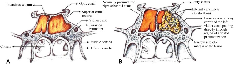
Arrested pneumatization can be diagnosed when a lesion fulfills Welker's criteria:8 (1) the lesion must be located at a site of normal pneumatization or a site of recognized accessory pneumatization; (2) the lesion should have sclerotic, well-circumscribed margins; (3) the lesion must be non-expansile; (4) the lesion should show fatty content; (5) on CT, internal curvilinear calcifications should be present; (6) any associated skull base foramina should retain a normal appearance.
The non-expansile nature of the lesion can best be evaluated at the inferior orbital fissure and vidian canal, which should not be displaced nor disrupted (Fig. 2B). The present case demonstrated all of the aforementioned features. Regarding the presence of adipose tissue, the gray-scale CBCT values of the hypodense portion of the mass were measured, and Hounsfield units were then derived using the comprehensive method described by Mah et al.11 and Reeves et al.12 The corrected Hounsfield units in the CBCT acquisition were characteristic of fatty tissue. Another approach to discern various tissue types on CBCT is to distinguish them based on density measurements acquired using pixel intensity values, which can then help distinguish benign lesions from possibly malignant lesions. The normal pixel intensity values for air in the sinuses are -700 to -1000 on the pixel intensity value scale. In this case, the area of the lesion measured approximately +185 to +200, which is considered higher than the normal values typically registered on the pixel intensity value scale for the sinuses. This pixel number range is typically associated with fatty tissue.
The formation of a pneumatized cavity in the sphenoid bone appears after a phase of bone marrow involution.13 The signs of arrested pneumatization delineated by Welker8 reflect the persistence of zones of bone marrow involution that failed to pneumatize. This process is different from hypoplasia of the sinus, in which the non-aerated segment of the sphenoid body displays a normal trabecular pattern. According to Kuntzler and Jankowski,14 the signs of arrested pneumatization may possibly reflect the mode of formation of the paranasal sinuses.
Differentiating arrested pneumatization from more threatening lesions that might involve the skull base is important to obviate the need for additional interventions such as biopsy or surgery. The presence of local fat in these cases can be misleading for radiologists unfamiliar with this entity. The low-density and heterogeneous appearance on CT/CBCT might prompt concern for a lytic process. The high signal from fat on T1-weighted MRI might be suggestive of an infiltrative lesion such as a chordoma. However, in some rare cases there might be no evidence of fat content. Some central skull base lesions that share similar imaging features with arrested pneumatization are intraosseous lipoma, chordoma, chondrosarcoma, fibrous dysplasia, intraosseous hemangioma, ossifying fibroma, and metastases. Table 1 outlines typical imaging hallmarks that help differentiate arrested pneumatization from the aforementioned lesions.
Intraosseous lipoma, a variant of classical lipoma, is composed of mature adipocyte proliferation within the marrow of normal trabecular bone.9 Although intraosseous lipoma can exhibit some overlapping characteristics with arrested pneumatization, such as internal mineralization, fat content, increased signal intensity on MRI-T1, and decreased intensity on MRI-T2 fat saturation; these lesions are commonly expansile, a characteristic which allows them to differentiated from arrested pneumatization. In addition, intraosseous lipomas in the maxillofacial region arise in the maxilla and the mandible17 more frequently than in the bones of the central skull base.
Chordomas and chondrosarcomas are other craniofacial lesions that should be differentiated from arrested pneumatization. Although chordomas more commonly develop in the vicinity of the sphenoid sinus, unlike arrested pneumatization, they are expansile and destructive and do not contain central regions containing fat.18 Their MRI features19 are presented in Table 1.
Instead of the fatty contents and curvilinear calcifications of arrested pneumatization, the internal matrix of fibrous dysplasia has an amorphous ground-glass appearance on CT imaging and contains no fat.20
Similar to arrested pneumatization, intraosseous hemangioma is generally asymptomatic unless it causes a mass effect on the surrounding structures. The CT and MRI features of hemangioma21 are presented in Table 1.
Arrested pneumatization can be differentiated from ossifying fibroma through careful observation of the internal contents. While ossifying fibroma exhibits a ground glass marrow pattern in CT images,22 the internal pattern of arrested pneumatization is distinct and is characterized by curvilinear calcification and fatty contents.
Central skull base metastasis most commonly results from breast, lung, and prostate cancers, which account for 40%, 14%, and 12% of cases, respectively.23 In CT images, metastatic disease can manifest as multifocal areas of dense sclerosis in the skull base, or may demonstrate lytic or permeative destruction of trabecular or cortical bone. Arrested pneumatization can be distinguished from skull base metastasis or osteomyelitis since it does not cause a permeative pattern of osseous destruction.24 The relevant MRI features are presented in Table 1.
We reviewed the pertinent imaging features of arrested pneumatization that can simulate the imaging characteristics of well-known lesions that can from the central skull base, including metastatic disease, intraosseous lipoma, chordoma and chondrosarcoma, intraosseous hemangioma, and fibro-osseous lesions. This comprehensive approach more realistically reflects the imaging challenges that occur when assessing whether imaging findings in this region indicate a benign anatomical variant or ominous lesions. Recognition of this variation and its typical imaging features is important for radiologists in order to avoid unnecessary further workups, biopsy, or surgery, which could potentially result in an adverse outcome. If the area of arrested pneumatization does not satisfy all of the aforementioned diagnostic criteria, serial imaging and watchful observation can help assess whether a lesion is benign.
In conclusion, the imaging features of arrested pneumatization can often resemble those of other lesions in the central skull base, including metastatic disease, intraosseous lipoma, chordoma and chondrosarcoma, intraosseous hemangioma, and fibro-osseous lesions. When encountering a non-expansile lesion with internal soft or fatty tissue, curvilinear calcifications, and sclerotic well-defined margins at a site of normal pneumatization or of recognized accessory pneumatization, a differential diagnosis of arrested pneumatization must be considered.

 XML Download