

 PDF
PDF ePub
ePub Citation
 Print
Print


Introduction
Maxillary third molars are generally less difficult to extract than mandibular third molars.1 The surgical removal of maxillary third molars is usually associated with low rates of complications and low morbidity.2 However, surgical extraction of the upper third molar can cause serious complications, such as displacement into adjacent anatomic spaces.34
The most frequent complications are fracture of the maxillary tuberosity, root fracture, and perforation of the maxillary sinus.56 The level of the occlusal plane, contact with the second molar, and the relationship of the molar to the maxillary sinus have been found to be significant predictors of surgical difficulty.1 An occlusal surface of the third molar deeper than the cementoenamel junction of the second molar can make maxillary third molar extraction more difficult.78 Contact of the third molar with the second molar root has been associated with difficulty in the surgical removal of impacted third molars.1 The possibility of accidental displacement was found to increase when the third molar was deeply impacted and when it was close to or in the maxillary sinus.9
Displacement of maxillary third molars has been associated with an inadequate clinical and radiographic examination, improper surgical technique, insufficient visibility, and excessive force during extraction.2310 Although iatrogenic tooth displacement was found to be rare during the extraction of maxillary third molars, maxillary third molars have been accidentally displaced into adjacent anatomic spaces.2 Depending on the direction of force application, the maxillary third molar can be displaced superiorly to the maxillary sinus,11 posteriorly to the infratemporal fossa,3410 posterolaterally to the buccal space,12 posterosuperiorly to the pterygopalatine space,13 or posteromedially into the lateral pharyngeal space.14 Inadequate bone height buccal and distal to the molar has been associated with a higher risk of displacement into the buccal space.12
Insufficient clinical and radiographic examination is an important factor that could lead to accidental tooth displacement.27 Accurate radiographic localization of the tooth is a prerequisite for both an initial extraction and the extraction of a displaced tooth.15 The preoperative assessment should include a detailed morphologic analysis of the third molar and its relationship to adjacent structures and surrounding tissues.16 The identification of predictive variables associated with adverse events may be helpful in reducing complications.17
Panoramic radiography is the standard preoperative imaging modality.818 However, it is difficult to precisely assess the angulation of the third molar and the position of the root relative to the maxillary sinus on panoramic radiographs. Computed tomography or cone-beam computed tomography (CBCT) is capable of providing more exact information regarding the position of the maxillary third molars.
This study was performed to investigate maxillary third molars and their relation to the maxillary sinus using panoramic radiography and CBCT.
Materials and Methods
The subjects of this retrospective study were randomly selected from patients who visited Pusan National University Dental Hospital for extraction of the upper third molars and who underwent panoramic radiography and CBCT imaging in 2013. The study sample consisted of 395 maxillary third molars from 234 patients aged 20 years and older, of which 196 were on the right and 199 were on the left. The patients comprised 120 males and 114 females with a mean age of 28.5 years (range, 20-67 years). Patients with pathology in the maxillary posterior teeth or who were missing the second molar were excluded from the study.
All panoramic radiographs were taken with a Proline XC machine (Planmeca Co., Helsinki, Finland). CBCT scans were acquired with a PaX-Zenith 3D scanner (Vatech Co., Hwasung, Korea). The scanning parameters were set at 110 kVp, 24 seconds, 5.7 mA, a voxel size of 0.2 mm, and field of view of 16 cm×14 cm. The CBCT volumetric data were reconstructed using the Ez3D Plus Professional CBCT software (Vatech Co., Hwasung, Korea).
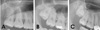
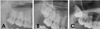
We examined the eruption level of the maxillary third molars, the available retromolar space, their relationship to the adjacent second molar, and their relationship to the maxillary sinus on panoramic radiographs. The eruption level of the maxillary third molars was assessed according to their relationship to the occlusal plane of the adjacent second molar using the Pell and Gregory classification system:19 level A; the occlusal plane of the third molar is at the same level as the adjacent tooth; level B, the occlusal plane of the third molar is between the occlusal plane and the cervical line of the adjacent tooth; and level C, the occlusal plane of the third molar is apical to the cervical line of the adjacent tooth (Fig. 1). The available retromolar space was measured as the distance between the distal surface of the second molar crown and the cortex of the maxillary tuberosity, and the space was categorized as sufficient (space greater than or equal to the mesiodistal length of the third molar), reduced (space greater than half and less than the entire mesiodistal length of the third molar), and insufficient (space less than half the mesiodistal length of the third molar) (Fig. 2). The relationship to the adjacent second molar was classified as no contact, contact with the crown, and contact with the root. The relationship of the maxillary third molar to the sinus on panoramic radiographs was classified into five types: class 1, the sinus floor is above the roots; class 2, the sinus floor touches the root tips; class 3, the sinus floor is superimposed on up to one third of the root; class 4, the sinus floor is superimposed on up to two thirds of the root; and class 5, the sinus floor extends up to the tooth cervix (Fig. 3).
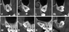
On CBCT images, the angulation of the maxillary third molars, the number of roots, and the horizontal relationship between the roots of the third molars and the sinus were investigated. The angulation of the maxillary third molar with respect to the long axis of the second molar was classified as vertical, buccoangular, linguoangular, buccolingual, mesioangular, distoangular, horizontal, and inverted using a modified version of Winter's classification (Fig. 4). The number of roots was classified into one fused root, two roots, three roots, and four roots. Class 3, class 4, and class 5 relationships on panoramic radiographs were subclassified into five types according to the horizontal relationship between the roots of the third molars and the sinus on CBCT images: type B, the lowest point of the maxillary sinus floor is located on the buccal side of the root; type C, the root projects into the sinus; type P, the lowest point of the sinus floor is located on the palatal side of the root; type M, the lowest point of the maxillary sinus floor is located on the mesial side of the maxillary third molar; and type D, the lowest point of the maxillary sinus floor is located on the distal side of the maxillary third molar (Fig. 5).
The statistical analysis consisted of descriptive crosstabulations showing frequency distributions among the selected categorical variables. Data were analyzed using the chi-square test and Fisher's exact test. P-values less than 0.05 were considered to indicate statistically significant differences. The statistical analyses were performed using SPSS version 21.0 (IBM Corp., Armonk, NY, USA).
Results
The eruption levels of the 395 maxillary third molars included in this study were classified as follows: level A for 183 (46.3%), level B for 50 (12.7%), and level C for 162 (41.0%). Level C was more common in females (49.0%) than in males (33.5%). The distribution of the eruption levels was significantly different between genders (P<0.01) (Table 1).
A total of 182 maxillary third molars with sufficient retromolar space were level A (94.3%), 105 third molars with reduced space were level C (71.9%), and all third molars with insufficient space were level C. The chisquare test showed statistically significant associations between the eruption level and the available retromolar space (P<0.01) (Table 2).
The most common angulation of the maxillary third molars was vertical (59.0%), followed by buccoangular (18.2%) and mesioangular (13.2%). Vertical angulation (85.0%) was most frequent in molars with sufficient retromolar space, as well as in molars with reduced space (40.4%). The buccoangular orientation was the second most common (29.5%) in molars with reduced space, and mesioangular angulation (39.3%) was the most common in molars with insufficient space (Table 3).
All level A maxillary third molars were in contact with the crown of the second molars, while 30% of level B molars and 99.4% of level C molars were in contact with the root of the second molar (Table 4). Maxillary third molars with three roots were most frequent in level A molars (44.8%), and the presence of one fused root was most frequent in level C molars (51.2%) (Table 5).
Class 3 was most common in level A and level B molars, and class 4 was most common in level C molars (Table 6). When the sinus floor was superimposed on the root of the maxillary third molars, type B was most frequent in class 3 and class 4 molars, and type M was most frequent in class 5 molars (Table 7).
Discussion
We investigated maxillary third molars and assessed their relationship to the maxillary sinus on panoramic radiographs and CBCT images. Careful radiographic localization of the third molar is a prerequisite for surgical extraction in order to prevent complications.15
Some studies have found impaction of the third molar to be common in females.20212223 Others have reported no gender predilection for third molar impaction.2425 The higher frequency reported in females has been explained by gender-based differences in growth patterns. Females usually stop growing when the third molars just begin to erupt, whereas in males, the jaws continue to grow while the third molars erupt, creating more space for third molar eruption.26 In this study, impacted third molars were more common in females than in males.
The etiology of third molar impaction has been investigated in many international studies. Several factors have been reported as possible causes of third molar impaction, including a lack of space distal to the permanent second molar, delayed third molar mineralization, and early physical maturation.26 Factors commonly associated with third molar impaction include a shortage of space available for eruption, and third molar impaction was found to be more likely to occur when the retromolar space was inadequate.27 These studies found that the retromolar space was closely related to the eruption level of maxillary third molars.
Increased third molar angulation has also been found to be significantly linked to third molar impaction.27 Hashemipour et al.20 evaluated the angulation of maxillary third molars using panoramic radiography, and the most common angulation was vertical, followed by distal. In our results, the most common angulation was vertical, followed by buccoangular. This could have been because we assessed the angulation of the maxillary third molars using CBCT images and added the categories of buccoangular, linguoangular, and buccolingual, which could not be assessed on panoramic radiographs. Buccoangular or buccolingual third molars could have been classified as distoangular on panoramic radiographs.
Greater proximity between the third and second molars has been associated with an additional risk of surgical difficulty.117 A close relationship between these teeth reduces the space between the distal surface of the second molar and the mesial surface of the third molar, impeding access to the tip of the elevator.17 Our results showed that most level C molars were in contact with the root of the adjacent second molar.
The root number and morphology have been found to be even more important, because these factors were associated with more issues in surgical management, although this trend did not reach statistical significance.1 In our results, one fused root was most frequent, followed by three roots, and four roots were rarely observed.
It is important to know the anatomical relationship between the maxillary sinus and the third molar in preoperative treatment planning for maxillary third molars.1 The more the sinus floor was projected onto the root on panoramic radiographs, the greater the relative probability of oroantral communication.18 If no superimposition of the root and sinus floor was visible on the panoramic radiographs, no oroantral perforation was to be expected.18 However, the relative probability of oroantral perforation increased in class 3 molars, and was significantly higher in class 4 and class 5 molars.18 In our results, the superimposition of the sinus and the root of the maxillary third molars was observed in more than two thirds of panoramic radiographs. The depth of the impaction of the maxillary third molars was associated with a greater likelihood of oroantral perforation.2829 In our results, eruption level C showed more superimposition between the sinus and root than levels A or B.
When the sinus floor is superimposed on the root on panoramic radiographs, it is necessary to obtain more information about the relationship between the sinus and the third molars in order to prevent sinus perforation.30 We used CBCT images to identify the horizontal relationship between the sinus and the third molars, and the sinus floor was most commonly found to be located on the buccal side of the root.
In order to minimize the risk of complications, surgical difficulty should not be underestimated. We should identify variables predictive of a higher risk of complications during removal of the maxillary third molars and take action accordingly. Further research is needed to evaluate the correlation of predictive radiographic factors with the occurrence and types of complications.
In conclusion, the eruption level showed a different distribution between males and females. A statistically significant association was found between the eruption level and the available retromolar space. Expansion of the sinus to the buccal side of the root was the most frequently seen pattern on CBCT when the panoramic radiographs showed a superimposition of the roots and the sinus floor.


 XML Download