

 PDF
PDF ePub
ePub Citation
 Print
Print


Introduction
Radiography plays an important role in endodontic treatment of teeth. The clinical assessment of an endodontic treatment is confirmed by a radiographic evaluation. Good-quality radiographs help endodontists to determine the treatment-need as well as the number, shape, size, and direction of roots and root canals. The ultimate goal of endodontics is to provide a proper seal at the apex, thus the knowledge of the exact location of the apex is critical.1 After the diagnosis, the next step in endodontic therapy is working length estimation in which radiography plays a critical role.2 The cleaning, shaping, and obturating of the root canal system cannot be accomplished accurately unless the working length is determined precisely.1
Different types of image receptors are available in dental radiography. These image receptors can be extensively classified into two categories: film based (or conventional) and digital image receptors. With several advantages of digital radiography such as less patient absorbed dose, the manipulation of image quality, and the noise reduction by image processing, it has been recommended in numerous fields of dentistry.3 The evaluation of digital radiography in endodontics initially had variable outcomes. Some researchers found that it was inferior to conventional radiography4-6 and others argued that it was more useful or at least similar to conventional radiography.7-9 The studies might have been reached by utilizing different digital devices.
The measurement of the canal length became more complicated as the degree of root curvature increased.10 There were limited studies8-11 about the accuracy of CMOS (complementary metal oxide semiconductor) digital sensor in the evaluation of working length of canals especially in curved canals.
The aim of this study was estimating the working length in a range of canal curvatures. In our study we tried to evaluate CMOS-based sensor in the measurement of root canal length in curved canals (slight to severe), because we thought that in clinical setting we might come across curved canal which may have affected the result of previous studies.
Materials and Methods
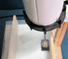
Sixty extracted first mandibular molars with varying mesial root curvature were obtained. The radiographs were taken from the teeth in a buccal to lingual orientation and tracings made on the projected mesiobuccal canal images. Then the canal curvature was determined using Schneider's method.12 The curvature of the roots was categorized to slight for curvature ranges of 0-15 degree, moderate 15-30 degree, and severe over 30 degree. There were 20 teeth in each group. Small occlusal reference point was established on each tooth with a 557 bur. A 15-k file with rubber stopper was inserted in the mesiobuccal canal and advanced until the file tip was visualized at the foramen. The stopper was set at the occlusal reference point and the file was removed. True canal length was measured for each tooth using a millimeter ruler (Faber-Castell, Sydney, Australia). The file was then returned to the canal and fixed in place with a light-cured composite resin. The teeth were mounted in opaque acrylic tray material. An apparatus was fabricated to allow for a constant spatial relationship among the x-ray source, the teeth and the receptor (Fig. 1). The angulation and position of the central ray entry was fixed in that way; the end of the PID (the position indicating device) brought the sensor against a ring of film holder (RWT, regular, Ellwangen, Germany). The distance from the source to the film/sensor was 40 cm. A 0.5 cm thick glass plate was used for the simulation of x-ray absorbing and scattering properties of the soft tissue. The samples were imaged with both conventional and digital techniques. The conventional radiographs were obtained using F speed film (Flow x-ray, FV-58, NY, USA) whereas the digital images were obtained using CMOS sensor (Schick, Long Island City, NY, USA). Both recording devices were exposed with x-rays generated by a Planmeca unit (Planmeca, Helsinki, Finland) operating at 70 kVp and 8 mA. The optimal exposure time for each radiographic method was established during a pilot study. The estimated canal length was then measured as the distance from the occlusal reference point to the most apical extent of the file tip. A maxillofacial radiologist and an endodontist with more than 6 years of professional work experience, familiar with digital radiographic methods served as the observers. They performed the following measurements of canal length under the ideal and same situations:
Estimation of canal length on conventional radiograph by a millimeter ruler.
Estimation of canal length by measuring tool of digital system with 2 clicks for the group of slight curves (first click at the reference point, second click at the file tip) and 3 clicks for groups of medium and severe curves (first click at the reference point, the second click at the tip of the angle along the canal curvature and the third click at the file tip).
The measurements were performed in a randomized sessions to decrease the risk of bias because it was not possible to blind the observers to the imaging techniques.
The correlation between the observers was measured by Pearson's correlation test. Average measurement of each examiner was compared with the true canal length according to root curvatures. Repeated measure analysis of variance and paired sample t-test with 95% confidence level were used to evaluate the results.
Results
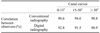
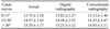
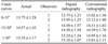
In this study, the working length was evaluated in 60 extracted teeth by two observers. There was no significant difference between the observers (Table 1). Thus the average measurement by the observers in each radiographic technique was compared with the average of actual measurement by repeated measure analysis of variance with 95% confidence level. No statistically significant differences were found between the experimental groups and the true canal lengths (Table 2), although conventional radiography underestimated the true canal in the range of 0.2-0.5 mm and digital radiography overestimated the true canal length in the range of 0.01-0.06 mm in the slight and moderate curvature groups and only underestimated the true canal length in severe curvature group (0.14 mm). The average measurements in conventional and digital radiography by each observer were compared with actual measurements as the basis of canal curves with paired sample t-test with 95% confidence level (Table 3). There was no significant difference between conventional and digital radiography with actual values in the basis of canal curves (p>0.05).
Discussion
Establishing an accurate working length is the first step toward successful non-surgical root canal therapy. However, it is clinically difficult to locate the apical constriction or dentino-cemental junction. The working length is commonly taken to be 0.5 to 1 mm short of the radiographic apex. The radiographic image of the apex seldom offers a clear view of the terminal part of the canal because radiographic magnification and distortion affects on root canal length estimation.13 Magnification can be minimized by keeping the object as close to the film as possible. Distortion can be minimized by using a paralleling technique to assist the positioning of the object in the central part of the x-ray beam.14 Therefore, the present study was done by using parallel technique. Our results indicated that there was no difference between the F-speed film and Schick CMOS sensor for working length determination. These findings were in agreement with the results of Lamus et al11 and there was no difference between direct digital and conventional film based radiographs in the estimation of working length but in contrast to their study the digital radiography was the most closely approaching method to the gold standard. They compared Schick CDR direct digital system and conventional E-speed film to estimate working Length.11 Other studies indicated that there were no differences among D-speed film, F-speed film, and Schick CDR system for working length determination.8 In their studies,8,11 the curvature of canals were not considered. However, in our study the accuracy of working length determination was done as the basis of the different categories of canal curvatures. In our study there was no significant difference between experimental groups, regardless of canal curvature. These findings were comparable with the results of Burger et al.9 They showed that there were no significant differences among true length, DR (two clicks, six clicks, and unlimited clicks) and conventional radiographs for estimation of canal length. They used D-speed film and Trophy's fourth-generation RVG system.
Mentes et al10 revealed that there was no significant difference between conventional and direct digital radiographs in the estimation of canal length as the basis of canal curves. They used E-speed film and Reveal sensor system (Welch Allyn Inc, Skaneateles Falls, NY, USA). In their study, both of the imaging modalities overestimated the true canal length and digital accuracy got better as the severity of curvature increased. In our study digital radiography overestimated the true canal length in the slight and moderate curvature groups.
We designed this study as an Invitro study because we were not able to achieve the aim of this study in a clinical setting. In an Invitro study, image quality can be standardized and ideal positioning of the anatomic specimen with exact reproducibility is possible.
In an Invivo study, we have the limitation in patient positioning and absolute reproducibility. The other limitations of Invivo study are comparing with actual anatomic structure and variation of image quality from one patient to another.
In conclusion, this study indicated that the accuracy of conventional and digital radiography in the evaluation of working length was acceptable and there was no difference in the accuracy of F-speed film and digital images for the determination of the endodontics file working length.
 XML Download