

 PDF
PDF ePub
ePub Citation
 Print
Print


INTRODUCTION
Hypertrophic cardiomyopathy (HCM) is a complex and relatively common genetic cardiac disease with an estimated prevalence of 0.2% (1). Frequently, patients with HCM have chest pain suggestive of angina pectoris, and the electrocardiogram (ECG) can resemble that of a myocardial infarction (MI) in the absence of coronary artery disease (CAD) (23). However, adult patients with HCM may also develop atherosclerotic CAD, and patients with HCM and coexistent CAD have an increased rate of morbidity compared to patients with HCM without CAD (45). Therefore, early detection of coexistent CAD in HCM is of high importance.
Myocardial bridging (MB) is more commonly associated with HCM than with non-HCM (6). Although most occurrences of MB are thought to be innocent, in some cases, they may cause angina pectoris, MI, life-threatening ventricular arrhythmias, or even sudden cardiac death (678).
Traditionally, coexistent CAD or MB in HCM was diagnosed with two different methods; the magnitude of myocardial hypertrophy was usually assessed with 2-dimensional echocardiography, while coexistent CAD or MB was evaluated with invasive coronary angiography (ICA) (57). Although pharmacologic stress echocardiography was used as a reasonable method for diagnosis of the occurrence of both coexistent CAD and HCM at the same time, the feasibility of dipyridamole-stress echocardiography was often limited, especially in patients with poor echo windows or chronic theophylline therapy (9). Currently, coronary computed tomography angiography (CCTA) with the ECG-gated technique is an attractive noninvasive imaging method for the evaluation of coronary arteries and myocardial hypertrophy at the same time (10). However, there is little data regarding the role of CCTA in the evaluation of patients with HCM.
Therefore, the purpose of this study was to investigate the prevalence and risk factors associated with coexistent CAD or MB in patients with HCM using CCTA. In addition, we assessed the feasibility and diagnostic accuracy of CCTA for the detection of coexistent CAD or MB in patients with HCM in comparison to ICA.
MATERIALS AND METHODS
Study Population
The Institutional Review Board approved the study protocol, and informed consent was waived. Among a registry of 19588 patients who underwent CCTA between January 2008 and December 2012 in Seoul National University Bundang Hospital, we retrospectively enrolled 150 adults with HCM [112 men; mean age ± standard deviation (SD): 62.4 ± 11.0 years; range: 36-90 years]. The diagnosis of HCM was based on clinical and ECG findings and echocardiographic features, with left ventricular hypertrophy (LVH), in the absence of another cardiac or systemic disease, known to cause hypertrophied myocardium (1). Clinical data including family history and ECG findings were retrieved through medical record review. LVH was assessed in the end-diastolic stage with M-mode and 2-dimensional transthoracic echocardiography by standard techniques.
According to morphological phenotype, septal HCM was defined as a septal thickness greater than 15 mm or a septal thickness to inferior LV wall thickness ratio of greater than 1.5 at the mid-ventricular level, and apical HCM was defined as an absolute apical wall thickness of greater than 15 mm or an apical to basal LV wall thicknesses ratio of 1.3-1.5 showing characteristic "spade-like configuration". The other types of HCM were named according to the hypertrophied location, such as midventricular type or concentric type HCM (10). The combination of two or more phenotypes was classified as combined or mixed type HCM.
Clinical Symptoms and Risk Factors
Clinical symptoms, basic demographic data, and clinical risk factors of each patient were obtained through the physician's interview and medical record. According to the Canadian Cardiac Society (CCS) angina classification, CCS III or higher was defined as severe chest pain (11). Body weight, height, and blood pressure were also measured at the time of CT scan. The traditional risk factors for CAD, such as hypertension, diabetes, smoking or hypercholesterolemia, and family history of premature coronary heart disease (FHx-CHD, CHD in a male first-degree relative less than 55 years of age; CHD in a female first-degree relative less than 65 years of age), were evaluated on the basis of the physician's interview and laboratory findings, and the details were published previously (12). The Framingham risk score (FRS) was calculated using risk factors including sex, age, total cholesterol, high-density lipoprotein cholesterol, smoking, systolic blood pressure, and antihypertensive medications and estimated to a 10-year risk of CAD (13).
Scan Protocol and Image Reconstruction of CCTA
Patients with a heart rate greater than 70 beats/min received intravenous esmolol 10-30 mg (Jeil Pharm. Co., Ltd., Seoul, Korea) before CCTA imaging. The cardiac CT examinations were performed using a 64-slice multidetector CT scanner (Brilliance 64, Philips Medical Systems, Best, the Netherlands) with 64 × 0.625 mm section collimation, 420-ms rotation time, 120-kV tube voltage, and 800-mA tube current. All scans were performed with ECG-gated dose modulation. A bolus of 80 mL iomeprol (Iomeron 400, Bracco, Milan, Italy) was intravenously injected (4 mL/s) followed by a 50-mL saline chaser. A region of interest (ROI) was placed in the descending thoracic aorta, and image acquisition was automatically initiated once a selected threshold (150 Hounsfield units) had been reached with bolus tracking. The patient's ECG was simultaneously recorded to allow for retrospective segmental data reconstruction. Images were initially reconstructed at the mid-diastolic phase (75% of R-R interval) of the cardiac cycle. If motion artifacts were present, additional reconstructions were performed for the motion-free phase.
CCTA Image Analysis
Two expert radiologists (SIC 10 years; EJC 8 years for interpretation of cardiac imaging) analyzed the CT images with a 3-dimensional workstation (Brilliance, Philips Medical Systems, Best, the Netherlands). They were blinded to the clinical information. After making independent evaluations, a consensus interpretation was achieved to obtain a final CCTA diagnosis. The contrast-enhanced portion of the coronary lumen was semi-automatically traced at the maximal stenotic site and compared with the mean value of the proximal and distal reference site. The severity of diameter stenosis was evaluated on a per-segment basis according to a 16-segment model (14), and graded as normal nonobstructive CAD (< 50% luminal stenosis), and obstructive CAD (≥ 50% luminal reduction). Image quality was evaluated on a per-segment basis using a 3-point grading scale (good, no artifacts; adequate, mild artifacts but fully evaluable; poor, noninterpretable). No segment was excluded from analysis.
MB was diagnosed when one of the vascular segments tunnels through the myocardium, causing the segment to be in contact with the left ventricular myocardium, without intervening fat (15). We divided instances of MB into two groups: 1) partial encasement, which was defined as the vascular segment partially embedded within the myocardium and 2) full encasement, which was defined as the vascular segment surrounded by the myocardium with measureable overlying muscle (15).
Invasive Coronary Angiography
ICA was performed in 37 of 150 patients within 1 month after the CCTA scan.
ICA was determined with a consideration of each patient's symptoms or subsequent diagnostic test such as single-photon emission computed tomography. ICA was performed using 5-French high-flow Judkins catheters (Cordis, Miami, FL, USA), and images were acquired in multiple projections.
An experienced cardiologist, blinded to the multidetector CT results, analyzed the coronary angiograms using a validated quantitative coronary angiographic system for determining the degree of coronary artery stenosis (Philips H5000, Philips Medical Systems, Andover, MA, USA; or Allula DCI program, Philips Medical Systems, Best, the Netherlands). The severity of coronary stenosis was quantified in two orthogonal views, and obstructive CAD was classified as significant if the lumen diameter reduction was greater than 50%. MB was diagnosed as a change in luminal narrowing that was more pronounced than that in neighboring normal vessels during systole (15).
Statistical Analysis
Continuous variables are expressed as mean ± SD, while categorical variables are presented using absolute value and percentage. Between two or more groups according to degree of stenosis (normal, nonobstructive, or obstructive CAD) or MB (none, partial encasement MB, or full encasement MB), differences in continuous variables were analyzed using the one-way analysis of variance test. Differences in categorical variables were analyzed using the chi-square test. A p value of < 0.05 was considered statistically significant, and all analyses were performed with the Statistical Package for the Social Sciences (SPSS) 20.0 statistical package (SPSS Inc., Chicago, IL, USA).
For determining the accuracy of CCTA to detect obstructive CAD or full encasement MB compared with ICA, estimations of sensitivity, specificity, positive predictive value (PPV), and negative predictive value (NPV) were calculated.
RESULTS
Baseline Characteristics of the Study Population
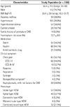
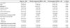
Table 1 demonstrates the baseline characteristics of the study population. In total, 56.7% of HCM patients complained of chest pain. Among them, 19 patients (12.7%) had severe chest pain with CCS III or higher. Although 36 patients (24.0%) with HCM were asymptomatic, we performed CCTA on these patients due to the presence of at least one risk factor such as diabetes, hypertension, or FHx-CHD.
Among the various morphological phenotypes, the septal type (38.0%) was most common, followed by the apical type (32.7%). Among 32 patients with combined or mixed type HCM, 25 patients (78.1%) had combined apical and septal type HCM.
Prevalence of CAD and MB in Patients with HCM
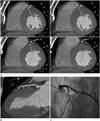
Ninety-six patients with HCM had coronary plaque (64.0%). Among them, 35 patients (23.3%) had obstructive CAD (Fig. 1). Table 2 demonstrates a comparison of the clinical characteristics and morphological phenotype of HCM according to stenosis degree of coexistent CAD. Increasing age and FRS showed a significant correlation to degree of stenosis in a stepwise fashion (p < 0.001). The prevalence of hypertension (p = 0.001) and FHx-CHD (p = 0.034) also tended to increase as severity of stenosis progressed. Although overall chest pain did not significantly differ according to stenosis degree, the prevalence of severe chest pain (CCS III-IV) was significantly higher in patients with obstructive CAD (p = 0.039).
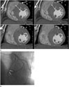
The prevalence of coexistent MB was 42.7% (64 of 150 patients). Partial encasement MB and full encasement MB were noted in 22 patients (14.7%) and 42 patients (28.0%), respectively. There were no significant differences in clinical characteristics among the degrees of MB besides male sex (p < 0.001). Among the morphological phenotypes of HCM, septal type HCM was significantly higher in full encasement MB (p = 0.001) (Fig. 2). A comparison of clinical characteristics according to the degree of the MB is summarized in Table 3.
Feasibility and Diagnostic Accuracy of CCTA in Comparison to ICA for Coexistent CAD and MB
The image quality of coronary arteries in 150 adults with HCM was classified as good in 2202 segments (91.8%), adequate in 171 segments (7.1%), and poor in 27 segments (1.1%). Therefore, overall feasibility of coronary artery visualization was 98.9%. Poor image quality was attributed to the following reasons: motion artifacts (12 segments), blooming artifacts related to calcification (11 segments), or small vessels (4 segments). Interobserver agreement for the degree of stenosis (Cohen's kappa = 0.89) was very good, and those of partial and full encasement MB (Cohen's kappa = 0.92 and 0.95, respectively) were excellent.
Among 37 patients who underwent ICA, 22 patients (59.5%) had obstructive CAD and 5 patients had MB. Three patients had both CAD and MB. With CCTA, per-patient sensitivity, specificity, PPV, and NPV for obstructive CAD were 86.4%, 93.3%, 95.0%, and 82.4%, respectively. Per-segment sensitivity, specificity, PPV, and NPV were 88.0%, 99.0%, 92.2%, and 98.9%, respectively. Among 37 patients with ICA, MB was found in 13 patients with CCTA (partial encasement MB and full encasement MB in 3 patients and 10 patients, respectively). However, five patients, all of whom had full encasement MB by CCTA, were finally confirmed by ICA. Therefore, the sensitivity, specificity, PPV, and NPV of CCTA for full encasement MB were 100%, 84.4%, 50.0%, and 100%, respectively.
DISCUSSION
The main findings of this study were as follows. 1) The prevalence of coexistent obstructive CAD and full encasement MB in patients with HCM were 23.3% and 28.0%, respectively. 2) Age, hypertension, FHx-CHD, FRS and severe chest pain were associated with CAD, whereas male gender and septal type were associated with MB. 3) In comparison to ICA, CCTA was a feasible and accurate noninvasive imaging modality for the evaluation of coexistent CAD or MB in patients with HCM.
Because the most common presentation of HCM is chest pain, which mimics angina, the diagnosis of coexistent CAD is very difficult (1617). The ECG pattern is also diverse and can resemble that of MI even in the absence of obstructive CAD (18). However, adult patients with HCM are known to have a higher incidence and severity of coexisting CAD as age increases (19). Ac-cording to earlier observations, the prevalence of CAD con-firmed by ICA is approximately 11-26% in patients with HCM (51920), which corresponds with the result of our investigation (23.3%).
In our study, age, hypertension, FHx-CHD, FRS and severe chest pain were associated with CAD in patients with HCM. FRS provided a 10-year risk of cardiovascular events regarding multiple clinical risk factors including age, sex, smoking history, blood pressure, and lipid profiles, which have been most widely used and validated as a prediction model for CAD (2122). Therefore, FRS may be a good screening tool for CAD in patients with HCM. Numerous studies reported that FHx-CHD had been associated with a persistent increase in mortality risk across long-term follow-ups (23). FHx-CHD was reported to be associated with enhanced development and progression of subclinical disease, independent of other risk factors, in a multiethnic, population-based study (24).
MB is a well-recognized phenomenon that has a prevalence of 1-3% in the general population (25), though it occurs far more frequently in patients with HCM with variable prevalence rates between 15 and 41% (681525). In the present study, the prevalence of coexistent MB was 42.7%, including partial encasement (14.7%) and full encasement (28.0%), in agreement with previous studies. The detection rate of MB by CCTA was significantly higher than that by CAG, due to the ability of CCTA to clearly show the anatomical structure and intramyocardial location of the involved coronary arterial segment, and to detect partial encasement MB, which cannot be detected by ICA (15). In our results, MB was frequently found in septal type HCM, indicating that hypertrophied septal myocardia have a higher chance of surrounding the mid portion of the left anterior descending artery. Whether MB in HCM is related to poor clinical outcomes has been controversial (2526). In studies of pediatric patients with HCM, MB has been associated with cardiac disease severity and an increased risk of sudden cardiac death, while no significant correlation was found in adult patients (625). In our study, the presence of MB was not associated with specific symptoms in adults with HCM. These results might reflect the generally benign nature of MB in adults, but future studies will be required to ascertain the correct prognosis of MB in HCM.
Although ICA is generally accepted as the gold standard for diagnosing CAD (17), it is difficult to use ICA as a screening test in patients with HCM because ICA is highly invasive. Studies of other noninvasive diagnostic tools, including pharmacological stress echocardiography or stress thallium myocardial perfusion scan, revealed relatively high false positive rates, possibly because of the innate vulnerability of hypertrophied myocardium for myocardial ischemia without significant CAD (117). With the advantage of detailed morphological assessment with 3-dimensional views, CCTA has been an effective alternative to ICA in patients with HCM, because it can evaluate both the coronary artery and the myocardium at the same time.
There were several limitations in this study. First, our study population was retrospectively analyzed and subject to bias. Second, as the decision for ICA was based on clinical reasons, not all patients with HCM underwent ICA. Thus, the diagnostic accuracy of CCTA for the evaluation of coexistent CAD or MB was not validated in all populations of our study, although CCTA showed high diagnostic accuracy in selected patients who underwent both CCTA and ICA. Finally, the lack of long-term follow-up results limited the clinical relevance of coexistent CAD or full encasement MB in patients with HCM. Further long-term follow-up studies will be needed to determine a more concrete clinical implication.
In conclusion, approximately one-quarter of patients with HCM had coexistent obstructive CAD or full encasement MB. CCTA can be a feasible and accurate noninvasive method for identification of coexistent CAD or MB in patients with HCM.
 XML Download