

 PDF
PDF ePub
ePub Citation
 Print
Print


Abstract
Purpose
To evaluate the changes of emphysema quantification in a follow-up low dose CT compared with pulmonary function test (PFT) results in asymptomatic smokers.
Materials and Methods
We selected 66 asymptomatic smokers (> 40 years old) who underwent a follow-up low dose CT at least one year after the first CT as well as PFT within the same time period. Emphysema quantification was performed using an automated measurement software and an emphysema index (EI) was calculated using multiple threshold values (-970--900 HU). The interval change of EI (ΔEI) was compared with the change in the PFT values.
Results
Mean follow-up %forced expiratory volume in 1 second (88.1), %forced vital capacity (FVC) (89.5) and forced expiratory flow between 25 and 75% of vital capacity (3.21) were significantly lower compared with the values of initial tests (93.3, 93.1, 3.48). The mean EIs (2.4-25.6%) increased on follow-up CTs compared with initial EIs (2.1-24.5%), though the increase was not statistically significant. In a group with a follow-up period of 2 years or more (n = 32), EI significantly increased when using -900 HU as the threshold. The ΔEIs were poorly correlated with the ΔPFT values, but significantly correlated with ΔFVC (r = -0.32--0.27).
Figures and Tables
Fig. 1
Example of emphysema quantification in a 47-year-old smoker.
A. Reconstructed coronal image shows highlighted pixels in both lungs indicating lower attenuated areas with the threshold of -950 HU.
B. Volume rendering image shows automatically segmented lungs and airways in different colors.
C. Histogram analysis demonstrates calculated lung volumes, emphysema volumes and emphysema indices.
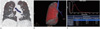
Fig. 2
Box plot of EIs with the threshold of -900 HU at initial and follow-up low dose CT in a group of patients with a follow-up period of 2 years or more. Follow-up mean EI (31.5%) are significantly increased comparing with initial mean EI (27.7%) (p = 0.02). Upper and lower margins of box plot indicate 75 and 25 percentile values of EI.
Note.-EI = emphysema index
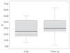


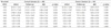
Table 3
Correlation Coefficients by Spearman Rank Correlation between Changes of PFT Parameters and EI in All Patients (n = 66)


References
1. American Thoracic Society. Standards for the diagnosis and care of patients with chronic obstructive pulmonary disease. Am J Respir Crit Care Med. 1995; 152:S77–S121.
2. The definition of emphysema. Report of a National Heart, Lung, and Blood Institute, Division of Lung Diseases workshop. Am Rev Respir Dis. 1985; 132:182–185.
3. Uppaluri R, Mitsa T, Sonka M, Hoffman EA, McLennan G. Quantification of pulmonary emphysema from lung computed tomography images. Am J Respir Crit Care Med. 1997; 156:248–254.
4. Coxson HO, Rogers RM. Quantitative computed tomography of chronic obstructive pulmonary disease. Acad Radiol. 2005; 12:1457–1463.
5. Litmanovich D, Boiselle PM, Bankier AA. CT of pulmonary emphysema--current status, challenges, and future directions. Eur Radiol. 2009; 19:537–551.
6. Nishimura K, Murata K, Yamagishi M, Itoh H, Ikeda A, Tsukino M, et al. Comparison of different computed tomography scanning methods for quantifying emphysema. J Thorac Imaging. 1998; 13:193–198.
7. Park KJ, Bergin CJ, Clausen JL. Quantitation of emphysema with three-dimensional CT densitometry: comparison with two-dimensional analysis, visual emphysema scores, and pulmonary function test results. Radiology. 1999; 211:541–547.
8. Revel MP, Faivre JB, Remy-Jardin M, Deken V, Duhamel A, Marquette CH, et al. Automated lobar quantification of emphysema in patients with severe COPD. Eur Radiol. 2008; 18:2723–2730.
9. Gierada DS, Pilgram TK, Whiting BR, Hong C, Bierhals AJ, Kim JH, et al. Comparison of standard- and low-radiation-dose CT for quantification of emphysema. AJR Am J Roentgenol. 2007; 188:42–47.
10. Horiuchi N, Fujita J, Suemitsu I, Yamasaki Y, Higa F, Tateyama M. Low-dose multislice CT and high-resolution CT assessment of pulmonary emphysema in public school teachers. Lung. 2007; 185:25–30.
11. Yuan R, Mayo JR, Hogg JC, Paré PD, McWilliams AM, Lam S, et al. The effects of radiation dose and CT manufacturer on measurements of lung densitometry. Chest. 2007; 132:617–623.
12. Gietema HA, Schilham AM, van Ginneken B, van Klaveren RJ, Lammers JW, Prokop M. Monitoring of smoking-induced emphysema with CT in a lung cancer screening setting: detection of real increase in extent of emphysema. Radiology. 2007; 244:890–897.
13. Bastarrika G, Wisnivesky JP, Pueyo JC, Díaz L, Arraiza M, Villanueva A, et al. Low-dose volumetric computed tomography for quantification of emphysema in asymptomatic smokers participating in an early lung cancer detection trial. J Thorac Imaging. 2009; 24:206–211.
14. Park SJ, Lee CH, Goo JM, Heo CY, Kim JH. Inter-scan repeatability of CT-based lung densitometry in the surveillance of emphysema in a lung cancer screening setting. Eur J Radiol. 2011; [Epub ahead of print].
15. Soejima K, Yamaguchi K, Kohda E, Takeshita K, Ito Y, Mastubara H, et al. Longitudinal follow-up study of smoking-induced lung density changes by high-resolution computed tomography. Am J Respir Crit Care Med. 2000; 161:1264–1273.
16. Dirksen A, Piitulainen E, Parr DG, Deng C, Wencker M, Shaker SB, et al. Exploring the role of CT densitometry: a randomised study of augmentation therapy in alpha1-antitrypsin deficiency. Eur Respir J. 2009; 33:1345–1353.
17. Boedeker KL, McNitt-Gray MF, Rogers SR, Truong DA, Brown MS, Gjertson DW, et al. Emphysema: effect of reconstruction algorithm on CT imaging measures. Radiology. 2004; 232:295–301.
18. Madani A, Zanen J, de Maertelaer V, Gevenois PA. Pulmonary emphysema: objective quantification at multi-detector row CT--comparison with macroscopic and microscopic morphometry. Radiology. 2006; 238:1036–1043.
19. Hochhegger B, Irion KL, Marchiori E, Moreira JS. Reconstruction algorithms influence the follow-up variability in the longitudinal CT emphysema index measurements. Korean J Radiol. 2011; 12:169–175.
20. Xu X, Dockery DW, Ware JH, Speizer FE, Ferris BG Jr. Effects of cigarette smoking on rate of loss of pulmonary function in adults: a longitudinal assessment. Am Rev Respir Dis. 1992; 146:1345–1348.
21. Kohansal R, Martinez-Camblor P, Agustí A, Buist AS, Mannino DM, Soriano JB. The natural history of chronic airflow obstruction revisited: an analysis of the Framingham offspring cohort. Am J Respir Crit Care Med. 2009; 180:3–10.
22. Wise RA. The value of forced expiratory volume in 1 second decline in the assessment of chronic obstructive pulmonary disease progression. Am J Med. 2006; 119:4–11.
23. Remy-Jardin M, Edme JL, Boulenguez C, Remy J, Mastora I, Sobaszek A. Longitudinal follow-up study of smoker's lung with thin-section CT in correlation with pulmonary function tests. Radiology. 2002; 222:261–270.
24. McGregor A, Roberts HC, Dong Z, Menezes R, Kauczor HU, Weinheimer O, et al. Repeated low-dose computed tomography in current and former smokers for quantification of emphysema. J Comput Assist Tomogr. 2010; 34:933–938.
25. Bellomi M, Rampinelli C, Veronesi G, Harari S, Lanfranchi F, Raimondi S, et al. Evolution of emphysema in relation to smoking. Eur Radiol. 2010; 20:286–292.
26. Kubo T, Lin PJ, Stiller W, Takahashi M, Kauczor HU, Ohno Y, et al. Radiation dose reduction in chest CT: a review. AJR Am J Roentgenol. 2008; 190:335–343.
27. Pontana F, Duhamel A, Pagniez J, Flohr T, Faivre JB, Hachulla AL, et al. Chest computed tomography using iterative reconstruction vs filtered back projection (Part 2): image quality of low-dose CT examinations in 80 patients. Eur Radiol. 2011; 21:636–643.
 XML Download