

 PDF
PDF ePub
ePub Citation
 Print
Print


Abstract
Recent advances in MRI have revealed congenital brain malformations and subtle developmental abnormalities of the cerebral and cerebellar cortical architecture. Typical cerebellar cortical dysplasia as a newly categorized cerebellar malformation, has been seen in patients with Fukuyama congenital muscular dystrophy. Cerebellar cortical dysplasia occurs at the embryonic stage and is often observed in healthy newborns. It is also incidentally and initially detected in adults without symptoms. To the best of our knowledge, cerebellar dysplasia without any related disorders is very rare. We describe the MRI findings in one patient with disorganized foliation of both cerebellar hemispheres without a related disorder or syndrome.
Figures and Tables
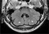
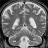
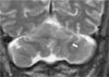
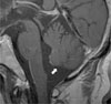
Fig. 1
Axial FLAIR image through the lower pons level. Mildly decreased volume of both cerebellar hemisphere are noted with diffusely multilobulated contour. Folia, fissures and white matter tracts of both cerebellar hemispheres run vertically (arrows) with cortical hypertrophy, resulting irregular corticomedullary junction of the affected cerebellar hemispheres.
References
1. Soto-Ares G, Delmaire C, Deries B, Vallee L, Pruvo JP. Cerebellar cortical dysplasia: MR findings in a complex entity. AJNR Am J Neuroradiol. 2000; 21:1511–1519.
2. Patel S, Barkovich AJ. Analysis and classification of cerebellar malformations. AJNR Am J Neuroradiol. 2002; 23:1074–1087.
3. Sasaki M, Oikawa H, Ehara S, Tamakawa Y, Tohgi H. Disorganised unilateral cerebellar folia: a mild form of cerebellar cortical dysplasia? Neuroradiology. 2001; 43:151–155.
4. Collin P. Embryology and development. In : Lawrence HB, Martin M, Patricia C, Mary D, Julian E, Mark WJF, editors. Gray's anatomy. 38th Ed. London: Churchill Livingstone;1995. p. 238–257.
5. Berry MM, Standring SM, Bannister LH. Nervous system. In : Lawrence HB, Martin M, Patricia C, Mary D, Julian E, Mark WJF, editors. Gray's anatomy. 38th Ed. London: Churchill Livingstone;1995. p. 1027–1065.
6. Demaerel P, Lagae L, Casaer P, Baert AL. MR of cerebellar cortical dysplasia. AJNR Am J Neuroradiol. 1998; 19:984–986.
 XML Download