

 PDF
PDF ePub
ePub Citation
 Print
Print


INTRODUCTION
Periodontal therapy aims at arresting periodontal infections and maintaining a healthy periodontium. This goal can be achieved by eliminating supra- and subgingival plaque and establishing destructive conditions because the essential characteristic in the treatment of periodontal diseases is the mechanical removal of bacterial deposits and calculus [1,2]. The techniques used for scaling, root planning, and curettage are hand instrumentation, sonic and ultrasonic instrumentation, laser scaling, demineralization, and chemical scaling. Currently, hand instruments and sonic and ultrasonic scalers are used most frequently [3]. Ultrasonic and sonic scalers are referred to as power-driven scalers. Ultrasonic and sonic scalers differ in their efficiency in removing calculus from tooth surfaces [4]. Ultrasonic instrumentation is as effective as hand scaling for plaque and calculus removal and the successful healing of diseased periodontal tissues [5,6]. Ultrasound can be produced by magnetostriction or piezoelectricity. Ultrasonic units in dentistry are currently available in two basic types: magnetostrictive and piezoelectric. Their mechanism of action is different. Magnetostrictive units operate between 18 kHz and 45 kHz using flat metal strips in a stack or a metal rod attached to a scaling tip, and the tip movement is elliptical. Piezoelectric units operate in the 25 kHz to 50 kHz range and are reactivated by dimensional changes in the crystals housed within the hand-piece as electricity passes over the surface of the crystals; tip movement is primarily linear in direction [7]. Tooth surface alterations produced using hand or ultrasonic instruments are of particular concern during periodontal therapy. An analysis of the literature on the aggressiveness of magnetostrictive and piezoelectric ultrasonic scaling devices on tooth substances showed varying results. Flemmig et al. [8] suggested that a magnetostrictive unit was more aggressive than a piezoelectric device for root substance removal. On the other hand, Busslinger et al. [9] reported that a piezoelectric device left a rougher surface than a magnetostrictive device after instrumentation. The roughness of the root surface after a scaling procedure is a factor to consider for maintenance because it has also been reported that bacterial plaque adheres easily to the rough root surfaces after treatment [10,11]. A comparison of different piezoelectric or magnetostrictive ultrasonic devices can be expected to produce differences in tooth surface roughness. The purpose of the present study was to compare the results of scaling with the use of magnetostrictive and piezoelectric devices on extracted teeth. Furthermore, the amount of time needed to clean the root surface and the effects of the lateral forces were noted.
MATERIALS AND METHODS
This study was conducted in vitro on 44 human tooth samples extracted due to severe periodontal disease. The tooth samples were evaluated both clinically and radiographically for periodontal disease involvement by expert periodontists. The samples had subgingival calculus, and they were divided into 4 experimental groups with each group containing 11 teeth. There were no significant differences between the initial calculus amounts among the groups.
For all the root experimental surfaces with subgingival calculus, scaling was performed on a 3×5 mm area. This area was selected and separated from other areas using a diamond-coated round bur. During the study, the teeth were stored in a formalin solution to prevent drying.
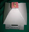
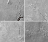
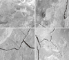
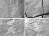
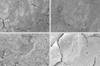
A standardized application of force for each treatment method was achieved by mounting the teeth in a specially pressure sensitive electronic device. The instrumentation with the ultrasonic devices was carried out under water-cooling. To prevent water accumulation over the pressure sensitive electronic device, we modified it and used a metal pyramidal cap. The metal pyramidal cap had a 3×3 cm area on which to mount and glue the teeth (Fig. 1). Before scaling, the extracted teeth were embedded in wax, except the scaling area (3×5 mm), and were fixed at the top of the pyramidal cap, and the lateral forces were calibrated by the operator. In the C100 and C200 groups, the teeth were scaled using a magnetostrictive device with 100 g and 200 g lateral force, respectively. In the P100 and P200 groups, the teeth were scaled with a piezoelectric device with 100 g and 200 g of lateral force, respectively. The magnetostrictive device (Dentin nL90, Isfahan, Faraz mehr, Iran) was used with a number 03 standard tip, and the piezoelectric device (Dentin nL90, Isfahan, Faraz mehr, Iran) was used with a 25 kHz Dentsply hand instrument. In each sample, the teeth were scaled until the area of interest was clean and smooth as determined visually and through tactile exploration with a sharp explorer. A digital stopwatch was used to measure the duration of the scaling procedures.. The tooth surface roughness was analyzed under a scanning electron microscope (SEM). The surfaces were sputtered with gold using a sputtering device for 240 seconds, and the surfaces were evaluated under the SEM (LEO 1450 VT) at 35 kV with a contrast of 2.5 nm at magnifications of ×30, ×200, and ×500. The ×200 magnification was used for comparing the groups to each other (Figs. 2-5). Two calibrated examiners compared the groups.
RESULTS
The scaling time, the time taken to clean the areas of interest, was the shortest in group C200. Other scaling times are shown in Table 1.
The mean scaling time for the magnetostrictive device in the C100 and C200 groups was 41.90 seconds, and for the piezoelectric device used in the P100 and P200 groups it was 50.54 seconds, but this difference was not statistically significant (P=0.171).
The mean scaling time of the C100 and P100 groups was 49.86 seconds, and the mean of the C200 and P200 groups was 42.58 seconds. This difference was not statistically significant (P=0.247).
The tooth surface roughness under SEM showed that the P100 group had a smoother surface than did the C100 group, but this difference was not significant (P=0.2).
The tooth surface roughness under SEM showed that the P200 group (8 of the 11 samples) had a smoother surface than did the C200 group, and this difference was significant (P=0.033).
The tooth surface roughness under SEM showed that the C100 group had a smoother surface than the C200 group, and that the P100 group had a smoother surface than the P200 group, but these differences were not significant (P=0.21 and P=0.66, respectively).
DISCUSSION
Many studies have demonstrated the effectiveness of initial periodontal therapy procedures for the improvement of clinical signs of periodontal diseases. In the present study, the results of the piezoelectric device were significantly smoother than those of the magnetostrictive device. According to a study by Flemming et al. [12], the magnetostrictive ultrasonic scaler that they assessed may be adapted to various clinical needs by adjusting the lateral force, tip angulation, and power setting to maintain its efficacy. Scaling with an ultrasonic instrument on high power has been less effective for calculus removal and surface roughness. In this study, we used the instruments on high power, and we maintained the tip angulation at 0 degrees to decrease this parameter.
The relationship between the force and defect volume caused by ultrasonic instruments was confirmed by Flemming et al. [12], and in this study we chose ultrasonic application forces of 100 g and 200 g. By controlling the force, the relationship between the force and tooth roughness could be evaluated using a digital pressure sensitive device [13]. A SEM was used to evaluate the tooth roughness. The field of scaling was 3×5 mm, and a magnification of ×200 was selected to compare the samples. The selections at this magnification caused an underestimate in our evaluation, but we randomized the selected samples to solve this problem. Several studies have investigated the time needed to reach the therapeutic endpoint of a clean and smooth root surface.
Time for scaling
The area and lateral forces were similar among the samples. This study did not demonstrate any differences between the times required using the magnetostrictive and piezoelectric devices. In contrast to our study, Busslinger et al. [9] compared magnetostrictive and piezoelectric devices and demonstrated that the difference in time required between them was significant. The difference between the two studies may be explained by differences in the power settings and/or other parameters between the devices in the two studies.
Type of devices
The SEM images after instrumentation were used to compare the 4 groups. The tooth surface roughness under SEM showed that the C100 group had a smoother surface than the C200 group and that the P100 group had a smoother surface than the P200 group, but this difference was not significant. Only the comparison between the C200 and P200 groups was significant. In contrast to our study, Santos et al. [13] showed that under SEM, there were no differences between the results of the magnetostrictive and piezoelectric devices. The main reasons for such differences are the methodology used such as an in vitro or in vivo study, the use of different tips, the power setting, and the time and load of the instrument.
In addition, the results were different than those obtained by Busslinger et al. [9], in that the magnetostrictive instrument produced a better surface finish than the piezoelectric manipulation, which likely corresponded to the different tip used.
Lateral forces
Standardization of the experimental conditions with respect to treatment modalities and surface analysis is important for such studies. Many studies have demonstrated that parameters such as the power setting, lateral force, and tip angulation determine the amount of root damage caused by ultrasonic instrumentation. Flemming et al. [12] demonstrated that lateral forces had a great effect in creating tooth roughness under magnetostrictive scaling and had another effect under piezoelectric scaling. Surprisingly, we found this effect in our study, and also found that the lateral forces from 100 g to 200 g caused poorer outcomes using the magnetostrictive device than the piezoelectric instrument.
Times needed to use each of the two instruments were similar, with increased forces creating more damage. We suggest using 100 g for scaling to decrease root and tooth damage and root sensitivity after scaling. In addition, the magnetostrictive device caused a rougher surface than the piezoelectric instrument. We suggest that scaling should be performed under 100 g and with a piezoelectric device.

 XML Download