

 PDF
PDF ePub
ePub Citation
 Print
Print


INTRODUCTION
Cytomegalovirus (CMV) is the most common and clinically significant virus in both adults and children after solid organ transplantation (SOT) [12]. CMV directly invades grafts and various organs, and CMV infections may be related with graft failure and mortality [3]. In addition, CMV infection is thought to indirectly increase the risk of acute rejection, opportunistic infections, and posttransplant lymphoproliferative disorder (PTLD) in adults with liver transplantation (LT) [4567]. In children with SOT, however, data on the direct and indirect effects of CMV remains limited [8].
The incidence of CMV infection generally depends on the CMV serostatus of the recipient and donor. Among adults receiving seropositive grafts, half of the CMV-naive recipients but only less than one-third of seropositive recipients suffer from CMV infection [9]. Recently introduced CMV prophylaxis has significantly decreased the incidence of CMV infection and disease in adults with LT [10]. However, there is little data available on CMV infection after pediatric LT [411121314]. In addition, CMV antibody by passive transfer of maternal CMV antibody is detected in children without effective CMV-specific immunity until 12-18 months after birth [8]. This confounding serostatus in the early age limits the risk analysis of CMV infection after pediatric LT.
Cadaveric organ donation for pediatric LT has not prevailed due to Confucian culture and liver grafts mainly come from the living donor pool, which generally consists of CMV-seropositive parents [15]. Korea is a CMV endemic area [3], which may result in the dominance of CMV-seropositive donors and potential early exposure to CMV after pediatric LT. We hypothesized that transplant children in Korea may have an increased likelihood of acquiring primary CMV infection after LT, and that a preventive strategy after LT should be intensified. However, the potential toxic effects and pharmacokinetics of prolonged antiviral therapy have not been well studied in developing children.
In this study, we regularly monitored CMV DNAemia using a CMV quantitative nucleic acid amplification testing (QNAT) assay and adopted a hydrid strategy for pediatric LT patients, which consisted of universal short-term prophylaxis and preemptive therapy of intravenous ganciclovir (GCV). The aim of the study was to evaluate the outcomes of this hybrid strategy in the prevention of CMV infection in children in the living donor-dominant LT program at a single center.
MATERIALS AND METHODS
Patients
From 2003 to 2012, 159 cases of LT were identified among 150 children <18 years of age with various etiologies at a tertiary hospial. To include only subjects who were immunosuppression-naïve before transplantation, seven children with retransplantation during the study period who had undergone primary LTs before 2003 were excluded. Although our center has been performing LTs since 1994, this specific time period was selected because the CMV QNAT assay and a hybrid strategy to prevent CMV infection were introduced in 2003. Subjects received follow-up for at least 48 months posttransplantation, and subjects who died within 48 months posttransplantation were not excluded. Medical data retrospectively extracted from digital records were as follows: age, gender, indications of LT, preoperative pediatric end-stage liver disease (PELD) score, CMV serostatus of the donor and recipient, immunosuppressants, and antiviral regimens for CMV prophylaxis and preemptive treatment. The study was approved by the institutional review board of Asan Medical Center (IRB no. 2015-0121).
Immunosuppression
Initial immunosuppression consisted of tacrolimus, high-dose steroids, and basiliximab during the study period. The target trough level of tacrolimus during the first 2 weeks was 10-15 ng/mL, while methyl-prednisolone (10 mg/kg) was intraoperatively administered and tapered off to 0.3 mg/kg on day 7. The maintenance of immunosuppression therapies consisted of low dosages of tacrolimus and steroids. Patients received oral tacrolimus with the dosages adjusted to maintain trough levels of 5-10 ng/mL after the first 2 weeks and <5 ng/mL during the next 2 months. Beginning on day 8, patients were also administered oral prednisolone for the first 3 months, starting at 0.3 mg/kg per day. For ABO-incompatible LT patients, the regimen included rituximab, plasma exchange, steroids, and cyclophosphamide. When rejection was histologically diagnosed, methyl-prednisolone pulse therapy (20 mg/kg on day 0, 2, and 4) was given.
Diagnosis of CMV infection and disease
The CMV viral load in peripheral blood was quantified using a commercially available QNAT using an automated nucleic extraction instrument, QIAamp Blood Mini Kit (Qiagen, Valencia, CA, USA), with a positivity cut-off value of more than 250 copies/mL whole blood. According to the International Consensus Guidelines [16], CMV infection was defined as detectable DNAemia on CMV regardless of symptoms. CMV disease was defined as symptomatic CMV infection with attributable symptoms such as fever, malaise, mayalgia, cytopenia, and organ invasion such as gastroenteritis, hepatitis, pneumonitis, and retinitis. Children with such symptoms also underwent Epstein-Barr virus (EBV) QNAT of peripheral blood and multiplex polymerase chain reaction (PCR) tests of nasal secretion for ten viruses such as adenovirus and enterovirus.
Hybrid strategy for CMV DNAemia
The hybrid strategy consisted of 1) universal short-term prophylaxis and 2) universal preemptive treatment of CMV infection. The universal prophylaxis using at least 2-week intravenous GCV (6 mg/kg once a day) was introduced regardless of CMV serostatus of the donor or recipient. Regardless of CMV serostatus of the donor or recipient, initial monitoring by QNAT for CMV DNAemia in all children consisted of weekly follow-ups for the first 2 months, biweekly for the next 3-4 months, and monthly thereafter. When the patients had fever, cytopenia, malaise, elevated liver function tests, and any viral manifestations, CMV DNAemia was measured. The QNAT results were obtained by physicians within 24 hours after the sample was taken.
When CMV DNAemia was noted during follow-up, immunosuppression was reduced regardless of preemptive treatment and DNAemia titer. The cut-off value of triggering preemptive treatment in this study was 1,000 copies/mL in QNAT [17]. Universal preemptive treatment of at least 2 weeks of intravenous GCV (10 mg/kg per a day) was given to all infected children, until detectable CMV DNAemia disappeared during weekly CMV QNAT follow-ups. The same initial monitoring was repeated after preemptive treatment. For asymptomatic patients with CMV titer <1,000 copies/mL in QNAT, preemptive treatments were delayed to allow for spontaneous resolution on the follow-up tests.
Outcome measures and risk stratification
The primary outcomes were incidence of CMV infection and CMV disease in the present study. The secondary outcomes were incidence of indirect effects from CMV infection, including acute/chronic rejection, fungal infection, EBV-related PTLD, loss of graft, and mortality. All rejections were histologically diagnosed based on the international Banff criteria [18]. Fungal infections were defined based on the proposed guidelines of the Centers for Disease Control [19]. PTLD was histologically confirmed. Pretransplant CMV serostatus was determined by CMV immunoglobulin G of enzyme immunoassay to evaluate the CMV serostatus mismatch such as donor-positivity/recipient-negativity (D+/R−).
Statistical analysis
For comparison between groups, chi-square test or Fisher's exact test were used for categorical variables, while the Mann-Whitney U test and Kruskal-Wallis test were used for the comparison of continuous variables. The cumulative survival rate of CMV infection was calculated using the Kaplan-Meier method, and differences in cumulative survival rates were assessed by the log-rank method. All statistical calculations were performed using PASW Statistics ver. 18.0 for Windows (IBM Co., Armonk, NY, USA). A p-value <0.05 was considered statistically significant.
RESULTS
Patient characteristics
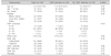
A total of 143 children underwent 143 primary LTs (living grafts, 86.0%; split grafts, 4.9%; deceased whole grafts, 9.1%) at our institution during the study period (Table 1). The median age of the subjects at the time of LT was 18.2 months (range, 4 months to 17 years). The most common indication for pediatric LT was biliary atresia (n=62, 43.4%), followed by acute liver failure (n=51, 35.7%) including fulminant Wilson's disease, liver tumors (n=10, 7.0%), liver cirrhosis (n=9, 6.3%), and others (n=11, 7.7%).
The mean follow-up duration was 91.6±37.7 months (range, 0.4-161 months), and all survivors were followed up for at least 4 years posttransplantation. Nine children died during follow-up, and all of these children died within 7 months after LT with the exception of one patient, who died of renal failure and bacterial infection at 2 years posttransplantation. Among the 143 children, eight children underwent re-LTs due to graft failure.
CMV DNAemia positivity and CMV disease
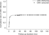
Of the 143 children, 69 (48.3%) experienced CMV infection defined as DNAemia positivity, with a median of 3,300 copies/mL (range, 370-655,500 copies/ mL) at the time of first detection. The median onset of CMV DNAemia was 1.9 months (range, 0.3-15.1 months). A rapid increase in CMV DNAemia was seen until 3 months posttransplantation (Fig. 1). Late-onset CMV DNAemia was noted among 7.2% (n=5/69) children at 12 months posttransplantation. The highest CMV DNAemia positivity was observed in 49.2% (n=60/122) of children in the D+/R+ group, followed by 46.7% (n=7/15) in the D+/R− group and 33.3% in both D−/R+ (n=1/3) and D−/R− (n=1/3) groups. Among the 69 children with CMV infection, 73.9% (n=51/69) were asymptomatic, while 26.1% (n=18/69) experienced symptomatic CMV infections (CMV disease), with the following symptoms noted: elevated liver enzymes (n=9), fever with malaise (n=7), and diarrhea (n=2). CMV disease was observed in 20.0% (n=3/15) of children in the D+/R− group and in 12.3% (n=15/122) in the D+/R+ group. Among children with CMV disease, no invasive organ disease was identified.
Preemptive therapy
Among the 69 children with CMV infection, 43 (62.3%) underwent preemptive therapy of intravenous GCV. Of the 69, 40 children had CMV titer ≥1,000 copies/mL and three had CMV titer <1,000 copies/mL. The three children with initial CMV titer <1,000 copies/mL had persistently DNAemia on the next follow-up and developed elevated liver enzyme levels. The remaining 26 children with CMV titer <1,000 copies/mL experienced spontaneous resolution of their low CMV DNAemia on follow-up tests. The mean duration of intravenous GCV treatment was 23.3±5.4 days. All children who underwent preemptive therapy showed no CMV DNAemia within 1 month after commencement of preemptive therapy, but 26% (n=18/69) of children developed more than two episodes of CMV DNAemia in a median of 2.1 months (range, 1.2-4.2 months). Among these, no patients developed tissue-invasive diseases.
Indirect effects of CMV infection and disease
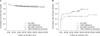
As no tissue-invasive CMV disease developed in this cohort group, no patients died of uncontrolled CMV disease. CMV infection did not affect survival rates of the graft (p=0.701 in log-rank test, Fig. 2A) or patient (p=0.238). Among the 93 children with biopsy-proven acute rejection during the follow-up period, 52 (55.9%) occurred after CMV DNAemia. There was a significant difference in the cumulative rate of acute rejection in patients with CMV infection compared to those with no CMV infection (p=0.027, Fig. 2B). However, no statistical analysis was performed to determine whether a reduction in immunosuppressive therapy or CMV infection induced acute rejection because a reduction in immunosuppression at this center is not standardized for cases of CMV infection in children. There were no differences in the cumulative rates of EBV-related PTLD, chronic rejection, and fungal infections according to CMV infection.
DISCUSSION
This retrospective pediatric study investigated the incidence of CMV infection and the outcomes of hybrid therapy in pediatric LT patients at a single center. CMV burden after pediatric SOT has not been fully elucidated due to limited data resulting from the various heterogeneous methodologies used to measure the incidence of CMV infection, such as monitoring (antigenemia vs. QNAT) and sampling (plasma vs. whole blood). In addition, preventive strategies in SOT (long-term prophylaxis of oral valganciclovir [VGC] and GCV vs. short-term intravenous GCV) affect the burden of CMV. In pediatric LT patients, the incidence of CMV antigenemia is 48-58% in children who do not receive universal prophylaxis [1220]. Although most centers in the United States use CMV QNAT for posttransplant CMV monitoring [21], only two centers have reported the incidence of CMV DNAemia, only one of which described the detailed method of measuring viral loading [411]. In a report from Cincinnati Children's Hospital, no CMV DNAemia was noted during 3 month oral prophylaxis, while late-onset CMV disease was noted in 8-22% of the population. In another report of a hybrid strategy similar to that used in the present study, the incidence of CMV DNAemia (plasma) without symptoms and disease were 34.4% and 9.8%, respectively. The corresponding findings in the present study were similar (35.6% and 12.6%, respectively), although the present study had a longer mean duration of intravenous GCV for prophylaxis and utilized whole blood for the tests. In pediatric kidney transplantation, which accounts for the majority of SOT, asymptomatic DNAemia was reported in 22% of patients who received long-term prophylaxis [22]. Currently, no outcomes of a hybrid strategy utilizing QNAT have been reported in pediatric kidney transplantation patients.
Although short-term prophylaxis was given to all patients in the present study, most episodes of DNAemia developed during the first 3 months posttransplantation, indicating a course similar to the natural course of CMV infection after SOT. This may mean that short-term prophylaxis is ineffective in preventing posttransplant CMV infection. By contrast, no early-onset CMV infection was noted in children who underwent 3-month prophylaxis of oral GCV or VGC [11]. The rationale of our center for the administration of short-term prophylaxis of intravenous GCV is as follows: firstly, it is given to at least minimally protect against CMV during the very early posttransplant period in patients who are under intensified immunosuppression. Secondly, toxicities and pharmacokinetics of long-term antiviral agents in developing children have not been fully studied. Prompt preemptive treatment of intravenous GCV in this cohort was initiated during the first 3 months with regular monitoring of DNAemia. The authors speculate that regular monitoring with QNAT led to early detection of CMV DNAemia before it could progress to a severe condition. However, one-fourth of the infected children experienced recurrence of DNAemia after preemptive therapy, showing that regular monitoring is still needed after preemptive therapy.
The incidence of CMV infection was higher in the D+/R+ group (49%) than the D+/R− group (46%). The risk stratification using the mismatch of CMV serostatus may not be predictive of CMV infection after LT in children. It is because CMV antibody by passive transfer of maternal CMV antibody is detected in children without effective CMV-specific immunity until 12-18 months after birth. This confounding serostatus in the early age limits the risk analysis of CMV infection after pediatric LT. It is well known that infants are at greater risk for CMV and other viral infections after LT, and most guidelines address approaches for risk stratification of infants [823].
Many transplantation centers have their own protocols for CMV prophylaxis based on their experience of CMV and types of immunosuppression [2124]. In addition, optimal selection and duration of the types of CMV prophylaxis and preemptive therapy are unclear in children because data are limited. Current international CMV consensus guidelines introduce three options for children at high risk for CMV: 3-month oral VGC/intravenous GCV, preemptive therapy only, and 2-week intravenous GCV followed by preemptive monitoring (in other words, a hybrid therapy) [8]. Oral VGC is another option for children. However, prophylaxis of oral VGC for pediatric LT is not allowed in Korea due to the inferiority of VGC in preventing CMV in LT patients and lack of pediatric data on the safety and efficacy of such therapy [25]. Patients pay approximately US$ 22 for oral VGC per day under the Korea government medical coverage guidelines, while they can pay approximately only US$ 5 of a total cost of US$ 50 for intravenous GCV per day. Therefore, prophylactic VGC is not strongly recommended both clinically and economically in Korea. Appropriate dosage (5 mg/kg/day vs. 10 mg/kg/day) and duration (2 weeks vs. >2 weeks) of intravenous GCV have not been determined [821]. In our study, 5 mg/kg per day of intravenous GCV for the first 3 weeks posttransplantation was universally administered regardless of CMV risk. Neutropenia was noted among 5 children who received intravenous GCV that complicated their clinical situations, especially in the children with bacterial infections and idiopathic fulminant hepatitis.
A higher incidence of acute rejection was noted among children after exposure to CMV in the present study. CMV infection in adults with SOT is thought to indirectly increase the risk of acute rejection [4567]. A study on pediatric LT also revealed a potential association between CMV infection and acute rejection [4]. However, this is still an area of significant controversy, and the indirect effects of CMV infection posttransplantation have not been well studied in children [8]. For example, in the present study, the routine process before preemptive therapy generally included reduction of immunosuppression, which may actually induce rejection. The degree of immunosuppression reduction was inconsistent, and further analysis was not available in this small population. Regardless of the reason for rejection in the infected children, the authors believe that acute rejection should be scrutinized after CMV infection.
In conclusion, in this study, we report the outcomes of a hybrid strategy to prevent CMV disease in pediatric LT patients. CMV infection still occurred at a similar rate as reported in previous studies. However, the preemptive treatments were effective, and no patient experienced tissue-invasive CMV disease, resulting in no CMV-associated mortality in this study. To use a hybrid strategy for pediatric LT patients, an intensified surveillance protocol by CMV QNAT is recommended for the first 3 months posttransplantation
 XML Download